Disease-Associated Neurotoxic Astrocyte Markers in Alzheimer Disease Based on Integrative Single-Nucleus RNA Sequencing
- PMID: 38345650
- PMCID: PMC10861702
- DOI: 10.1007/s10571-024-01453-w
Disease-Associated Neurotoxic Astrocyte Markers in Alzheimer Disease Based on Integrative Single-Nucleus RNA Sequencing
Abstract
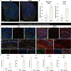
Alzheimer disease (AD) is an irreversible neurodegenerative disease, and astrocytes play a key role in its onset and progression. The aim of this study is to analyze the characteristics of neurotoxic astrocytes and identify novel molecular targets for slowing down the progression of AD. Single-nucleus RNA sequencing (snRNA-seq) data were analyzed from various AD cohorts comprising about 210,654 cells from 53 brain tissue. By integrating snRNA-seq data with bulk RNA-seq data, crucial astrocyte types and genes associated with the prognosis of patients with AD were identified. The expression of neurotoxic astrocyte markers was validated using 5 × FAD and wild-type (WT) mouse models, combined with experiments such as western blot, quantitative real-time PCR (qRT-PCR), and immunofluorescence. A group of neurotoxic astrocytes closely related to AD pathology was identified, which were involved in inflammatory responses and pathways related to neuron survival. Combining snRNA and bulk tissue data, ZEP36L, AEBP1, WWTR1, PHYHD1, DST and RASL12 were identified as toxic astrocyte markers closely related to disease severity, significantly elevated in brain tissues of 5 × FAD mice and primary astrocytes treated with Aβ. Among them, WWTR1 was significantly increased in astrocytes of 5 × FAD mice, driving astrocyte inflammatory responses, and has been identified as an important marker of neurotoxic astrocytes. snRNA-seq analysis reveals the biological functions of neurotoxic astrocytes. Six genes related to AD pathology were identified and validated, among which WWTR1 may be a novel marker of neurotoxic astrocytes.
Keywords: Alzheimer disease; Biomarkers; Bulk RNA-seq; Neurotoxic astrocytes; snRNA-seq.
© 2024. The Author(s).
Conflict of interest statement
The authors declare no competing interests.
Figures


Similar articles
-
Regulatory T cells decrease C3-positive reactive astrocytes in Alzheimer-like pathology.J Neuroinflammation. 2023 Mar 8;20(1):64. doi: 10.1186/s12974-023-02702-3. J Neuroinflammation. 2023. PMID: 36890536 Free PMC article.
-
Astrocytic autophagy plasticity modulates Aβ clearance and cognitive function in Alzheimer's disease.Mol Neurodegener. 2024 Jul 23;19(1):55. doi: 10.1186/s13024-024-00740-w. Mol Neurodegener. 2024. PMID: 39044253 Free PMC article.
-
Quantitative proteomics reveals that PEA15 regulates astroglial Aβ phagocytosis in an Alzheimer's disease mouse model.J Proteomics. 2014 Oct 14;110:45-58. doi: 10.1016/j.jprot.2014.07.028. Epub 2014 Aug 7. J Proteomics. 2014. PMID: 25108202
-
Glial cells in Alzheimer's disease: From neuropathological changes to therapeutic implications.Ageing Res Rev. 2022 Jun;78:101622. doi: 10.1016/j.arr.2022.101622. Epub 2022 Apr 12. Ageing Res Rev. 2022. PMID: 35427810 Review.
-
Comprehensive analyses of brain cell communications based on multiple scRNA-seq and snRNA-seq datasets for revealing novel mechanism in neurodegenerative diseases.CNS Neurosci Ther. 2023 Oct;29(10):2775-2786. doi: 10.1111/cns.14280. Epub 2023 Jun 2. CNS Neurosci Ther. 2023. PMID: 37269061 Free PMC article. Review.
Cited by
-
Comparative Analysis of Human Brain RNA-seq Reveals the Combined Effects of Ferroptosis and Autophagy on Alzheimer's Disease in Multiple Brain Regions.Mol Neurobiol. 2025 May;62(5):6128-6149. doi: 10.1007/s12035-024-04642-2. Epub 2024 Dec 23. Mol Neurobiol. 2025. PMID: 39710824
-
Glial Contribution to the Pathogenesis of Post-Operative Delirium Revealed by Multi-omic Analysis of Brain Tissue from Neurosurgery Patients.bioRxiv [Preprint]. 2025 Mar 14:2025.03.13.643155. doi: 10.1101/2025.03.13.643155. bioRxiv. 2025. PMID: 40161597 Free PMC article. Preprint.
-
Mechanisms of astrocyte aging in reactivity and disease.Mol Neurodegener. 2025 Feb 21;20(1):21. doi: 10.1186/s13024-025-00810-7. Mol Neurodegener. 2025. PMID: 39979986 Free PMC article. Review.
-
The dopamine analogue CA140 alleviates AD pathology, neuroinflammation, and rescues synaptic/cognitive functions by modulating DRD1 signaling or directly binding to Abeta.J Neuroinflammation. 2024 Aug 11;21(1):200. doi: 10.1186/s12974-024-03180-x. J Neuroinflammation. 2024. PMID: 39129007 Free PMC article.
-
Astrocytes phenomics as new druggable targets in healthy aging and Alzheimer's disease progression.Front Cell Neurosci. 2025 Jan 6;18:1512985. doi: 10.3389/fncel.2024.1512985. eCollection 2024. Front Cell Neurosci. 2025. PMID: 39835288 Free PMC article. Review.
References
-
- Andersen JV, Markussen KH, Jakobsen E, Schousboe A, Waagepetersen HS, Rosenberg PA, Aldana BI (2021) Glutamate metabolism and recycling at the excitatory synapse in health and neurodegeneration. Neuropharmacology 196:108719. 10.1016/j.neuropharm.2021.108719 - PubMed
-
- Andl T, Zhou L, Yang K, Kadekaro AL, Zhang Y (2017) YAP and WWTR1: New targets for skin cancer treatment. Cancer Lett 396:30–41. 10.1016/j.canlet.2017.03.001 - PubMed
-
- Balu DT, Pantazopoulos H, Huang CCY, Muszynski K, Harvey TL, Uno Y, Rorabaugh JM, Galloway CR, Botz-Zapp C, Berretta S, Weinshenker D, Coyle JT (2019) Neurotoxic astrocytes express the d-serine synthesizing enzyme, serine racemase, in Alzheimer’s disease. Neurobiol Dis 130:104511. 10.1016/j.nbd.2019.104511 - PMC - PubMed
-
- Barres BA (2008) The mystery and magic of glia: a perspective on their roles in health and disease. Neuron 60(3):430–440. 10.1016/j.neuron.2008.10.013 - PubMed
MeSH terms
Substances
Grants and funding
LinkOut - more resources
Full Text Sources
Medical
Miscellaneous

