

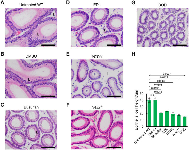
Busulfan administration replicated the characteristics of the epididymal initial segment observed in mice lacking testis-epididymis lumicrine signaling
- PMID: 38346723
- PMCID: PMC11017096
- DOI: 10.1262/jrd.2023-102
Busulfan administration replicated the characteristics of the epididymal initial segment observed in mice lacking testis-epididymis lumicrine signaling
Abstract
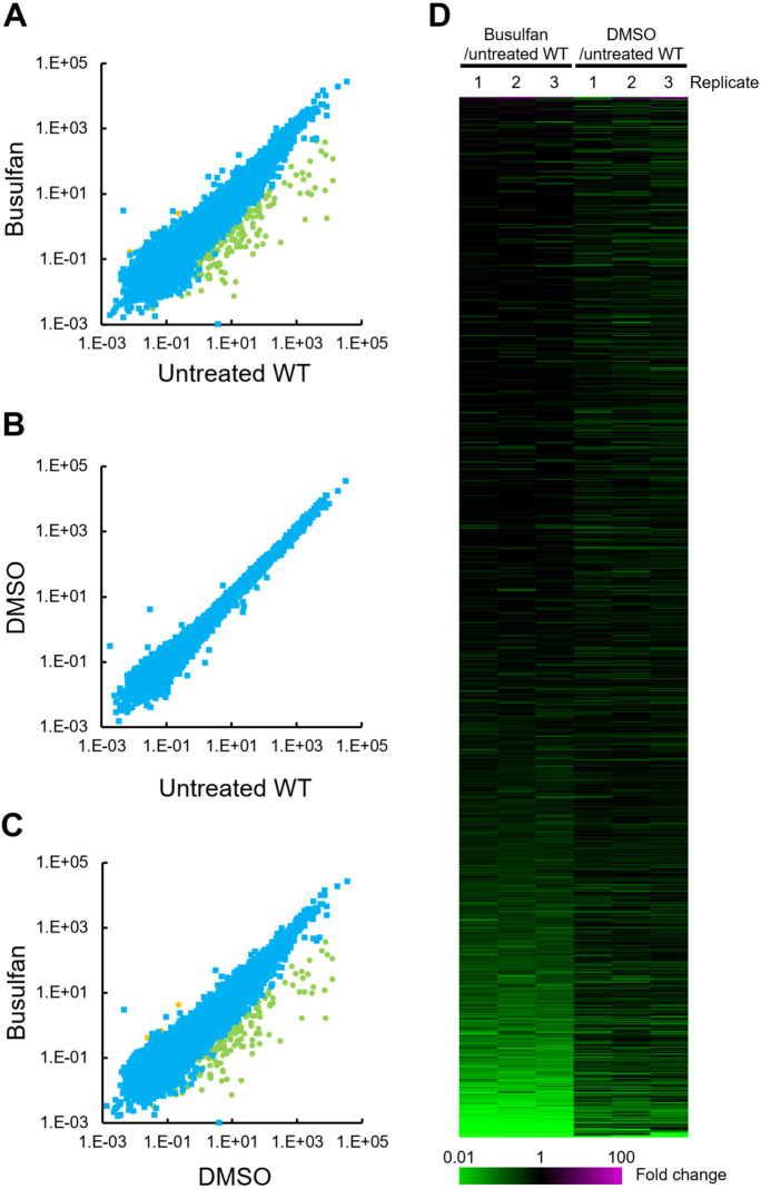
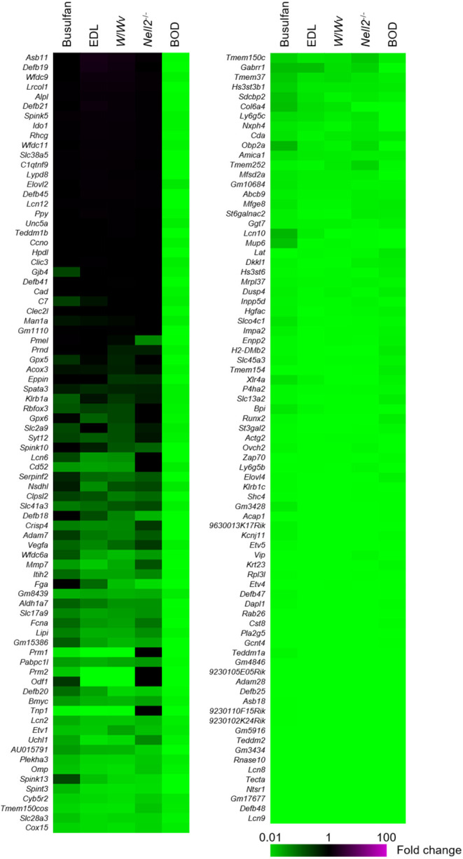
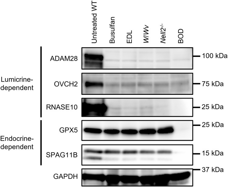
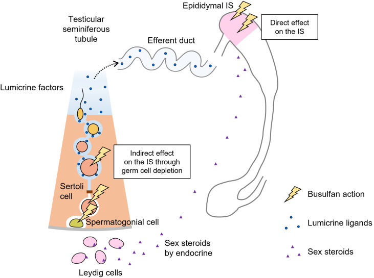
The physiological functions of the mammalian epididymis are typically regulated by the testes. In addition to sex steroids secreted by testicular Leydig cells, which act on the epididymis in an endocrine manner, there is a non-sex-steroidal signaling pathway known as the lumicrine pathway. This lumicrine signaling pathway involves ligand proteins secreted from germ cells within the testicular seminiferous tubules traversing the male reproductive tract, which induce epithelial differentiation in the epididymis. These findings prompted an inquiry into whether treatments influencing testis physiology can disrupt epididymal function by interfering with testis-epididymis communication. Busulfan, an alkylating agent commonly used to deplete testicular germ cells in reproductive biology, has not been sufficiently explored because of its effects on the epididymis. This study investigated the effects of busulfan administration on the proximal epididymis using histological and transcriptomic analyses. Notably, busulfan, as opposed to the vehicle dimethyl sulfoxide (DMSO), altered the morphology of the initial segment of the epididymis, leading to a reduction in the cell height of the luminal epithelium. RNA sequencing identified 185 significantly downregulated genes in the proximal epididymis of busulfan-administered mice compared to DMSO-administered mice. Comparative transcriptome analyses revealed similarities between the epididymal transcriptome of busulfan-administered mice and lumicrine-deficient mice, such as efferent-duct-ligated W/Wv and Nell2-/- mice. However, this differed from that of bilaterally orchidectomized mice, in which both the endocrine and lumicrine signaling pathways were simultaneously ablated. Collectively, these results suggested that the harmful effects of busulfan on the proximal epididymis are secondary consequences of the ablation of testis-epididymis lumicrine signaling.
Keywords: Busulfan; Epididymis; Gene expression; Initial segment; Lumicrine.
Conflict of interest statement
The author declares no competing interests.
Figures
References
-
- Robaire B, Hinton BT, Orgebin-Crist M-C. The epididymis. Knobil and Neill’s Physiology of Reproduction. 2006;1071–148.
-
- O’Hara L, Welsh M, Saunders PTK, Smith LB. Androgen receptor expression in the caput epididymal epithelium is essential for development of the initial segment and epididymal spermatozoa transit. Endocrinology 2011; 152: 718–729. - PubMed