. 2024 Feb 12;15(1):1310.
doi: 10.1038/s41467-024-45632-1.

Poly-γ-glutamylation of biomolecules

Affiliations

Poly-γ-glutamylation of biomolecules

Ghader Bashiri et al. Nat Commun. .

Abstract

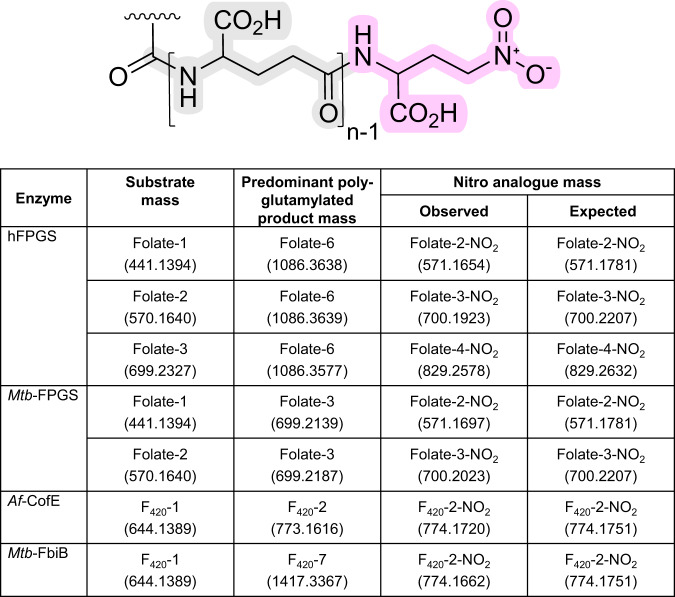
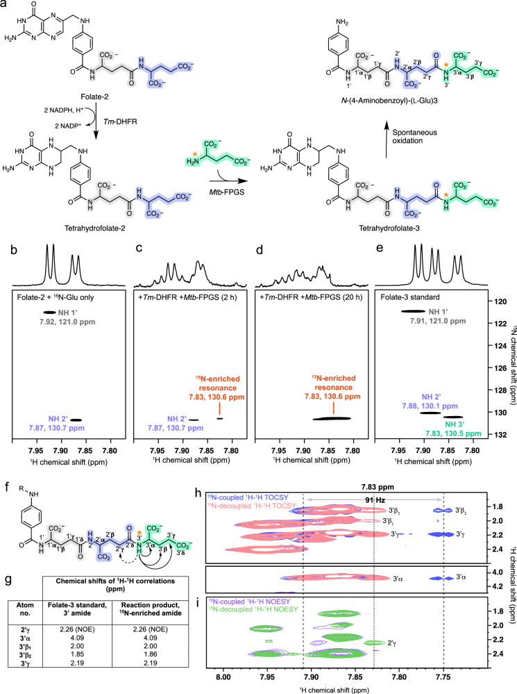
Poly-γ-glutamate tails are a distinctive feature of archaeal, bacterial, and eukaryotic cofactors, including the folates and F420. Despite decades of research, key mechanistic questions remain as to how enzymes successively add glutamates to poly-γ-glutamate chains while maintaining cofactor specificity. Here, we show how poly-γ-glutamylation of folate and F420 by folylpolyglutamate synthases and γ-glutamyl ligases, non-homologous enzymes, occurs via processive addition of L-glutamate onto growing γ-glutamyl chain termini. We further reveal structural snapshots of the archaeal γ-glutamyl ligase (CofE) in action, crucially including a bulged-chain product that shows how the cofactor is retained while successive glutamates are added to the chain terminus. This bulging substrate model of processive poly-γ-glutamylation by terminal extension is arguably ubiquitous in such biopolymerisation reactions, including addition to folates, and demonstrates convergent evolution in diverse species from archaea to humans.


Conflict of interest statement

The authors declare no competing interests.

Figures

Fig. 1
Fig. 1. Cofactor substrates of folylpolyglutamate synthase (folate) or γ-glutamyl ligase (F420) bearing variable length poly-γ-glutamate tails.
The glutamyl residues in green indicate the terminus of the growing tail in both molecules.
Fig. 2
Fig. 2. 4-Nitro-L-2-aminobutyric acid as a chain terminator of poly-γ-glutamylation in folates and F420.
A poly-γ-glutamate tail is shown with variable L-glutamate numbers and terminated by a non-reactive nitro functional group. Calculated masses of nitro-terminated folate and F420 molecules are provided for comparison to experimental results – the central number indicates the total residue count in the tail including the chain terminator residue. The masses take into account functional group protonation within the mass spectrometer operating in positive ion mode and are provided to 4 decimal places to match the precision of the high-resolution LC-MS experiment. The table of data provides the identity and experimentally measured masses of substrate, predominant polyglutamylated product, and nitro-terminated product. Enzymes systems investigated are human FPGS (hFPGS), Mycobacterium tuberculosis FPGS (Mtb-FPGS), Archaeoglobus fulgidus CofE (Af-CofE), and Mycobacterium tuberculosis FbiB (Mtb-FbiB).
Fig. 3
Fig. 3. Mtb-FPGS-dependent glutamylation of folate-2 with 15N-glutamate monitored by NMR spectroscopy.
a Reaction scheme for Mtb-FPGS/Tm-DHFR coupled conversion of folate-2 to tetrahydrofolate-3 and subsequent oxidation to N-(4-aminobenzoyl)-(L-Glu)3. The orange asterisk indicates the 15N-enriched site. Panels b to e show the amide region of 1D 1H and 2D 1H-15N HSQC spectra for samples as follows. b Reaction sample containing folate-2 and 15N-glutamate prior to addition of enzyme. c Reaction sample following a 2 h preincubation with Tm-DHFR, followed by incubation with both Tm-DHFR and Mtb-FPGS for 2 h and d 20 h. e Folate-3 standard. Panels f to i show the results of 1H-1H correlation experiments confirming addition of 15N-glutamate at the terminus of the Mtb-FPGS/Tm-DHFR-catalyzed reaction product. f Arrows indicate through-bond (solid line) and through-space (dashed line) correlations observed between the 15N-labelled amide and other 1H nuclei in the polyglutamate chain, based on folate-3 assignments. g Comparison of the chemical shifts of 1H-1H correlations observed for the 15N-enriched amide and the 3′ glutamyl amide of folate-3. h Overlay of 15N-coupled (blue), and 15N-decoupled (pink) 1H-1H TOCSY experiments for the 15N-enriched amide highlighting correlations with the 3′α, 3′β, and 3′γ protons. i Overlay of 15N-coupled (purple), and 15N-decoupled (green) 1H-1H NOESY experiments for the 15N-enriched amide highlighting a correlation with the 2′γ proton.
Fig. 4
Fig. 4. High-resolution structures of reaction ligands in the active site of Archaeoglobus fulgidus γ-glutamyl ligase CofE.
a An overlay of F420-1 (yellow) and F420-2 (cyan) molecules highlighting the bulge in the tail of F420-2. GTP/GDP (pink stick model), manganese and sodium cations (white spheres) are located adjacent to the terminal glutamyl binding site. b The F420-1 phospholactyl moiety binds in a secondary glutamate binding pocket and its carbonyl closely approaches the γ-phosphate atom of a modelled GTP. c Free L-glutamate binding in the primary glutamate pocket. Extensive hydrogen bonding between the co-substrate and two oxyanion-like holes via α- and γ-carboxylates locks the ligand in place. d The first, basal glutamyl of F420-2 occupies the secondary glutamate pocket and projects the carbonyl oxygen of the following cis-amide bond toward the GTP γ-phosphate atom. Polar interactions are shown as dashed white lines with distances labelled in Å units.
Fig. 5
Fig. 5. Schematic of the proposed poly-γ-glutamylation mechanism in CofE and its homologue FbiB.
Isoalloxazine and primary glutamate binding sites are depicted as bold, dotted lines, with glutamate or glutamyl residues colored green or blue by their order of addition. In STEP 1, F420-0 effects nucleophilic attack on the GTP γ-phosphate to form an acyl intermediate. The phosphate abstracts a proton from the free glutamate amino group and is eliminated from GTP. STEP 2 shows an additional proton abstraction, nucleophilic attack of the glutamate on the acyl phosphate, and the consequent and concerted loss of phosphate as H2PO4. STEP 3 depicts the crystal structure model of F420-1 binding. STEP 4 and STEP 5 show a second cycle of phosphorylation/activation, proton abstraction, nucleophilic attack, and elimination of H2PO4. STEP 6 depicts the crystal structure model of F420-2. STEPS 4–6 highlight the bulge in the growing poly-glutamyl chain. The schematic has been simplified for clarity and multiple binding, bond making, and bond breaking events have been combined – this does not imply a specific order of binding nor concerted reactions.

References

    1. Janke C, Rogowski K, van Dijk J. Polyglutamylation: a fine-regulator of protein function? ‘Protein Modifications: beyond the usual suspects’ review series. EMBO Rep. 2008;9:636–641. doi: 10.1038/embor.2008.114. - DOI - PMC - PubMed
    1. Fox JT, Stover PJ. Folate-mediated one-carbon metabolism. Folic Acid Folates. 2008;79:1–44. doi: 10.1016/S0083-6729(08)00401-9. - DOI - PubMed
    1. Ducker GS, Rabinowitz JD. One-carbon metabolism in health and disease. Cell Metab. 2017;25:27–42. doi: 10.1016/j.cmet.2016.08.009. - DOI - PMC - PubMed
    1. Shane B. Folylpolyglutamate synthesis and role in the regulation of one-carbon metabolism. Vitam. Hormones. 1989;45:263–335. doi: 10.1016/S0083-6729(08)60397-0. - DOI - PubMed
    1. Bashiri G, et al. A revised biosynthetic pathway for the cofactor F420 in prokaryotes. Nat. Commun. 2019;10:1558. doi: 10.1038/s41467-019-09534-x. - DOI - PMC - PubMed