. 2024 Feb 12;14(1):3488.
doi: 10.1038/s41598-024-53941-0.

Significance of melanin distribution in the epidermis for the protective effect against UV light

Affiliations

Significance of melanin distribution in the epidermis for the protective effect against UV light

Daniela F Zamudio Díaz et al. Sci Rep. .

Abstract

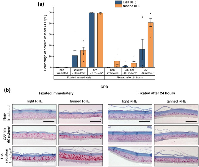
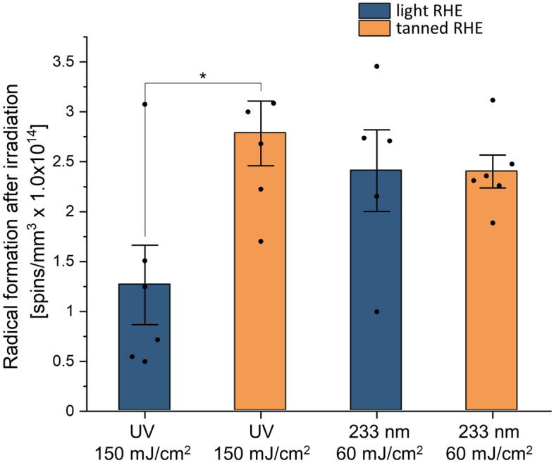
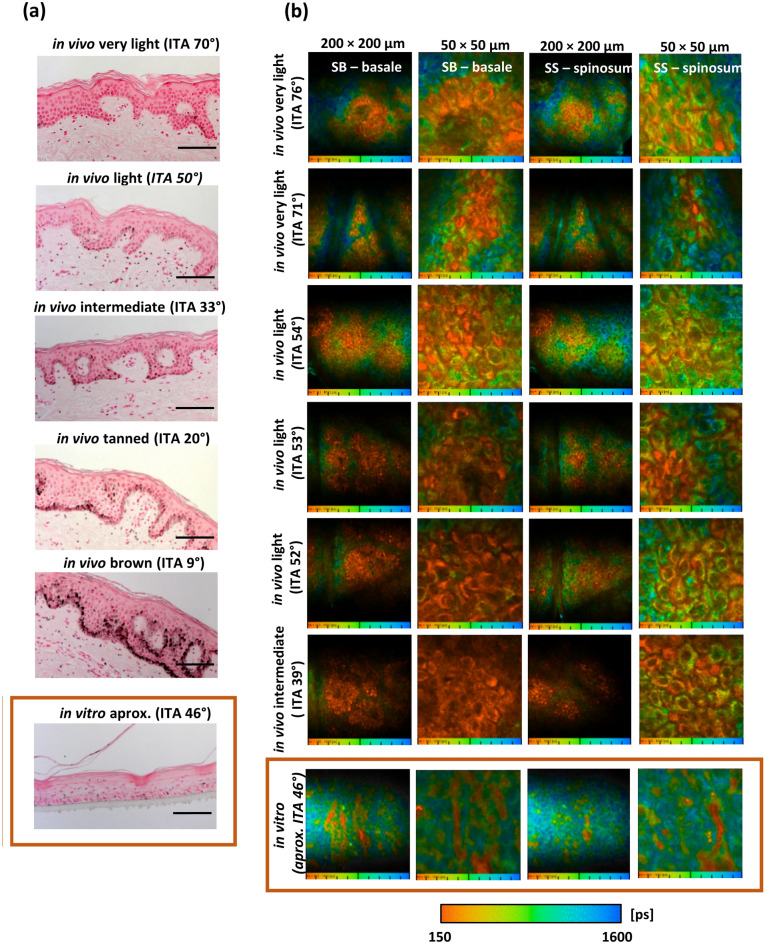
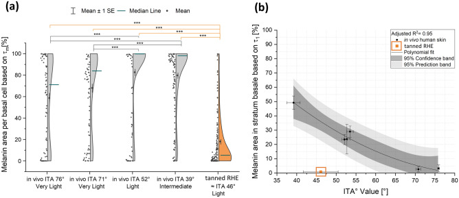
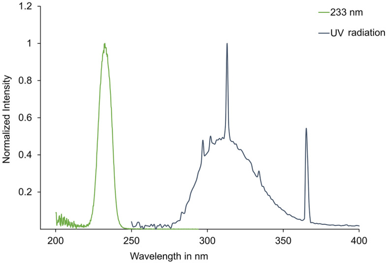
Melanin, the most abundant skin chromophore, is produced by melanocytes and is one of the key components responsible for mediating the skin's response to ultraviolet radiation (UVR). Because of its antioxidant, radical scavenging, and broadband UV absorbing properties, melanin reduces the penetration of UVR into the nuclei of keratinocytes. Despite its long-established photoprotective role, there is evidence that melanin may also induce oxidative DNA damage in keratinocytes after UV exposure and therefore be involved in the development of melanoma. The present work aimed at evaluating the dependence of UV-induced DNA damage on melanin content and distribution, using reconstructed human epidermis (RHE) models. Tanned and light RHE were irradiated with a 233 nm UV-C LED source at 60 mJ/cm2 and a UV lamp at 3 mJ/cm2. Higher UV-mediated free radicals and DNA damage were detected in tanned RHE with significantly higher melanin content than in light RHE. The melanin distribution in the individual models can explain the lack of photoprotection. Fluorescence lifetime-based analysis and Fontana-Masson staining revealed a non-homogeneous distribution and absence of perinuclear melanin in the tanned RHE compared to the in vivo situation in humans. Extracellularly dispersed epidermal melanin interferes with photoprotection of the keratinocytes.


Conflict of interest statement

Karsten R. Mewes, Lars Vierkotten are employees of Henkel AG & Co. KGaA. The authors declare no competing interests.

Figures

Figure 1
Figure 1
Melanin content of ex vivo skin samples and reconstructed human epidermis (RHE). (a) Correlation between skin melanin content [µg melanin/mg epidermis] (after an isolation procedure from ex vivo skin) and skin color measured as ITA [°] from ex vivo skin. The linear regression can be described by the functional equation y =  − 0.068x + 6.460 with R2 = 0.89. An increase in melanin content is reflected in a decrease in ITA° value. A statistically significant relationship exists between skin melanin content and ITA° as determined by ANOVA test (p < 0.05). Plot of linear regressions with respective 95% confidence band and prediction band. Data represents mean ± standard deviation. (b) Melanin content [µg epidermis/mg epidermis] after isolation procedure from RHE. An estimate of skin color according to the ITA° value classification system was calculated using the linear regression between melanin content and ITA° for ex vivo human skin. Tanned RHE achieved significantly higher melanin content compared to light RHE (Student’s t test), ***p < 0.001. For each ex vivo skin sample (n = 13 skin donors) n = 5 epidermis samples were analyzed (a), n = 5 for light RHE, and n = 16 tanned RHE (b). (c) Macroscopic view of tanned and light RHE after 15 days of culture at air–liquid interphase.
Figure 2
Figure 2
UV-induced cyclobutane pyrimidine dimers (CPD) in reconstructed human epidermis (RHE). (a) DNA damage, as percentage of cells with CPD formation, of light RHE (blue) and tanned RHE (orange), determined by immunofluorescence, fixated directly and 24 h after UV irradiation (233 nm–60 mJ/cm2 and UV—3 mJ/cm2). The non-irradiated sample serves as negative control and shows no damage when the samples were immediately fixated. Irradiation with 3 mJ/cm2 of UV induced the most DNA damage, significantly higher than damage after irradiation with 60 mJ/cm2 of 233 nm. Assessed immediately and 24 h post-irradiation, there is a reduction in CPD formation. Tanned RHE showed higher, although not significant, DNA damage compared to light RHE when fixated immediately after 233 nm irradiation and when fixating 24 h independent of the applied irradiation. Data of n = 6 biopsies (n = 3 models for each type of RHE). Data represents mean ± SEM. (b) Representative images of immunohistochemical detection of CPD positive cells in light and tanned RHE. Positive cells are stained in dark red. Samples irradiated with UV—3 mJ/cm2 resulted in stronger damage throughout the whole epidermis than those irradiated with 233 nm–60 mJ/cm2, where damage was concentrated on the uppermost layer of the epidermis. Scale bar: 100 μm.
Figure 3
Figure 3
UV-induced free radical formation in reconstructed human epidermis (RHE). Radical formation directly after irradiation with 150 mJ/cm2 of UV radiation and 60 mJ/cm2 of far UV-C at 233 nm in light and tanned RHE. Data show the spin concentration (spins/mm3 × 1014) measured by electron paramagnetic resonance (EPR) spectroscopy. Significantly higher radical formation is induced in tanned RHE after UV irradiation compared to light RHE, while the formation after far UV-C exposure was similar for both types of RHEs (one way ANOVA with Bonferroni correction for post hoc test). n = 6 biopsies (n = 3 for each type of RHE). Data represents mean ± SEM, *p < 0.05.
Figure 4
Figure 4
Epidermal melanin distribution by Fontana–Masson staining and two-photon excited fluorescence lifetime imaging (TPE-FLIM). (a) Representative images of Fontana–Masson staining of human skin (in vivo) and tanned RHE (in vitro). An example of each skin type is shown with the corresponding Individual Typology Angle (ITA°): “very light” skin, “light” skin, “intermediate” skin, “tanned” skin and “brown” skin. Melanin content increases with lower ITA°. Independent of the skin type, melanin in vivo is located perinuclear within basal cells, whereas melanin in tanned RHE remains extracellular. Scale bar: 100 μm. (b) TPE-FLIM (transversely) of in vivo human skin and tanned RHE. Melanin distribution in the stratum basale (SB) and the stratum spinosum (SS) is visualized continuously in false colors for lifetimes between 150 (orange) and 1600 ps (blue). Melanin-containing cells (stained orange) were masked by the characteristic short fluorescence mean lifetime of melanin (Tm < 480 ps). Both general (200 × 200 µm) and magnified (50 × 50 µm) images were acquired. The distribution of melanin in different skin types is shown with the corresponding individual typological angle (ITA°) and contrasted with the distribution of melanin in tanned RHE. In in vivo human skin, melanin-containing cells are concentrated in the basal cells and increase with decreasing ITA°. In volunteers with lighter skin, an increase in non-melanized (stained blue) or moderately melanized cells was evident, although a homogeneous distribution was maintained, shown in the images as a smooth transition between lower mean lifetime τm and higher mean lifetime τm. In contrast, in tanned RHEs, melanin is concentrated in a limited number of extracellular sites, while most cells do not contain melanin. n = 6 spots were acquired from ventral forearms of volunteers with “very light” (n = 2), “light” (n = 3) and “intermediate” skin (n = 1) and from tanned RHE (n = 3).
Figure 5
Figure 5
Intracellular and stratum basale melanin distribution using multiphoton FLIM analysis. (a) Modulation of intracellular melanin distribution in basal cells with skin color ITA° classification system for in vivo human skin and tanned RHE. Intracellular melanin distribution, the cell area (excluding nuclei) covered by melanin in each defined basal cell (extracted from magnified FLIM images at 50 × 50 µm, Fig. 4) was evaluated for volunteers with “very light” (n = 2), “light” (n = 1) and “intermediate” skin (n = 1) and for tanned RHE (n = 3). Melanin mask of τm < 480 ps. Regardless of skin type, melanin in vivo is homogeneously distributed: while darker skin types contain a high melanin area in most cells, lighter skin expresses a lower melanin area, but melanin is still present in most basal cells. In contrast, in tanned RHEs, the pigment is concentrated in a limited number of extracellular sites, while most cells do not exhibit melanin. Significant differences were found between the distribution of melanin in RHE and in all the human skin samples analyzed (Kruskal–Wallis-Test with Bonferroni correction), ***p < 0.001. (b) Correlation between melanin area in stratum basale [%] and skin color measured as ITA° [°] from measurements on in vivo human skin and tanned RHE. Melanin area in stratum basale is defined as the ratio between melanin pixels to stratum basale pixels (extracted from FLIM images at 200 × 200 µm, Fig. 4b). Melanin mask of τ1 < 150 ps and a1 [%] > 92%. The melanin area was evaluated for volunteers with “very light” (n = 2), “light” (n = 3) and “intermediate” skin (n = 1) and from tanned RHE (n = 3). A statistically significant relationship exists between melanin area and ITA° value as determined by ANOVA test (p < 0.05) and can be described by a second order polynomial fit with intercept = 164.09, B1 = -3.76 and B2 = 0.02, and R2 = 0.95. Plot of polynomial regression with respective 95% confidence band and prediction band. Data represents mean ± standard error of the mean.
Figure 6
Figure 6
Emission spectra of applied UV light sources. The normalized spectra are shown for 233 nm (green), broadband UV-radiation (blue).

Similar articles

Cited by

References

    1. Nguyen NT, Fisher DE. MITF and UV responses in skin: From pigmentation to addiction. Pigment Cell Melanoma Res. 2019 doi: 10.1111/pcmr.12726. - DOI - PMC - PubMed
    1. Fajuyigbe D, Douki T, van Dijk A, Sarkany RPE, Young AR. Dark cyclobutane pyrimidine dimers are formed in the epidermis of Fitzpatrick skin types I/II and VI in vivo after exposure to solar-simulated radiation. Pigment Cell Melanoma Res. 2021 doi: 10.1111/pcmr.12956. - DOI - PubMed
    1. Svobodová A, Vostálová J. Solar radiation induced skin damage: Review of protective and preventive options. Int. J. Radiat. Biol. 2010;86:999–1030. doi: 10.3109/09553002.2010.501842. - DOI - PubMed
    1. Tran TNT, Schulman J, Fisher DE. UV and pigmentation: Molecular mechanisms and social controversies. Pigment Cell Melanoma Res. 2008;21:509–516. doi: 10.1111/j.1755-148X.2008.00498.x. - DOI - PMC - PubMed
    1. Cadet J, Douki T. Formation of UV-induced DNA damage contributing to skin cancer development. Photochem. Photobiol. Sci. 2018;17:1816–1841. doi: 10.1039/c7pp00395a. - DOI - PubMed