

Stable Levels of Antibodies Against Unrelated Toxoid Vaccines After COVID-19: COVID-19 Infection Does Not Affect Toxoid Vaccine Antibody Levels
- PMID: 38347963
- PMCID: PMC10860543
- DOI: 10.20411/pai.v8i2.627
Stable Levels of Antibodies Against Unrelated Toxoid Vaccines After COVID-19: COVID-19 Infection Does Not Affect Toxoid Vaccine Antibody Levels
Abstract
Background: Lymphopenia is common in COVID-19. This has raised concerns that COVID-19 could affect the immune system akin to measles infection, which causes immune amnesia and a reduction in protective antibodies.
Methods: We recruited COVID-19 patients (n = 59) in Helsinki, Finland, and collected plasma samples on 2 to 3 occasions during and after infection. We measured IgG antibodies to diphtheria toxin, tetanus toxoid, and pertussis toxin, along with total IgG, SARS-CoV-2 spike protein IgG, and neutralizing antibodies. We also surveyed the participants for up to 17 months for long-term impaired olfaction as a proxy for prolonged post-acute COVID-19 symptoms.
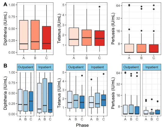
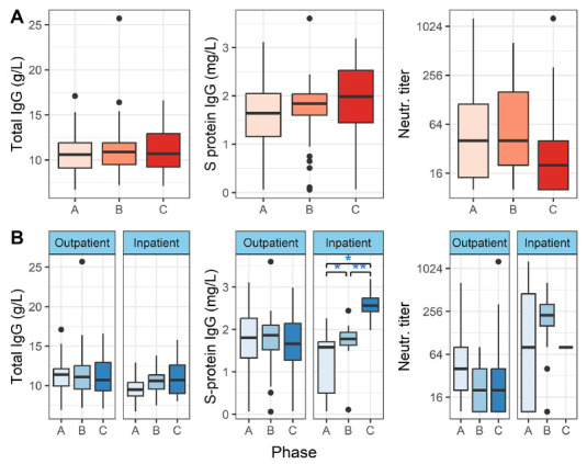
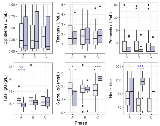
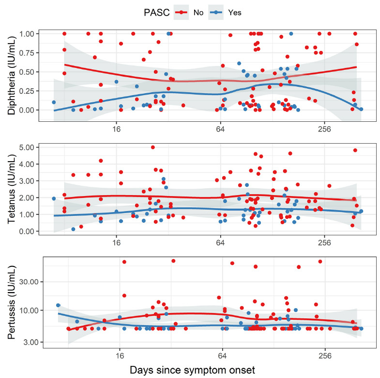
Results: No significant differences were found in the unrelated vaccine responses while the serological response against COVID-19 was appropriate. During the acute phase of the disease, the SARSCoV-2 IgG levels were lower in outpatients when compared to inpatients. SARS-CoV-2 serology kinetics matched expectations. In the acute phase, anti-tetanus and anti-diphtheria IgG levels were lower in patients with prolonged impaired olfaction during follow up than in those without.
Conclusions: We could not detect significant decline in overall humoral immunity during or after COVID-19 infection. In severe COVID-19, there appears to be a temporary decline in total IgG levels.
Keywords: Antibodies; COVID-19; Humoral; Immunity; Post-acute COVID-19 syndrome; Vaccines.
Copyright © 2024 Pathogens and Immunity.
Conflict of interest statement
All authors declare that they have no conflicts of interest.
Figures
Similar articles
-
The long term vaccine-induced anti-SARS-CoV-2 immune response is impaired in quantity and quality under TNFα blockade.J Med Virol. 2022 Dec;94(12):5780-5789. doi: 10.1002/jmv.28063. Epub 2022 Aug 19. J Med Virol. 2022. PMID: 35945627 Free PMC article.
-
Real-world serological responses to extended-interval and heterologous COVID-19 mRNA vaccination in frail, older people (UNCoVER): an interim report from a prospective observational cohort study.Lancet Healthy Longev. 2022 Mar;3(3):e166-e175. doi: 10.1016/S2666-7568(22)00012-5. Epub 2022 Feb 23. Lancet Healthy Longev. 2022. PMID: 35224524 Free PMC article.
-
Seroprevalence of antibodies against diphtheria, tetanus, and pertussis among healthy Thai adolescents.Int J Infect Dis. 2020 Jul;96:422-430. doi: 10.1016/j.ijid.2020.04.088. Epub 2020 May 6. Int J Infect Dis. 2020. PMID: 32387447
-
Heterologous boosting of nonrelated toxoid immunity during acute Puumala hantavirus infection.Vaccine. 2021 Mar 26;39(13):1818-1825. doi: 10.1016/j.vaccine.2021.02.046. Epub 2021 Mar 4. Vaccine. 2021. PMID: 33678453
-
IgG Anti-Spike Antibodies and Surrogate Neutralizing Antibody Levels Decline Faster 3 to 10 Months After BNT162b2 Vaccination Than After SARS-CoV-2 Infection in Healthcare Workers.Front Immunol. 2022 Jun 15;13:909910. doi: 10.3389/fimmu.2022.909910. eCollection 2022. Front Immunol. 2022. PMID: 35784321 Free PMC article.
Cited by
-
Whole Blood as a Sample Matrix in Homogeneous Time-Resolved Assay-Förster Resonance Energy Transfer-Based Antibody Detection.Diagnostics (Basel). 2024 Mar 29;14(7):720. doi: 10.3390/diagnostics14070720. Diagnostics (Basel). 2024. PMID: 38611633 Free PMC article.
References
-
- WHO Coronavirus (COVID-19) Dashboard. Accessed May 17, 2022. https://covid19.who.int/
-
- Al-Saadi EAKD, Abdulnabi MA. Hematological changes associated with COVID-19 infection. J Clin Lab Anal. 2022;36(1):e24064. doi: 10.1002/jcla.24064; PMCID: PMC8646489. - DOI - PMC - PubMed
-
- Wilk AJ, Rustagi A, Zhao NQ, Roque J, Martínez-Colón GJ, McKechnie JL, Ivison GT, Ranganath T, Vergara R, Hollis T, Simpson LJ, Grant P, Subramanian A, Rogers AJ, Blish CA. A single-cell atlas of the peripheral immune response in patients with severe COVID-19. Nat Med. 2020;26(7):1070–6. doi: 10.1038/s41591-020-0944-y; PMCID: PMC7382903. - DOI - PMC - PubMed
-
- Mina MJ, Kula T, Leng Y, Li M, de Vries RD, Knip M, Siljander H, Rewers M, Choy DF, Wilson MS, Larman HB, Nelson AN, Griffin DE, de Swart RL, Elledge SJ. Measles virus infection diminishes preexisting antibodies that offer protection from other pathogens. Science. 2019;366(6465):599–606. doi: 10.1126/science.aay6485; PMCID: PMC8590458. - DOI - PMC - PubMed
-
- Petrova VN, Sawatsky B, Han AX, Laksono BM, Walz L, Parker E, Pieper K, Anderson CA, de Vries RD, Lanzavecchia A, Kellam P, von Messling V, de Swart RL, Russell CA. Incomplete genetic reconstitution of B cell pools contributes to prolonged immuno-suppression after measles. Sci Immunol. 2019;4(41). doi: 10.1126/sciimmunol.aay6125. - DOI - PubMed
LinkOut - more resources
Full Text Sources
Miscellaneous