Listeria monocytogenes Brain Abscess Presenting With Stroke-Like Symptoms: A Case Report
- PMID: 38347999
- PMCID: PMC10859722
- DOI: 10.7759/cureus.52216
Listeria monocytogenes Brain Abscess Presenting With Stroke-Like Symptoms: A Case Report
Abstract
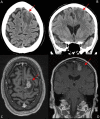
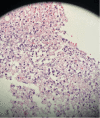
We present a case of Listeria monocytogenes brain abscess in an immunocompromised patient admitted for stroke-like symptoms of headache and aphasia. Computerized tomography of the head revealed a 1.7 x 1.3 cm left frontal lobe lesion with surrounding edema, secondary to stroke, tumor, or abscess. Magnetic resonance imaging brain revealed a ring-enhancing lesion and a small contralateral area of restricted diffusion. Two of the two blood cultures grew an organism identified as L. monocytogenes using matrix-assisted laser desorption ionization time-of-flight mass spectrometry. Treatment with ampicillin and trimethoprim-sulfa yielded marked symptomatic improvement. A brain biopsy was consistent with bacterial abscess. The patient's clinical course was favorable, with improved aphasia and negative follow-up blood cultures. A literature review found a limited number of L. monocytogenes abscess cases and none had clear guidelines for diagnosis. Recent studies have proposed five criteria for diagnosis. Our patient fulfilled three of these proposed guidelines.
Keywords: brain abscess; case report; diagnosis; listeria; treatment.
Copyright © 2024, Dragomir et al.
Conflict of interest statement
The authors have declared that no competing interests exist.
Figures


References
-
- Making sense of the biodiversity and virulence of Listeria monocytogenes. Disson O, Moura A, Lecuit M. Trends Microbiol. 2021;29:811–822. - PubMed
-
- Listeria monocytogenes infections: presentation, diagnosis and treatment. Valenti M, Ranganathan N, Moore LS, Hughes S. Br J Hosp Med (Lond) 2021;82:1–6. - PubMed
-
- What is your diagnosis? Cerebrospinal fluid from a goat. Lanier CJ, Fish EJ, Stockler JW, Newcomer BW, Koehler JW. Vet Clin Pathol. 2019;48:358–360. - PubMed
-
- Listeriosis. Lorber B. Clin Infect Dis. 1997;24:1. - PubMed
Publication types
LinkOut - more resources
Full Text Sources
