. 2024 Jan 29:15:1340467.
doi: 10.3389/fimmu.2024.1340467. eCollection 2024.

Roles of tumor necrosis factor-like ligand 1A in γδT-cell activation and psoriasis pathogenesis

Affiliations

Roles of tumor necrosis factor-like ligand 1A in γδT-cell activation and psoriasis pathogenesis

Shangyi Wang et al. Front Immunol. .

Abstract

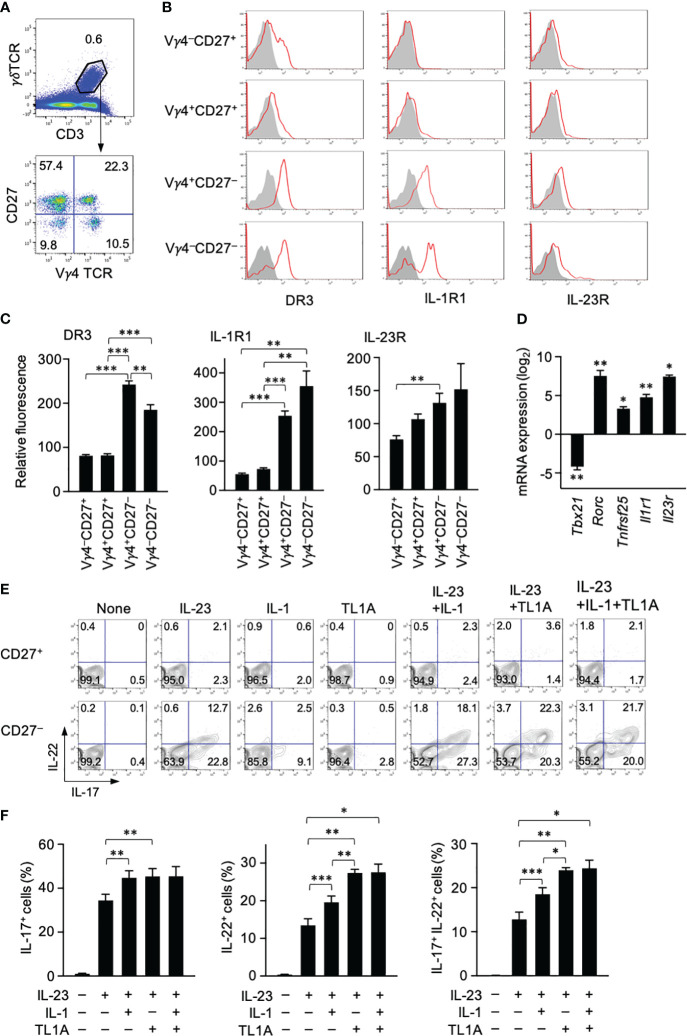
Background: Interleukin (IL)-17-producing γδT (γδT17) cells mediate inflammatory responses in barrier tissues. Dysregulated γδT17 cell activation can lead to the overproduction of IL-17 and IL-22 and the development of inflammatory diseases, including psoriasis. IL-23 and IL-1β are known to synergistically activate γδT17 cells, but the regulatory mechanisms of γδT17 cells have not been fully elucidated. This study aimed to reveal the contribution of the inflammatory cytokine tumor necrosis factor-like ligand 1A (TL1A) to γδT17 cell activation and psoriasis development.

Methods: Anti-TL1A antibody was injected into an imiquimod (IMQ)-induced murine psoriasis model. TL1A receptor expression was analyzed in splenic and dermal γδT cells. γδT cells were tested for cytokine production in vitro and in vivo under stimulation with IL-23, IL-1β, and TL1A. TL1A was applied to a psoriasis model induced by intradermal IL-23 injection. Mice deficient in γδT cells were intradermally injected with IL-23 plus TL1A to verify the contribution of TL1A-dependent γδT-cell activation to psoriasis development.

Results: Neutralization of TL1A attenuated γδT17 cell activation in IMQ-treated skin. TL1A induced cytokine production by splenic γδT17 cells in synergy with IL-23. Dermal γδT17 cells constitutively expressed a TL1A receptor at high levels and vigorously produced IL-22 upon intradermal IL-23 and TL1A injection but not IL-23 alone. TL1A exacerbated the dermal symptoms induced by IL-23 injection in wild-type but not in γδT cell-deficient mice.

Conclusion: These findings suggest a novel regulatory mechanism of γδT cells through TL1A and its involvement in psoriasis pathogenesis as a possible therapeutic target.

Keywords: IL-17; IL-22; TL1A; cytokine; psoriasis; γδ T cells.


Conflict of interest statement

The authors declare that the research was conducted in the absence of any commercial or financial relationships that could be construed as a potential conflict of interest.

Figures

Figure 1
Figure 1
Imiquimod (IMQ) treatment activates dermal γδT cells through TL1A. (A–D) Mouse ears were smeared with IMQ cream or control Vaseline for 3 consecutive days. One day after the last treatment, single-cell suspensions from ears were analyzed for cytokine production (n = 5). (A, B) Frequencies of cytokine-producing cells in the CD45+ gate. (C, D) TCR usage of IL-17+ and IL-22+ lymphocytes in IMQ-treated mice. DN, double negative. (E–I) Ears were smeared with IMQ for 3 (E, F) or 4 (G–I) consecutive days. Anti-TL1A antibodies were injected from day −1 until the day before the analysis. (E, F) CD3intγδTCRint cells from ears on day 3 were analyzed for cytokine production (n = 5). (G) Ear swelling was calculated as changes in thickness from day 0 (IMQ-treated n = 6; Vaseline-treated n = 3). (H) H&E staining sections were prepared from ears on day 4. Scale bar, 100 μm. (I) Epidermal keratinocyte layers and epidermal and dermal thicknesses were measured in the sections. Representative of three (A, C, E) and two (H) independent experiments. Accumulated from three (B, D, F) and two (G, I) independent experiments. Error bars, mean ± standard error (SE). *P < 0.05; **P < 0.01; ***P < 0.001.
Figure 2
Figure 2
TL1A activates splenic γδT17 cells synergistically with IL-23. (A) CD3+γδTCR+ cells were subdivided into four fractions based on CD27 and Vγ4 expression. (B) Expression of DR3, type I IL-1 receptor (IL1-R), and IL-23R in indicated γδT-cell subsets. (C) Expression levels of cytokine receptors in each γδT-cell subset are shown as relative fluorescence intensities normalized with the mean fluorescence intensity in total γδT cells defined as 100 (n = 6). (D) CD27+ and CD27 γδT cells isolated by sorting were analyzed by qPCR for the expression of the indicated genes (n = 3). mRNA expression in CD27 γδT cells is shown as relative levels in CD27+ γδT cells defined as 1. The statistical significance of the expression levels between CD27+ and CD27 cells is shown by asterisks. (E, F) Spleen cells were cultured with the indicated cytokines for 24 (h) A protein transport inhibitor was added for the last 4 (h) IL-17 and IL-22 production was analyzed by intracellular staining. (E) Representative flow cytometry profiles of γδT cells at the end of culture. (F) Frequencies of IL-17+, IL-22+, and IL-17+IL-22+ γδT cells (n = 5). Representative of three (A, B) and four (E) independent experiments. Cumulative results from three (C, D) and four (F) independent experiments. Error bars, mean ± SE. *P < 0.05; **P < 0.01; ***P < 0.001.
Figure 3
Figure 3
Dermal γδT cells are ready to respond to IL-23 and TL1A. (A) CD45+ gate in single-cell suspensions from ears contained CD3hiγδTCRhi dendritic epidermal T cells (DETCs) and CD3intγδTCRint conventional dermal γδT cells. CD3intγδTCRint dermal γδT cells were divided into Vγ4+ and Vγ4 cells. (B) Expression of DR3, type I IL-1R, and IL-23R in DETCs and dermal γδT cells. (C) Expression levels of cytokine receptors in Vγ4+ and Vγ4 γδT cells are shown as relative fluorescence intensities normalized with the mean fluorescence intensity in DETCs defined as 100 (n = 5). (D, E) Mouse ears were intradermally injected with IL-23 alone or combined with TL1A. Twenty-two hours later, DETCs and γδT cells were analyzed for IL-17 (D) and IL-22 (E) by intracellular staining (n = 7 per group). Representative data of three independent experiments (A, B). Cumulative results from four (C–E) independent experiments. Error bars, mean ± SE. *P < 0.05; **P < 0.01; ***P < 0.001.
Figure 4
Figure 4
TL1A exacerbates symptoms in the IL-23-induced murine psoriasis model. Mouse ears were intradermally injected with PBS, IL-23 alone, or the combination of IL-23 and TL1A for 4 consecutive days from days 0 to 3. (A) Thickness of ears was monitored (n = 4). Swelling was calculated as changes in thickness before the treatments. (B–E) H&E staining sections were prepared on day 4. (B) Representative view with low magnification. Scale bar, 100 μm. (C) Epidermal layers of keratinocytes and epidermal and dermal thicknesses were measured (n = 6). (D) Magnified view of the epidermis. Scale bar, 50 μm. (E) Number of microabscesses per section (n = 6). nd, not detected. (F) Immunohistochemical analysis. Scale bar, 25 μm (top), 50 μm (middle), and 100 μm (bottom). (G) PCNA+ and Gr1+ cells were counted in epidermal and dermal areas, respectively (n = 6). Cumulative results from two (A) and three (C, E, G) independent experiments. Representative data of three independent experiments (B, D, F). Error bars, mean ± SE. *P < 0.05; **P < 0.01; ***P < 0.001.
Figure 5
Figure 5
Involvement of TL1A-mediated γδT-cell activation in early psoriasis. Mouse ears were intradermally injected with indicated cytokines. One day later, ears were harvested. (A–D) Single-cell suspensions were prepared for flow cytometry analysis (n = 7). (A, B) Frequencies of cytokine-producing cells in the CD45+ gate. (C, D) IL-17+ and IL-22+ lymphocytes were analyzed for CD3 and γδTCR expression. (E–G) Comparison of wild-type and Tcrd-knockout mice. (E) Ear thickness was measured before and a day after the treatments (n = 4–5). (F, G) Histological analysis of H&E sections. (F) Representative views from mice injected with IL-23 plus TL1A. Scale bar, 50 μm. (G) Epidermal layers and epidermal and dermal thicknesses (n = 4–5). (H) Ear samples were subjected to qPCR analysis (n = 4–6). mRNA expression is shown relative to the average values in wild-type mice injected with IL-23 alone defined as 1. Representative of two (F) and four (A, C) independent experiments. Accumulated from two (E, G, H) and four (B, D) independent experiments. Error bars, mean ± SE. *P < 0.05; **P < 0.01; ***P < 0.001; ns, not significant.

Similar articles

Cited by

References

    1. Ribot JC, Debarros A, Pang DJ, Neves JF, Peperzak V, Roberts SJ, et al. . CD27 is a thymic determinant of the balance between interferon-γ-and interleukin 17–producing γδ T cell subsets. Nat Immunol (2009) 10:427–36. doi: 10.1038/ni.1717 - DOI - PMC - PubMed
    1. Chien Y, Meyer C, Bonneville M. γδ T cells: first line of defense and beyond. Annu Rev Immunol (2014) 32:121–55. doi: 10.1146/annurev-immunol-032713-120216 - DOI - PubMed
    1. Jensen KDC, Su X, Shin S, Li L, Youssef S, Yamasaki S, et al. . Thymic selection determines γδ T cell effector fate: antigen-naive cells make interleukin-17 and antigen-experienced cells make interferon γ. Immunity (2008) 29:90–100. doi: 10.1016/j.immuni.2008.04.022 - DOI - PMC - PubMed
    1. Haas JD, González FHM, Schmitz S, Chennupati V, Föhse L, Kremmer E, et al. . CCR6 and NK1. 1 distinguish between IL-17A and IFN-γ-producing γδ effector T cells. Eur J Immunol (2009) 39:3488–97. doi: 10.1002/eji.200939922 - DOI - PubMed
    1. Cua DJ, Tato CM. Innate IL-17-producing cells: the sentinels of the immune system. Nat Rev Immunol (2010) 10:479–89. doi: 10.1038/nri2800 - DOI - PubMed

Publication types