Review
. 2024 Jan 29:11:1327227.
doi: 10.3389/fcell.2023.1327227. eCollection 2023.

Species-specific roles of the Notch ligands, receptors, and targets orchestrating the signaling landscape of the segmentation clock

Affiliations
Review

Species-specific roles of the Notch ligands, receptors, and targets orchestrating the signaling landscape of the segmentation clock

Pranav S Ramesh et al. Front Cell Dev Biol. .

Abstract

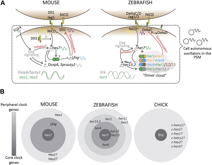
Somitogenesis is a hallmark feature of all vertebrates and some invertebrate species that involves the periodic formation of block-like structures called somites. Somites are transient embryonic segments that eventually establish the entire vertebral column. A highly conserved molecular oscillator called the segmentation clock underlies this periodic event and the pace of this clock regulates the pace of somite formation. Although conserved signaling pathways govern the clock in most vertebrates, the mechanisms underlying the species-specific divergence in various clock characteristics remain elusive. For example, the segmentation clock in classical model species such as zebrafish, chick, and mouse embryos tick with a periodicity of ∼30, ∼90, and ∼120 min respectively. This enables them to form the species-specific number of vertebrae during their overall timespan of somitogenesis. Here, we perform a systematic review of the species-specific features of the segmentation clock with a keen focus on mouse embryos. We perform this review using three different perspectives: Notch-responsive clock genes, ligand-receptor dynamics, and synchronization between neighboring oscillators. We further review reports that use non-classical model organisms and in vitro model systems that complement our current understanding of the segmentation clock. Our review highlights the importance of comparative developmental biology to further our understanding of this essential developmental process.

Keywords: Notch signailing pathway; gene oscillation; presomitic mesoderm (PSM); segmentation clock; somite; somitogenesis.


Conflict of interest statement

The authors declare that the research was conducted in the absence of any commercial or financial relationships that could be construed as a potential conflict of interest.

Figures

FIGURE 1
FIGURE 1
Overview of the Delta-Notch signaling pathway depicting trans-activation and cis-inhibition. Trans-activation is initiated when the DSL domain of the Delta/Serrate/Jagged ligands binds to the extracellular domain of Notch receptors. This initiates two crucial cleavages-the S2 cleavage by the ADAM protease; and subsequently an S3 cleavage by gamma-secretase. The two cleavages result in the production of free NICD that translocates into the nucleus to initiate transcription of target genes. Notch receptors, with are also a target of Notch signaling are subject to a wide range of post-translational modifications in the trans ER-Golgi space by Protein O-Fucosyltransferase (POFUT), Protein O-glucosyltransferase (POGLUT), Lunitic fringe (Lfng), etc., which subsequently affect the type of ligand-receptor interactions and downstream signaling. Illustrations adapted and modified from (Zhou et al., 2022).
FIGURE 2
FIGURE 2
A molecular prepattern underlies the specification of PSM cells from the tail bud (A). Inversion of the unsegmented chick PSM results in this tissue forming somites in the opposite direction (B). An explant of chick segmental plate will form 10 somites irrespective of the site/timing of cutting (C). A smaller mouse embryo will form shorter somites, but maintain the mouse-specific somite number. Illustrations adapted and modified from (Pourquié, 2004).
FIGURE 3
FIGURE 3
Schematic representation of the ‘Clock-and-wavefront’ model that shows the existence of a clock travelling from the posterior to the anterior PSM (in grey) coupled with a regressing wavefront (in yellow) which interact precisely with another T-box TF, Tbx6 (in cyan) to specify anterior PSM cells to differentiate into a somite. S-1: unsegmented PSM that will form the next somite; S0- The somite that is being specified; S1- The most recently formed somite; S2- The earliest formed somite that has completed rostro-caudal patterning (blue-red gradient). Illustrations adapted and modified from (Pourquié, 2011).
FIGURE 4
FIGURE 4
Clustal-based phylogenetic tree of mice (m) and zebrafish (z) Hes1/5/7 orthologs. Amino acid sequences of mHes7 (NP_149030.2), mHes5 (NP_034549.1), mHes1 (P35428) zher1 (Q90463), zher7 (Q9I9K1), zher11 (Q6W4T8), zher2 (Q90464), zher4 (Q90466), zher12 (Q6TA36), zher15 (Q7T3J0), and zher6 (Q6PBX3) were first aligned in the FASTA format using MUltiple Sequence Comparison by Log- Expectation (MUSCLE). The FASTA alignment file was input into the EMBL-EBI Simple Phylogeny tool. The tree was generated in a Clustal format with distance corrections and a UPGMA (Unweighted Pair Group Method with Arithmetic Mean) clustering method. The phylogenetic tree depicts the separation of the Hes1, Hes5, and Hes7 clusters. The symbol beside each gene shows their expression dynamics in the PSM, which is unclear specifically for her6 as it is expressed only during the 3-5 somite stage during fish segmentation.
FIGURE 5
FIGURE 5
Complexities beyond the clock and wavefront (A). The regressing Wnt/FGF-based gradients from the posterior to anterior PSM specify the determination front, however, targets of these signaling pathways (green-Wnt; yellow-FGF) oscillate in the PSM, with changing phase relationships to Notch signaling targets (grey) (B). The period and the phase of cyclic gene oscillations change along the length of the PSM (C). The period of the segmentation clock changes temporally as somitogenesis proceeds (D). The anterior and posterior body segmentation are plausibly regulated by separate mechanisms. Illustrations adapted and modified from (Schröter et al., 2008; Lauschke et al., 2012).
FIGURE 6
FIGURE 6
Transcriptional landscape of the segmentation clock. (A) A simplified schematic of the complex regulatory landscape underlying the mouse and zebrafish segmentation clocks. (B) “Core” and “Peripheral” bHLH genes and Lfng, amongst many others, occupy this regulatory landscape. Based on mutant phenotypes, these clock genes regulate the segmentation clock to varying degrees. Zebrafish consists of many more her/hey genes as a part of its segmentation clock, as compared to mouse or chick, plausibly making the zebrafish clock more robust to genetic or environmental perturbations compared to other species. Finally, we know very little about similar core vs. peripheral clock genes in other species including higher vertebrates like humans, resulting in a knowledge gap that needs to be studied to fully understand this biological clock.
FIGURE 7
FIGURE 7
Species-specific differences in clock dynamics and Notch mutant phenotypes. Elevated levels of the Notch signaling pathway opposingly affect the mouse and zebrafish clock periods, and as a result, the final number of somites/vertebrae formed (Upper panel). Species-specific differences in the requirement of Delta ligands during embryonic development. Mouse-red; zebrafish-blue (Lower panel)..

Similar articles

Cited by

References

    1. Acar M., Jafar-Nejad H., Takeuchi H., Rajan A., Ibrani D., Rana N. A., et al. (2008). Rumi is a CAP10 domain glycosyltransferase that modifies Notch and is required for Notch signaling. Cell 132, 247–258. 10.1016/J.CELL.2007.12.016 - DOI - PMC - PubMed
    1. Akiyama R., Masuda M., Tsuge S., Bessho Y., Matsui T. (2014). An anterior limit of FGF/Erk signal activity marks the earliest future somite boundary in zebrafish. Development 141, 1104–1109. 10.1242/DEV.098905 - DOI - PubMed
    1. Alpert T., Herzel L., Neugebauer K. M. (2017). Perfect timing: splicing and transcription rates in livingcells. Wiley Interdiscip. Rev. RNA 8. 10.1002/WRNA.1401 - DOI - PMC - PubMed
    1. Anderson M. J., Magidson V., Kageyama R., Lewandoski M. (2020). Fgf4 maintains Hes7 levels critical for normal somite segmentation clock function. Elife 9, e55608–e55622. 10.7554/ELIFE.55608 - DOI - PMC - PubMed
    1. Andrawes M. B., Xu X., Liu H., Ficarro S. B., Marto J. A., Aster J. C., et al. (2013). Intrinsic selectivity of notch 1 for delta-like 4 over delta-like 1. J. Biol. Chem. 288, 25477–25489. 10.1074/jbc.M113.454850 - DOI - PMC - PubMed