. 2024 Jan 29:16:1353562.
doi: 10.3389/fnmol.2023.1353562. eCollection 2023.

Human iPSC-derived neurons reveal NMDAR-independent dysfunction following HIV-associated insults

Affiliations

Human iPSC-derived neurons reveal NMDAR-independent dysfunction following HIV-associated insults

Alexander Starr et al. Front Mol Neurosci. .

Abstract

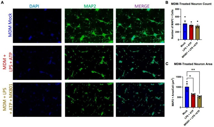
The central nervous system encounters a number of challenges following HIV infection, leading to increased risk for a collection of neurocognitive symptoms clinically classified as HIV-associated neurocognitive disorders (HAND). Studies attempting to identify causal mechanisms and potential therapeutic interventions have historically relied on primary rodent neurons, but a number of recent reports take advantage of iPSC-derived neurons in order to study these mechanisms in a readily reproducible, human model. We found that iPSC-derived neurons differentiated via an inducible neurogenin-2 transcription factor were resistant to gross toxicity from a number of HIV-associated insults previously reported to be toxic in rodent models, including HIV-infected myeloid cell supernatants and the integrase inhibitor antiretroviral drug, elvitegravir. Further examination of these cultures revealed robust resistance to NMDA receptor-mediated toxicity. We then performed a comparative analysis of iPSC neurons exposed to integrase inhibitors and activated microglial supernatants to study sub-cytotoxic alterations in micro electrode array (MEA)-measured neuronal activity and gene expression, identifying extracellular matrix interaction/morphogenesis as the most consistently altered pathways across HIV-associated insults. These findings illustrate that HIV-associated insults dysregulate human neuronal activity and organization even in the absence of gross NMDA-mediated neurotoxicity, which has important implications on the effects of these insults in neurodevelopment and on the interpretation of primary vs. iPSC in vitro neuronal studies.

Keywords: HIV; NMDA; antiretroviral therapy; iPSC; microglia; neuron.


Conflict of interest statement

The authors declare that the research was conducted in the absence of any commercial or financial relationships that could be construed as a potential conflict of interest.

Figures

FIGURE 1
FIGURE 1
Human i3 cortical neurons are resistant to cytotoxicity from HIV-ADA-infected myeloid cell supernatants. (A) Representative images of i3 neurons following 96-h exposure to HIV-iMg supernatants (n = 3 donors, mean ± s.e.m.) and (D) HIV-MDM supernatants (n = 5 donors, mean ± s.e.m.) (B,E) Quantification of average number of MAP2+ cells and (C,F) average MAP2+ area per cell.
FIGURE 2
FIGURE 2
LPS + ATP-treated myeloid cell supernatants reduce neuronal area in an NMDAR-independent manor. (A) Representative images of i3 neurons following 96-h exposure to LPS-MDM supernatants (n = 4 donors, mean ± s.e.m., One-way ANOVA, *p < 0.05, **p < 0.01). (B) Quantification of average number of MAP2+ cells and (C) average MAP2+ area per cell.
FIGURE 3
FIGURE 3
Human i3 neurons are resistant to NMDA-mediated cytotoxicity. (A) Representative images of i3 neurons following 24-h exposure to NMDA (n = 3 neuronal differentiations, mean ± s.e.m.). (B) Quantification of average number of MAP2+ cells and (C) average MAP2+ area per cell. (D) RNA-Seq read counts per million of glutamate receptor subunit genes expressed by i3 neurons with and without iMg supernatant supplement (crossed out box = not detected, minimum CPM cutoff = 5).
FIGURE 4
FIGURE 4
Integrase inhibitors elvitegravir and raltegravir are not cytotoxic to i3 neurons. (A) Representative images of i3 neurons following 9 days of exposure to 3 doses of elvitegravir (EVG) or (B) raltegravir (RAL, n = 3 neuronal differentiations mean ± s.e.m). (C,D) Quantification of average number of MAP2+ cells. Staining for Beta III tubulin (Red) is present in merged images but is not part of the analysis.
FIGURE 5
FIGURE 5
Elvitegravir and raltegravir differentially alter i3 neuron spontaneous activity. MEA analysis of i3 neurons throughout 9 days of exposure to 3 doses of elvitegravir [EVG, (A–C)] or raltegravir [RAL, (D–F), n = 3 neuronal differentiations, mean ± s.e.m, one-way ANOVA, *p < 0.05]). (A,D) Viability and cell area as measured by conductance. (B,E) Spontaneous firing rate and rate normalized to total cellular material. (C,F) Synchronous activity as measured by number of local and network burst events.
FIGURE 6
FIGURE 6
Integrase inhibitors similarly alter i3 neuron gene expression. Analysis of significantly differentially expressed genes (DEGs) from RNA-seq on neurons post-MEA analysis. (A) Clustered heatmap of log2 normalized counts per million for top 30 most significant DEGs between vehicle and 1 μM elvitegravir and (B) 1 μM raltegravir. (C) Top 10 enriched KEGG pathways containing significant DEGs induced by EVG and (E) RAL. (D) Venn diagram showing commonalities among significant DEGs induced by integrase inhibitors.
FIGURE 7
FIGURE 7
LPS-iMg supernatants alter i3 neuron activity and transcription. MEA analysis of i3 neurons throughout 4 days of exposure to supernatants from LPS + ATP treated iMg (n = 3 neuronal differentiations, mean ± s.e.m, one-way ANOVA with Dunnett’s correction for multiple comparisons, *p < 0.05). (A) Viability and cell area as measured by conductance. (B) Spontaneous firing rate and rate normalized to total cellular material. (C) Synchronous activity as measured by number of local and network burst events. (D) Clustered heatmap of log2 normalized counts per million for top 30 most significant DEGs between neurons exposed to iMg and LPS-iMg supernatants. (E) Top 10 enriched KEGG pathways containing significant DEGs induced by LPS-iMg supernatants.

Similar articles

Cited by

References

    1. Akay C., Cooper M., Odeleye A., Jensen B. K., White M. G., Vassoler F., et al. (2014). Antiretroviral drugs induce oxidative stress and neuronal damage in the central nervous system. J. Neurovirol. 20 39–53. - PMC - PubMed
    1. Akay C., Lindl K. A., Shyam N., Nabet B., Goenaga-Vazquez Y., Ruzbarsky J., et al. (2012). Activation status of integrated stress response pathways in neurones and astrocytes of HIV-associated neurocognitive disorders (HAND) cortex. Neuropathol. Appl. Neurobiol. 38 175–200. 10.1111/j.1365-2990.2011.01215.x - DOI - PMC - PubMed
    1. Alakkas A., Ellis R. J., Watson C. W., Umlauf A., Heaton R. K., Letendre S., et al. (2019). White matter damage, neuroinflammation, and neuronal integrity in HAND. J. Neurovirol. 25 32–41. - PMC - PubMed
    1. Alvarez-Carbonell D., Ye F., Ramanath N., Garcia-Mesa Y., Knapp P. E., Hauser K. F., et al. (2019). Cross-talk between microglia and neurons regulates HIV latency. PLoS Pathog. 15:e1008249. 10.1371/journal.ppat.1008249 - DOI - PMC - PubMed
    1. Ambrosius B., Gold R., Chan A., Faissner S. (2019). Antineuroinflammatory drugs in HIV-associated neurocognitive disorders as potential therapy. Neurol. Neuroimmunol. Neuroinflamm. 6:e551. 10.1212/NXI.0000000000000551 - DOI - PMC - PubMed