. 2023 Oct;6(10):969-977.
doi: 10.1038/s41929-023-01031-1. Epub 2023 Oct 2.

Coupled catalytic states and the role of metal coordination in Cas9

Affiliations

Coupled catalytic states and the role of metal coordination in Cas9

Anuska Das et al. Nat Catal. 2023 Oct.

Abstract

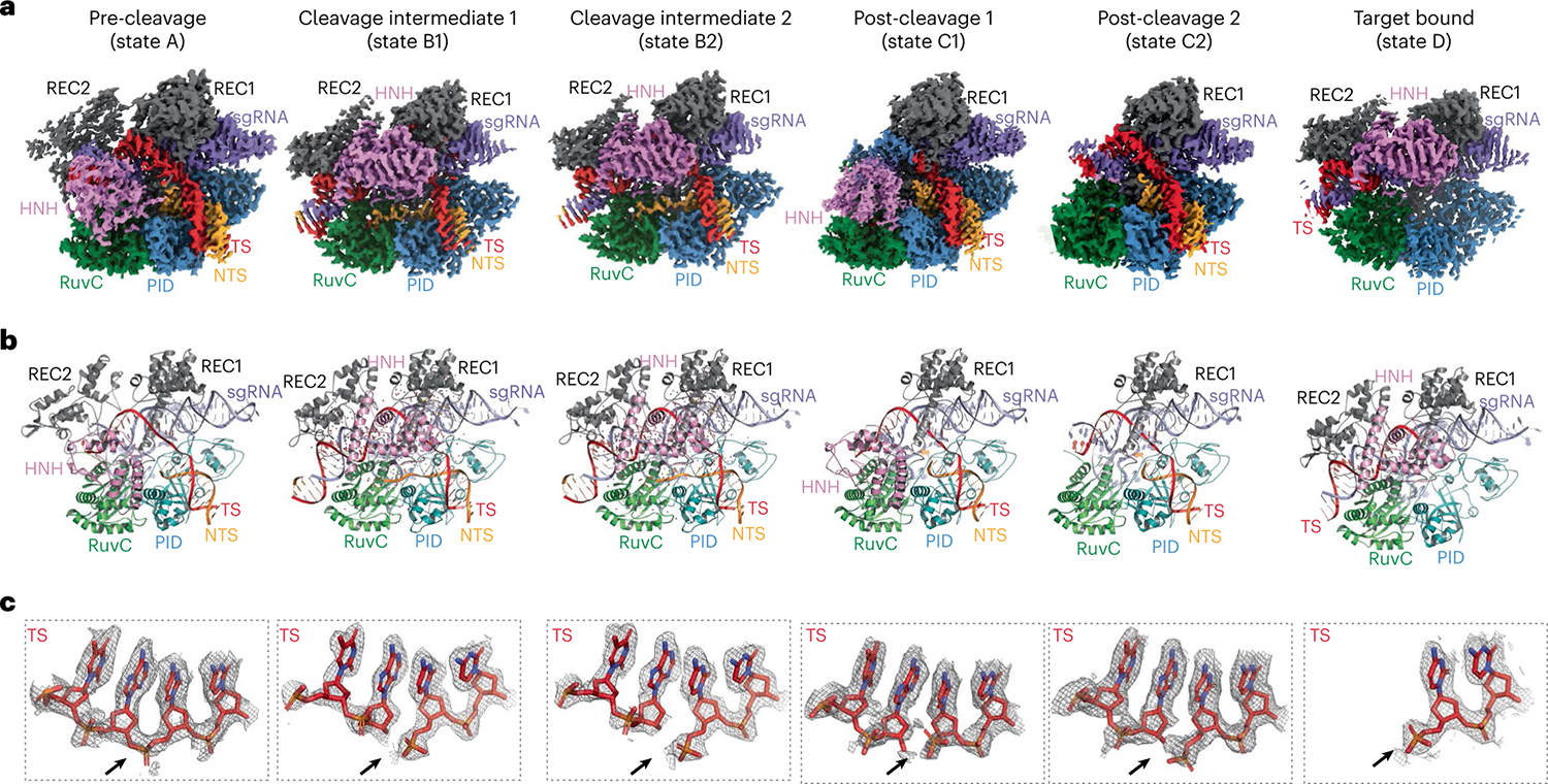
Controlling the activity of the CRISPR-Cas9 system is essential to its safe adoption for clinical and research applications. Although the conformational dynamics of Cas9 are known to control its enzymatic activity, details of how Cas9 influences the catalytic processes at both nuclease domains remain elusive. Here we report five cryo-electron microscopy structures of the active Acidothermus cellulolyticus Cas9 complex along the reaction path at 2.2-2.9 Å resolution. We observed that a large movement in one nuclease domain, triggered by the cognate DNA, results in noticeable changes in the active site of the other domain that is required for metal coordination and catalysis. Furthermore, the conformations synchronize the reaction intermediates, enabling coupled cutting of the two DNA strands. Consistent with the roles of conformations in organizing the active sites, adjustments to the metal-coordination residues lead to altered metal specificity of A. cellulolyticus Cas9 and commonly used Streptococcus pyogenes Cas9 in cells.


Figures

Fig. 1 |
Fig. 1 |. AceCas9 and its metal dependence.
a, Top: domain organization of AceCas9 shown as coloured blocks in the direction from the N terminus to the C terminus. The regions corresponding to the structural domains are coloured and labelled, and the relevant residues are labelled. RuvC-I–RuvC-III, discontinuous segments of the RuvC domain; BH, bridge helix; REC1, nucleic acid-recognition domain 1; REC2, nucleic acid-recognition domain 2; HNH, HNH nuclease domain; PID, PAM interaction domain. Bottom: schematic diagram of the nucleic acids used in this study, shown as nucleotides in the predicted secondary structures. Cleavage sites for the NTS DNA by the RuvC domain and the TS DNA by the HNH domain are indicated by the green and purple downtriangles, respectively. The PAM and the guide region are highlighted in grey.The TS and NTS are numbered sequentially with NTS numbers denoted with asterisks. b, Overlay of the gel filtration profiles of the AceCas9 protein and its ribonucleoprotein (RNP) complex assembled with the sgRNA shown in a. Samples collected for biochemistry and cryo-EM analysis are highlighted by the grey shaded area. c, Cleavage results of double-stranded DNA (dsDNA) assembled with either TS DNA labelled with hexachlorofluorescein (HEX) (red) or the NTS oligonucleotide labelled with fluorescein amidites (FAM) (green) at 10 nM by AceCas9 or its catalytic mutants at 1 μM in the presence of various divalent ions at 10 mM. WT, wild-type AceCas9; U, uncleaved DNA substrate; C cleaved DNA substrate; dHNH, AceCas9 with deactivated HNH; dRuvC, AceCas9 with deactivated RuvC.
Fig. 2 |
Fig. 2 |. The observed cryo-EM structures of six reaction states.
a, Electron potential density maps corresponding to the pre-cleavage (A), cleavage intermediates (B1 and B2), post-cleavage (C1 and C2) and target-bound (D) states. RuvC, RuvC nuclease domain. b, Cartoon representations of the structural models corresponding to the maps in a, where each domain or nucleic acid molecule is labelled in the same colour. c, Close-up views of the cleavage site of the TS DNA overlaid with the density for each of the corresponding states shown in a and b. The arrow indicates the position of the scissile phosphate bond.
Fig. 3 |
Fig. 3 |. HNH conformation-linked changes in the RuvC active centre.
Domain conformations are compared when the sgRNA between the two compared complexes is aligned. In a,c, for the two compared AceCas9 structures the red lines indicate the pair-wise displacement of Cα atoms within the HNH domain, and the green lines indicate the pair-wise displacement of Cα atoms within the REC2 domain. a, Comparison between the pre-cleavage structure (state A) and the catalytic intermediate structure (state B1). Curved arrows indicate the overall rotations of the HNH (red) and REC2 (green) domains from state A to state B1. The two-fold rotation symbol (in black) indicates the location of the axis of the two-fold rotation for the HNH domain. b, Comparison of β4 and Glu516 of the RuvC domain (shown as a stick model) between the pre-cleavage structure (state A, coloured in grey and pink) and the catalytic intermediate structure (state B1, coloured in green and pink). Metal ions A and B from intermediate state B1 are shown as gold spheres. c, Comparison between the two cleavage intermediate structures (states B1 and B2). Curved arrows indicate the overall rotations of the HNH (red) and REC2 (green) domains from state B1 to state B2. d, Comparison of β4 and Glu516 of the RuvC domain between the two catalytic intermediates (state B1, coloured in green and pink, and state B2, coloured in grey). Metal ions A and B from intermediate state B1 are shown as gold spheres, and those from B2 are shown as grey spheres. Dashed lines indicate the distance between the two metal ions in the two different states, as labelled.
Fig. 4 |
Fig. 4 |. Structural changes of metal coordination and their impact on activity.
ad, Density and structures of the HNH catalytic centre of the intermediate B1 (a), the RuvC catalytic centre of the intermediate B1 (b), the HNH catalytic centre of the intermediate B2 (c) and the RuvC catalytic centre of the intermediate B2 (d). Density is shown via the grey mesh. Protein residues and DNA nucleotides are shown as coloured stick models; metal ions are shown as gold spheres; and water molecules are shown as red spheres. The insets in dashed boxes show close-up views of the metal-coordination environment, in which grey dotted lines indicate coordination distances of 1.7–2.2 Å and red dotted lines indicate the distances notably varied between the B1 and B2 intermediates. DNA residues are labelled by their names and corresponding numbers. e, In vitro DNA-cleavage activities on a dsDNA oligonucleotide substrate by the His750Asn (H750N) and His750Asp (H750D) variants in the presence of various divalent ions. The NTS DNA is labelled with the FAM fluorescence probe (green dot) at its 5′ end. The stick models represent putative structures of the mutated residues (teal) in comparison with wild-type His750 (grey). f, Comparison of DNA plasmid cleavage activities of AceCas9 (WT), H750N and H750D in the presence of Mg2+ or Mn2+ ions. RNP (500 nM) and DNA plasmid (6 nM) were used, and the reactions were incubated for 15 min at 50 °C. N, nicked; L, linearized; S, supercoiled plasmid DNA. g, Cell survival assay results of the H750N (for AceCas9) and H983N (for SpyCas9) variants in the presence of Mg2+ or Mn2+ ions. The Val709 to Ala mutation (V709A) of AceCas9 and the Lys918 to Asn mutation (K918N) of SpyCas9 are catalytically enhanced (CE) variants of the two Cas9 proteins and were used to assist stronger survival in non-optimal targeting (Supplementary Fig. 7). Raw images showing the formation of colonies are included in Supplementary Fig. 7. Survival rates are calculated from ratios of the colony-forming units on the arabinose–chloramphenicol plates to those of the chloramphenicol-only plates, as shown in Supplementary Fig. 3. Each experiment was performed in triplicate and the data are plotted with error bars that show the standard deviation. For AceCas9, n = 2 biologically independent trials were examined. For SpyCas9, n = 3 biologically independent trials were examined. Individual rates of survival are plotted as open circles, and the vertical bars show the mean ± standard deviation.
Fig. 5 |
Fig. 5 |. AceCas9 reaction scheme derived from the observed reactive states.
From left to right, the correctly placed HNH domain from the OPEN (state A) to the CLOSED conformation initiates catalysis that begins by nucleophilic attack, followed by cleavage intermediates and, ultimately, relaxation back to the OPEN conformation (states C1 and C2). The same colouring scheme for domains and nucleic acids as that in Figs. 1 and 2 is used for the cartoon. The use of wild-type enzyme under reaction-prone conditions prevented capture of the substrate–enzyme complex, although two intermediates (states B1 and B2), as shown in square brackets, were trapped immediately after nucleophilic attack. ‘B’ and ‘P’ in nucleotide structures indicate base and phosphate, respectively. The grey-coloured leaving nucleotide at the RuvC site reflects the fact that it is not captured in the structures, possibly due to the known 3′–5′ exonuclease activity of RuvC or its disorder. A slight but noticeable rotation of the HNH domain from that in the first intermediate B1 to that in the second intermediate B2 (conformation adjustment) relaxes the HNH and RuvC metal-coordination spheres towards the enzyme–product state. The green arrow indicates the conformation-dependent glutamate of the RuvC centre. Red dashed lines with marked distances highlight key changes in the reactive states. M, metal; MA, metal A; MB, metal B.

References

    1. Hsu PD, Lander ES & Zhang F Development and applications of CRISPR–Cas9 for genome engineering. Cell 157, 1262–1278 (2014). - PMC - PubMed
    1. Sternberg SH & Doudna JA Expanding the biologist’s toolkit with CRISPR–Cas9. Mol. Cell 58, 568–574 (2015). - PubMed
    1. Jiang F & Doudna JA CRISPR–Cas9 structures and mechanisms. Annu. Rev. Biophys. 46, 505–529 (2017). - PubMed
    1. Sun W et al. Structures of Neisseria meningitidis Cas9 complexes in catalytically poised and anti-CRISPR-inhibited states. Mol. Cell 76, 938–952 (2019). - PMC - PubMed
    1. Zhu X et al. Cryo-EM structures reveal coordinated domain motions that govern DNA cleavage by Cas9. Nat. Struct. Mol. Biol. 26, 679–685 (2019). - PMC - PubMed