. 2024 May 8;52(8):4604-4626.
doi: 10.1093/nar/gkae081.

RIP-seq reveals RNAs that interact with RNA polymerase and primary sigma factors in bacteria

Affiliations

RIP-seq reveals RNAs that interact with RNA polymerase and primary sigma factors in bacteria

Viola Vaňková Hausnerová et al. Nucleic Acids Res. .

Abstract

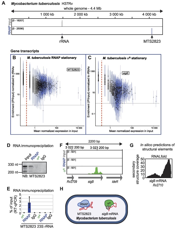
Bacteria have evolved structured RNAs that can associate with RNA polymerase (RNAP). Two of them have been known so far-6S RNA and Ms1 RNA but it is unclear if any other types of RNAs binding to RNAP exist in bacteria. To identify all RNAs interacting with RNAP and the primary σ factors, we have established and performed native RIP-seq in Bacillus subtilis, Corynebacterium glutamicum, Streptomyces coelicolor, Mycobacterium smegmatis and the pathogenic Mycobacterium tuberculosis. Besides known 6S RNAs in B. subtilis and Ms1 in M. smegmatis, we detected MTS2823, a homologue of Ms1, on RNAP in M. tuberculosis. In C. glutamicum, we discovered novel types of structured RNAs that associate with RNAP. Furthermore, we identified other species-specific RNAs including full-length mRNAs, revealing a previously unknown landscape of RNAs interacting with the bacterial transcription machinery.


Figures

Graphical Abstract
Graphical Abstract
Figure 1.
Figure 1.
RIP-seq in Mycobacterium smegmatis. (A) The primary RNAP holoenzyme consists of RNAP core with the primary σ factor and associates with 6S RNA. The secondary structure of 6S RNA from E. coli is shown. (B) Ms1 (predicted secondary structure from M. smegmatis) binds to core RNAP lacking a σ factor. (C) RIP-seq detects both RNA molecules binding to RNAPs or σ factors or RNAP-σ holoenzymes and also nascent RNAs that are connected with the transcribing RNAP-σ holoenzymes or RNAPs. (D) The immunoprecipitated proteins visualized by silver-stained SDS-PAGE. The anti-RNAP antibody that recognizes the β subunit precipitates mainly the core RNAP and additional RNAP-associated proteins (HelD, MSMEG_2174 (52) and RbpA, MSMEG_3858 (98,99)). The anti-σ70 antibody binds both σA and σB and also their RNAP holoenzyme complexes, because of very similar protein sequences (E). (FI) Quantification of RIP-seq data in M. smegmatis in exponential and stationary phase for RNAP (F, G) and σAB (H, I) for each annotated gene. For intergenic regions, please see Supplementary Figure S2. The horizontal axis (log scale) represents the mean of the normalized counts in the input, vertical axis (log scale) shows the estimated fold change in the ratio of read counts in immunoprecipitated to input samples, each normalized to the read counts for rRNA. Estimates (points) and 95% confidence intervals (lines) are shown for all transcripts. Transcripts to the left of the vertical dashed line had zero reads in all replicates of the input material. The horizontal gray line marks a fold change of 1, i.e. no enrichment or depletion of the respective RNA transcript after immunoprecipitation.
Figure 2.
Figure 2.
The interaction between σAB protein and sigB mRNA is affected by MMC. (A) Stationary phase RIP-seq and ChIP-seq data from M. smegmatis for the sigB gene. RNA-seq data was published previously (34). ChIP-seq detects genomic DNA sequences where the transcribing or stalled RNAP-σ holoenzymes or RNAPs are enriched. The position of primers used in RT-PCR in H and I are shown. (B) σAB peaks detected by ChIP-seq in stationary phase. (C) The level of σA and σB in total protein lysates after NaCl, heat and mitomycin C (MMC) stresses in M. smegmatis as detected by western blotting. The level of σA and σB protein changes after treating cells with NaCl and heat but not after MMC treatment. The amounts of σA and σB were detected with the anti-σ70 antibody, purified σA and σB were used as positive control. The anti-RNAP antibody was used to detect the level of RNAP. (D) mRNA levels of sigA, sigB andrecO in NaCl, heat and MMC treated cells. The y-axis shows the mRNA level in stressed cells that was normalized to the control cells, which were set as 1. Spike-in RNAs were added at the beginning of the RNA isolation (see Materials and methods) and RT-qPCR was normalized to spike-in RNAs. recO (MSMEG_4491) mRNA was used as a positive control for MMC treatment (67). The data shows the average value from three independent replicates, error bars represent SEM. (E) Growth curves of M. smegmatis in the presence or absence of mitomycin C (MMC), the data shows average values from three independent replicates, error bars represent SEM. (F) In MMC-treated cells, no σA was pulled down with the anti-σ70 antibody. The pulled-down proteins were resolved on SDS-PAGE, silver-stained and their identities were confirmed by MALDI mass spectrometry. σA and σB were detected also by western blotting with the anti-σ70 antibody. (G) sigB transcript was enriched in RNA immunoprecipitations with anti-σ70 antibody after MMC treatment. The data shows the average values from three independent replicates, error bars represent SEM. The asterisk indicates a significant difference as detected by paired t-test (P-value 0.03). (H, I) The presence of long sigB RNAs in MMC-treated cells was confirmed by RT-PCR with different primer pairs (shown in A). (J) Stable secondary structures were detected at the 3′end of sigB RNA by RNALfold. The secondary structure coverage displays the numbers of detected short, stable secondary structures (y-axis) along the sigB mRNA sequence. (K) The secondary structure of a fragment of sigB mRNA predicted by RNAfold (Vienna RNAfold webserver). The parts of the secondary structure with the highest base pair probabilities are highlighted in red. The locally stable secondary structures detected by RNALfold are marked by rectangles. The presented fragment of sigB mRNA was selected based on RIP-seq enrichment and has the following genome coordinates: NC_008596.1: 2822589–2822224.
Figure 3.
Figure 3.
The interaction between sigB mRNA and σA and σB proteins is not enhanced in NaCl and heat stress and sigB mRNA co-sediments with σB protein. (A) sigB transcript was enriched in RNA immunoprecipitations with anti-σ70 antibody after MMC treatment, while in NaCl and heat stress, sigB mRNA interaction with σA and σB proteins did not increase. The y-axis shows the % of input in control and stressed cells that was normalized to the control cells, set as 1. The data shows the average values from three independent replicates, error bars represent SEM. (B) Western blotting of the immunoprecipitated σA and σB proteins from control cells and cells subjected to NaCl and heat stress, detected by the anti-σ70 antibody. (C) Total protein lysates from M. smegmatis stationary phase cells were separated by glycerol gradient ultracentrifugation. The amounts of RNAP and σA and σB in individual fractions were detected by western blotting using anti-RNAP and anti-σ70 antibodies, respectively. The amount of sigB mRNA in individual fractions was quantified using RT-qPCR. rplM (MSMEG_1556) and hsp20 (MSMEG_0424) mRNAs were used as negative controls. Note that compared to negative controls, sigB mRNA was enriched in fractions 5 and 6, co-sedimenting with the free σB protein.
Figure 4.
Figure 4.
Ms1 is the major RNAP-interacting RNA in M. smegmatis. (A) Example of RIP-seq and ChIP-seq data from Mycobacterium smegmatis stationary phase, whole genome view. In the RNAP RIP-seq sample, most of the reads mapped to Ms1. (B) Quantification of RIP-seq data. In stationary phase, >60% of reads mapped to Ms1, while in exponential phase, RNAP binds mainly to rRNAs or other mRNAs that either represent actively transcribed nascent RNAs or background of non-specifically bound RNAs. (C) recO RNA fragments specifically bind RNAP. recO RNA was detected by northern blotting, for the position of the recO probe, see Supplementary Figure S3D. (D) Summary of RNAs that bind to the transcription machinery in M. smegmatis that we detected by RIP-seq. Question marks indicate that the exact composition of individual RNA-protein complexes is not known. (E) RNAP peaks detected by ChIP-seq in stationary phase. RNAP signal was detected on recO gene, but the exact position of RIP-seq peaks along the gene differs from the ChIP-seq peaks position (Supplementary Figure S3D), indicating that free and not co-transcriptionally associated recO RNAs are bound to RNAP.
Figure 5.
Figure 5.
RIP-seq in Mycobacterium tuberculosis. (A) RIP-seq data for Mycobacterium tuberculosis H37Rv, whole genome view. In the RNAP RIP-seq sample, most of the reads mapped to MTS2823 RNA. (B, C) Quantification of RIP-seq data from M. tuberculosis in stationary phase for RNAP (B) and σA (C) for each annotated gene. For further details, see legend to Figure 1. For intergenic regions, please see Supplementary Figure S4. (D, E) The binding of MTS2823 RNA to RNAP was confirmed by northern blotting and RT-qPCR. (F) High amounts of the sigB transcript were detected in the σA RIP-seq sample. (G) Stable secondary structures were detected at the 3′end of sigB mRNA by RNALfold. (H) Summary of RNAs that bind to the transcription machinery in M. tuberculosis as detected by RIP-seq. Question marks indicate that the exact composition of individual RNA-protein complexes is not known. Although σB has not been detected in the immunoprecipitations with anti-σ70 antibody (see Supplementary Figure S4A), we cannot rule out the possibility that it is present among the pulled-down proteins in low amount.
Figure 6.
Figure 6.
RIP-seq in Streptomyces coelicolor. Quantification of RIP-seq data for RNAP (A, B) and HrdB (C, D) for each annotated gene in S. coelicolor in exponential and stationary phase. For intergenic regions, please see Supplementary Figure S5. For further details, see legend to Figure 1. (E) RIP-seq using S. coelicolor cells in stationary phase revealed the scr3559 transcript (Ms1 homolog) to strongly enrich with RNAP, but there are also some reads mapping to scr3559 RNA in HrdB RIP-seq samples (in green). (F) The first nucleotide of scr3559 RNA that associates with RNAP in stationary phase of growth corresponds to the first nucleotide of the scr3559 transcript as defined by 5′RACE using total RNA and published previously (37). (G) scr0792, a novel transcript from the intergenic region between the genes SCO0791 and SCO0792 binds to RNAP in stationary phase of growth in S. coelicolor. The position of the two probes that were used for northern blotting are shown. (H) The two independent probes complementary to scr0792 RNA detected a ∼85 nt long RNA in stationary S. coelicolor cells. (I) Summary of RNAs that bind to the transcription machinery in S. coelicolor as detected by RIP-seq. Question marks indicate that the exact composition of individual RNA-protein complexes is not known.
Figure 7.
Figure 7.
CoRP RNA associates with the transcription machinery in C. glutamicum. (A) RIP-seq using C. glutamicum cells in stationary phase. In stationary phase σA and RNAP RIP-seq samples, most of the reads mapped to the 1366239–1366719 intergenic region. The new RNA bound to σA and RNAP was named CoRP RNA. (BE) Quantification of RIP-seq data for RNAP (B, C) and σAB (D, E) for each intergenic region in the genome of in C. glutamicum in exponential and stationary phase. For further details, see legend to Figure 1. For annotated genes, please see Supplementary Figure S7.
Figure 8.
Figure 8.
CoRP RNAs associate with RNAP holoenzyme and core in exponential and stationary phase. (A) Detailed view of the mapped reads at the CoRP RNA locus. 5′ and 3′flanking genes encode tRNAs. (B) CoRP RNA expression in C. glutamicum cells in exponential and stationary phase of growth. Based on 5′ and 3′RACE, the full-length CoRP RNA is cleaved into two fragments (5′ fragment 318 nt and 3′ fragment 198 nt). Two northern blot probes were used to detect the 5′ and 3′ fragments and the sizes of both fragments correspond to the cleavage site detected by RACE. (C) In both exponential and stationary phases, the full-length CoRP RNA interacts with σAB and RNAP. The positions of primers used for RT-PCR are indicated in (A). (D) The association of the full-length CoRP RNA with σAB and RNAP was also confirmed by northern blotting, although the majority of CoRP RNA bound to σAB and RNAP is fragmented. Both 5′ and 3′ fragments interact with σAB and RNAP in stationary phase. (E) Total protein lysates from C. glutamicum stationary phase cells were separated by glycerol gradient ultracentrifugation. The RNAP and σA and σB profiles across individual fractions were detected by western blotting using anti-RNAP and anti-σ70 antibodies, respectively. (F) The CoRP RNA was detected in individual fractions by northern blotting. The majority of the RNAP-σA holoenzyme sedimented in fractions 7–9, while fractions 10–11 (marked by a violet rectangle) contained both the RNAP-σA holoenzyme and CoRP RNA. No CoRP RNA was detected in the upper fractions of the gradient, where free proteins (including free σA and σB) sediment. Most of the CoRP RNA was detected in fractions 12–18, accompanied by RNAP without σA or σB, respectively. (G) The protein pull-down was performed using the full-length 516 nt CoRP RNA. In vitro transcribed CoRP RNA was coupled to streptavidin coated beads via a biotinylated antisense oligonucleotide and incubated with the lysates from exponential and stationary phase cells. The pulled-down proteins were resolved on SDS-PAGE and detected by Coomassie and silver staining. The identity of RNAP subunits was confirmed by western blotting using the anti-RNAP antibody. (H) CoRP RNAs bind both RNAP core and RNAP–σAB holoenzyme in C. glutamicum. Question marks indicate that the exact composition of individual RNA-protein complexes is not known.
Figure 9.
Figure 9.
RIP-seq in Bacillus subtilis. Quantification of RIP-seq data for RNAP (A, B) and σA (C, D) for each annotated gene in B. subtilis in exponential and stationary phase. For intergenic regions, please see Supplementary Figure S10. For further details, see legend to Figure 1.
Figure 10.
Figure 10.
RNA interactions of σA-RNAP, RNAP and σA in B. subtilis. (A) Amounts of immunoprecipitated σA and RNAP β subunit from B. subtilis exponential and stationary phase lysates examined by western blotting. The arrow indicates the position of σA bands. (B) The lysates from B. subtilis exponential phase cells were incubated with anti-σA and anti-RNAP antibodies, co-immunoprecipitated RNAs were isolated and the amounts of 6S-1, 6S-2 and 5S rRNA (negative control) detected by specific probes and northern blotting (B) or the level of co-immunoprecipitated 6S-1, 6S-2, rpoC and sigA mRNA was measured by RT-qPCR (C). (D) A lysate from E. coli mid-exponential phase was incubated with anti-σ70 and anti-RNAP antibodies, co-immunoprecipitated RNA isolated and the amount of 6S RNA detected by northern blotting. (E) Detailed view on RIP-seq mapped reads at the RNase Y (rny) locus and in flanking genes. RIP-seq was performed for exponential and stationary phase B. subtilis cells, the positions of primers used for RT-PCR validation are shown. (F) RT-PCR using RNA associated with RNAP in exponentially growing B. subtillis cells. RNAP binds intact rny mRNA in exponential phase. (G) Summary of RNAs that were detected by RIP-seq in B. subtilis. Question marks indicate that the exact composition of individual RNA-protein complexes is not known.
Figure 11.
Figure 11.
Overview of RNAs identified by RIP-seq. Group 1 represents abundant RNAs that associate with either RNAP (Ms1 homologs) or σA-RNAP (6S RNAs) or both (CoRP RNAs). Group 2 includes mRNAs interacting with RNAP or σ factors. Group 3 contains sRNAs or fragments of mRNAs associated with RNAP. Question marks indicate that the exact composition of individual RNA-protein complexes is not known.

Similar articles

Cited by

References

    1. Sensi P. History of the development of rifampin. Rev. Infect. Dis. 1983; 5:S402–S406. - PubMed
    1. Chen J., Boyaci H., Campbell E.A.. Diverse and unified mechanisms of transcription initiation in bacteria. Nat. Rev. Micro. 2021; 19:95–109. - PMC - PubMed
    1. Gomez M., Doukhan L., Nair G., Smith I.. sigA is an essential gene in Mycobacterium smegmatis. Mol. Microbiol. 1998; 29:617–628. - PubMed
    1. Brown K.L., Wood S., Buttner M.J.. Isolation and characterization of the major vegetative RNA polymerase of Streptomyces coelicolor A3(2); renaturation of a sigma subunit using GroEL. Mol. Microbiol. 1992; 6:1133–1139. - PubMed
    1. Browning D.F., Busby S.J.. Local and global regulation of transcription initiation in bacteria. Nat. Rev. Micro. 2016; 14:638–650. - PubMed

Publication types

MeSH terms

LinkOut - more resources