. 2024 Feb 13;73(3):45.
doi: 10.1007/s00262-023-03619-9.

Treatment of secondary CNS lymphoma using CD19-targeted chimeric antigen receptor (CAR) T cells

Affiliations

Treatment of secondary CNS lymphoma using CD19-targeted chimeric antigen receptor (CAR) T cells

Kathryn Kline et al. Cancer Immunol Immunother. .

Abstract

Background: Aggressive B cell lymphoma with secondary central nervous system (CNS) involvement (SCNSL) carries a dismal prognosis. Chimeric antigen receptor (CAR) T cells (CAR-T) targeting CD19 have revolutionized the treatment for B cell lymphomas; however, only single cases with CNS manifestations successfully treated with CD19 CAR-T have been reported.

Methods: We prospectively enrolled 4 patients with SCNSL into our study to assess clinical responses and monitor T cell immunity.

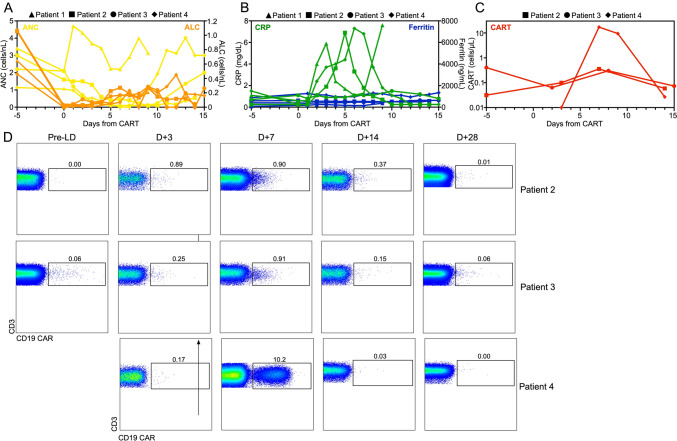
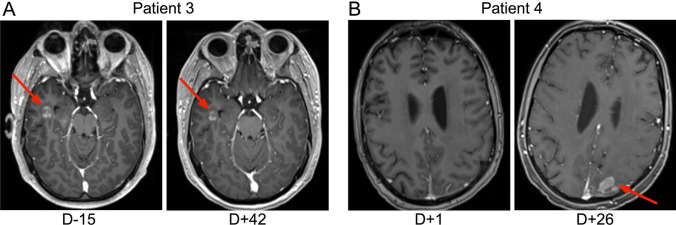
Results: Two of four SNCSL patients responded to the CD19-targeted CAR-T. Only one patient showed a substantial expansion of peripheral (PB) CAR-T cells with an almost 100-fold increase within the first week after CAR-T. The same patient also showed marked neurotoxicity and progression of the SNCSL despite continuous surface expression of CD19 on the lymphoma cells and an accumulation of CD4+ central memory-type CAR-T cells in the CNS. Our studies indicate that the local production of chemokine IP-10, possibly through its receptor CXCR3 expressed on our patient's CAR-T, could potentially have mediated the local accumulation of functionally suboptimal anti-tumor T cells.

Conclusions: Our results demonstrate expansion and homing of CAR-T cells into the CNS in SNCSL patients. Local production of chemokines such as IP-10 may support CNS infiltration by CAR-T cells but also carry the potential of amplifying local toxicity. Future studies investigating numbers, phenotype, and function of CAR-T in the different body compartments of SNSCL patients receiving CAR-T will help to improve local delivery of "fit" and highly tumor-reactive CAR-T with low off-target reactivity into the CNS.

Keywords: B cell lymphoma; CAR-T cells; CNS lymphoma; Cytokines; Neurotoxicity; T cells.


Conflict of interest statement

SD serves on advisory boards for Bristol-Myers Squibb, Incyte, and Atara Biotherapeutics. NMH serves on advisory boards for InCyte and Kite-Gilead and is a member of the DSMB for American Gene Technologies. The remaining authors declare that they do not have any competing interests.

Figures

Fig. 1
Fig. 1
Antigen expression on tumor tissue. A Immunohistochemical analyses performed on tissue from the mandibular buccal mucosa of patient 2 at approximately 2 years prior to CAR-T cell therapy. Hematoxylin- and eosin-stained sections (left; 400 × magnification) are shown. Arrows indicate areas with large cells with frequent mitoses. The tumor cells were stained for expression of CD19 (right; 400 × magnification). B Analysis of CNS-infiltrating B cell lymphoma cells by flow cytometry in patient 3 at approximately two months prior to CAR-T cell therapy. The infiltrating tumor cells were stained for surface expression of CD19 antigen. C Analysis of CNS-infiltrating B cell lymphoma cells by flow cytometry in patient 4 at day + 9 (left) and day + 21 (right) after CAR-T cell therapy. Again, the infiltrating tumor cells were stained for surface expression of CD19 antigen
Fig. 2
Fig. 2
CAR-T cell expansion and persistence in patients with secondary CNS lymphoma post CD19 CAR-T cells. Time course of A reconstitution of absolute neutrophil counts (ANC) and absolute lymphocyte counts (ALC), B serum C-reactive protein (CRP) and ferritin levels, and C CAR-T cell numbers after lymphodepleting chemotherapy and CAR-T cell infusion. D Dot plots showing peripheral blood CAR-T cells in 3 of 4 patients at different timepoints post CAR-T infusion. CAR-T cells were identified by staining of the expression of the CAR on the cell surface and costaining with anti-CD3 and other T cell markers (Supplemental Table 1)
Fig. 3
Fig. 3
CNS infiltration by CD19-targeted CAR-T cells in a SCNSL patient with ICANS. A Proportions of CD4+ and CD8+ CAR-T cells were determined in the CSF (upper panel) and in the PB (lower panel) of patient 4 shortly after onset of potentially immune-mediated CNS toxicity using flow cytometry. Dot plots show CNS-infiltrating CAR-T cells at day + 8 (CSF) and day + 9 (PB), respectively. CAR-T cells were identified by staining of the expression of the CAR on the cell surface and costaining with anti-CD3, anti-CD4, anti-CD8, and other T cell markers (Supplemental Table 1). CAR-T cell memory subtypes were determined by costaining for CD45RA and CD62L. Central memory (CM) CAR-T cells are shown in the right lower quadrant. B Concentrations of 22 different T cell-related cytokines/chemokines were determined in our patient on days + 8 (left) and + 9 (right), respectively, at the onset of potentially immune-mediated CNS toxicity using CodePlex Secretome technology. Results are shown as absolute concentrations in pg/mL. C Histograms show surface expression of receptors involved in CNS-directed homing of T cells on PB CAR-T cells (red histograms) and non-CAR-T (gray histograms) from our patient measured on day + 7. The dot plot on the right shows proportions of peripheral blood CAR-T cells expressing α4β1 integrin required for the entry of T cells into the CNS. D Increased levels of CD27 and CD127 were found on PB CAR-T cells (red histograms) vs. non-CAR-T (gray histograms) as measured on day + 9. In addition, cytoplasmic granzyme B and surface levels of CXCR3 were determined on day + 14 post CAR-T cell treatment in PB CAR-T cells. E Expression of exhaustion markers on PB CAR-T cells (red histogram) from our patients compared to their own non-CAR-T (gray histogram) on day + 9 post CAR-T cell treatment
Fig. 4
Fig. 4
CNS imaging results post CD19 CAR-T cells. MRIs were taken on A patient 3 and B patient 4 at the timepoints indicated. In the case of patient 3, repeat imaging showed a decrease in the size of the brain lesion post CAR-T. For patient 4, imaging showed a new lesion in the left posterior parietal lobe

References

    1. Bromberg JE, et al. Central nervous system recurrence of systemic lymphoma in the era of stem cell transplantation–an International Primary Central Nervous System Lymphoma Study Group project. Haematologica. 2013;98(5):808–813. doi: 10.3324/haematol.2012.070839. - DOI - PMC - PubMed
    1. Ghose A, et al. Influence of rituximab on central nervous system relapse in diffuse large B-cell lymphoma and role of prophylaxis–A systematic review of prospective studies. Clin Lymphoma Myeloma Leuk. 2015;15(8):451–457. doi: 10.1016/j.clml.2015.02.026. - DOI - PubMed
    1. El-Galaly TC, et al. Treatment strategies, outcomes and prognostic factors in 291 patients with secondary CNS involvement by diffuse large B-cell lymphoma. Eur J Cancer. 2018;93:57–68. doi: 10.1016/j.ejca.2018.01.073. - DOI - PMC - PubMed
    1. Tang K, Nastoupil LJ. Real-world experiences of CAR T-cell therapy for large B-cell lymphoma: How similar are they to the prospective studies? J Immunother Precis Oncol. 2021;4(3):150–159. doi: 10.36401/JIPO-21-2. - DOI - PMC - PubMed
    1. Abramson JS, et al. Anti-CD19 CAR T cells in CNS diffuse large-B-cell lymphoma. N Engl J Med. 2017;377(8):783–784. doi: 10.1056/NEJMc1704610. - DOI - PubMed