Review
. 2025 Mar;35(3):1205-1240.
doi: 10.1007/s10895-023-03561-0. Epub 2024 Feb 13.

The Role of Anions in Rare-earth Activated Inorganic Host Materials for its Luminescence Characteristics

Affiliations
Review

The Role of Anions in Rare-earth Activated Inorganic Host Materials for its Luminescence Characteristics

Leelakrishna Reddy. J Fluoresc. 2025 Mar.

Abstract

This work is inspired from the comprehensive work done by our research team aimed at improving the efficiency of white light emitting diodes (LEDs) through improvements in the colour rendering index of the red light (CRI), one of the primary colours of white light. Such work is triggered through the incorporation of anions (BO33-, PO43-, SO42-), either individually or as an integral part of dopant activated inorganic phosphor host materials. Numerous host materials such as ZnO, Y2O3, Ca3(PO4)2, CaMoO4, ABPO4, ABSO4 (where A represents alkali metals and B alkaline earth metals) have been considered ideal hosts materials for studying luminescence properties of materials (including other phosphors). In addition, red emitting dopants such as Sm3+, Eu3+ and Ce3+ have been incorporated into these host materials to achieve a higher CRI of red colour, an essential component of white light. The role anions in various materials is multifaceted; firstly, it acts as sensitizer whereby it absorbs excitation energy and transfers it non-radiatively to the dopants, secondly, it acts as a charge compensator to dopants with a charge of + 3, thirdly, it creates crystal fields that affects the electronic transitions of the dopants and fourthly, it creates a stable crystal structure that allows for dopant embedding. By understanding the exact role of these anions and their interactions with the host lattice and dopant ions, we could further optimize the luminescent properties of these activated host materials, which leads to higher efficiencies and performances in white light-emitting diodes and other lighting technologies. This work is a comprehensive review of the work undertaken by our research team aimed at enhancing the luminescent properties of WLEDs.

Keywords: Dopants; Energy transfer; Luminescence; Phosphor; WLEDs.


Conflict of interest statement

Declarations. Ethics Approval: N/A. Competing Interests: The authors declare no competing interests.

Figures

Fig. 1
Fig. 1
XRD patterns of the NaMPO4-Eu3+ phosphor materials with M2+ = Ca, Ba, Sr, and Mg [15] (Reused with permission from [15])
Fig. 2
Fig. 2
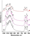
FTIR spectra of the NaMPO4-Eu3+ phosphor materials with M2+ = Ca, Ba, Sr, and Mg. The inset show the various series doped with Eu3+ ions [15] (Reused with permission from [15])
Fig. 3
Fig. 3
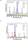
PLE and PL spectra of a sampler NaBaPO4:Eu3+ phosphor material is shown in figure (a) above, while the entire PL emission series for NaMPO4:Eu3+phosphors is shown in (b) [15] (Reused with permission from [15])
Fig. 4
Fig. 4
XRD patterns of the CaMoO4-SO4-xEu3+ (x = 0 to 2.5 mol%) phosphor materials [17] (Reused with permission from [17])
Fig. 5
Fig. 5
FTIR spectra of the CaMoO4-SO4-Eu3+ phosphor material [17] (Reused with permission from [17])
Fig. 6
Fig. 6
PLE spectra of the CaMoO4-SO4-xEu3+ phosphor material is shown in (a), while the PL emission spectra of the sample for concentration variations is shown in (b) [17] (Reused with permission from [17])
Fig. 7
Fig. 7
XRD patterns of the NaMPO4:Ce3+ (M2+ = Mg, Ca, Sr, and Ba) phosphor materials [19] (Reused with permission from [19])
Fig. 8
Fig. 8
FTIR spectra of the NaMPO4:Ce3+ (M2+ = Mg, Ca, Sr, and Ba) phosphor materials [19] (Reused with permission from [19])
Fig. 9
Fig. 9
PLE excitation spectra for NaMPO4:Ce3+ (M2+ = Mg, Ca, Sr, and Ba) phosphor materials are shown in (a), while the PL emission spectra is shown in (b) as a function of binding energy for different wavelengths [19] (Reused with permission from [19])
Fig. 10
Fig. 10
XRD patterns of the Ca3(PO4)2: Ce3+phosphor material co-doped with Gd3+ ions [20] (Reused with permission from [20])
Fig. 11
Fig. 11
PLE excitation and PL emission spectra of Gd3+ co-doped Ca3(PO4)2: Ce3+phosphor material, excited at different wavelengths in respective pairs at (a) and (b) [20] (Reused with permission from [20])
Fig. 12
Fig. 12
XRD patterns of the ZnO: Ce3+phosphor material with SO42- anionic incorporation in (a) and enlarged section in (b) showing the relative shifts in diffraction peaks [21] (Reused with permission from [21])
Fig. 13
Fig. 13
FTIR spectra of the ZnO: Ce3+ phosphor materials with SO42− incorporation [21] (Reused with permission from [21])
Fig. 14
Fig. 14
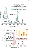
PL emission spectra of the ZnO: Ce3+ phosphor materials, with SO42- incorporations in (a), and (b) showing enlarged sections of (a), and (c) shows the trends of PL emission when SO42- or Ce3+ ions are added to ZnO [21] (Reused with permission from [21])
Fig. 15
Fig. 15
XRD spectra of the CaMoO4:Eu3+ phosphor material with BO33− incorporations [22] (Reused with permission from [22])
Fig. 16
Fig. 16
FTIR spectra of the CaMoO4:Eu3+ phosphor material with BO33- incorporation [22] (Reused with permission from [22])
Fig. 17
Fig. 17
PLE excitation spectra of the CaMoO4:Eu3+ phosphor material is shown in (a), while the PL emission spectra of the same sample is shown in (b) with BO33- incorporations [22] (Reused with permission from [22])
Fig. 18
Fig. 18
XRD pattern of the Y2O3-AG-Eu3+ phosphor materials, with AG = (BO33−), (PO43−), and (SO42−) additions, and transformed structures for borate and phosphate additions [23] (Reused with permission from [23])
Fig. 19
Fig. 19
FTIR spectra of the Y2O3-AG-Eu3+ phosphor materials, with AG = (BO33-), (PO43-), and (SO42-) [23] (Reused with permission from [23])
Fig. 20
Fig. 20
PL excitation and emission spectra of the Y2O3-AG-Eu3+ phosphor materials, with AG =(BO33-), (PO43-), and (SO42-) [23] (Reused with permission from [23])
Fig. 21
Fig. 21
XRD patterns of the Na2Ca (SO4)2: Sm3+/Eu3+ phosphor material is shown in (a), while figure (b) is an enlarged section of the (-2 2 1) plane [24] (Reused with permission from [24])
Fig. 22
Fig. 22
FTIR spectra of the co-doped Na2Ca (SO4)2: Sm3+/Eu3+ phosphor materials [24] (Reused with permission from [24])
Fig. 23
Fig. 23
PL excitation and emission spectra of the co-doped Na2Ca (SO4)2: Sm3+/Eu3+ phosphor materials [25] (Graph taken from reference [25], published in the Journal of Molecular Structure)
Fig. 24
Fig. 24
XRD patterns of the Na6Mg (SO4)4: Sm3+ phosphor material, co-doped with Eu3+ ions [25] (Graph taken from reference [25], published by Inorg. Nano-Metal Chem., 2022)
Fig. 25
Fig. 25
FTIR measurements of the Na6Mg (SO4)4: Sm3+ phosphor material, co-doped with Eu3+ ions [25] (Graph taken from reference [25], published by Inorg. Nano-Metal Chem., 2022)
Fig. 26
Fig. 26
Full PLE and PL spectrum of the Na6Mg (SO4)4: Sm3+ phosphor material is shown in (a), figure (b) shows the PLE spectrum of the phosphor material and c shows the PL emission spectra of the material, co-doped with Eu3+ ions [25] (Graph taken from reference [25], published by Inorg. Nano-Metal Chem., 2022)
Fig. 27
Fig. 27
XRD spectra of the CaMoO4-AG: Sm3+ phosphor materials, with AG = BO33−, PO43− and SO42− ions [3] (Reused with permission from [3])
Fig. 28
Fig. 28
FTIR spectra of CaMoO4 -AG: Sm3+ (1 mol%) phosphor materials with AG = BO33-, PO43- and SO42- ions [3] (Reused with permission from [3])
Fig. 29
Fig. 29
Figure a shows the PLE excitation spectra and figure b shows the PL emissions spectra of the CaMoO4 -AG: Sm3+ (1 mol%) phosphor materials with AG = BO33-, PO43- and SO42- ions [3] (Reused with permission from [3])
Fig. 30
Fig. 30
XRD spectra of the Ca3-xLix(PO4)2-x(SO4)x:Dy3+, Sm3+ phosphor materials [14] (Reused with permission from [14])
Fig. 31
Fig. 31
PLE excitation and PL emission spectra of the Ca3-xLix(PO4)2-x(SO4)x:Dy3+, Sm3+ phosphor materials [14] (Reused with permission from [14])
Fig. 32
Fig. 32
XRD spectra of the Ca10.5–0.5x(PO4)7-x(SO4)x and Ca9.5–0.5xMg(PO4)7-x(SO4)x phosphor material for x = 1 mol% [26] (Reused with permission from [26])
Fig. 33
Fig. 33
FTIR spectra of the Ca10.5-0.5x(PO4)7-x(SO4)xand Ca9.5-0.5xMg(PO4)7-x(SO4)x phosphor material for x = 1 mol% [26] (Reused with permission from [26])
Fig. 34
Fig. 34
PLE excitations spectra of the prepared material is shown in (a), while the PL emission spectra is shown in figure (b) [26] (Reused with permission from [26])
Fig. 35
Fig. 35
XRD spectra of the Eu3+ activated LiY(PO3)4(1-x)SO4x phosphor materials for varying concentrations of SO42- ions [13] (Graph taken from reference [13], published by ECS J Solid State Sci. Technol., 2023)
Fig. 36
Fig. 36
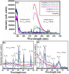
PLE (first) and PL spectra (second) of the Eu3+ activated LiY(PO3)4(1-x)SO4x phosphor materials for varying concentrations of SO42- ions [13] (Graphs taken from reference [13], published by ECS J Solid State Sci. Technol., 2023)

References

    1. Xiao F, Xue YN, Ma YY, Zhang QY (2010) Ba2Ca(B3O6)2:Eu2+, Mn2+: A potential tunable blue-white-red phosphor for white light-emitting diodes. Phys B 405(3):891–895
    1. Hu SH, Lin YS, Su SH, Dai H, He JS (2022) Study on the enhancement of light intensity and high color rendering index of a white light emitting diode. J Electron Mater 52:270–275
    1. Balakrishna A, Swart HC, Ramaraghavulu R, Bedyal AK, Kroon RE, Ntwaeaborwa OM (2017) Structural evolution induced by substitution of designated molybdate sites (MoO42-) with different anionic groups (BO33-, PO43- and SO42-) in CaMoO4:Sm3+ phosphors – A study on color tunable luminescent properties. J Alloy Compd 727:224–237
    1. Caisheng X, Shikao S, Huili G, Ji Z (2010) Preparation and luminescent properties of Na5Eu(MoO4)4–x(PO4)x red phosphors for white-emitting diodes application. Mater Sci Forum 654–656:2025–2028
    1. Yan Z, Shikao S, Jing G, Ji Z (2010) Effects of SO42- or SiO32- doping on the photoluminescence of NaEu(MoO4)2 nanophosphors for light-emitting diodes. J Nanosci Nanotechnol 10(3):2156–3160 - PubMed

LinkOut - more resources