. 2024 Feb 15;151(4):dev202214.
doi: 10.1242/dev.202214. Epub 2024 Feb 13.

A mutation in F-actin polymerization factor suppresses the distal arthrogryposis type 5 PIEZO2 pathogenic variant in Caenorhabditis elegans

Affiliations

A mutation in F-actin polymerization factor suppresses the distal arthrogryposis type 5 PIEZO2 pathogenic variant in Caenorhabditis elegans

Xiaofei Bai et al. Development. .

Abstract

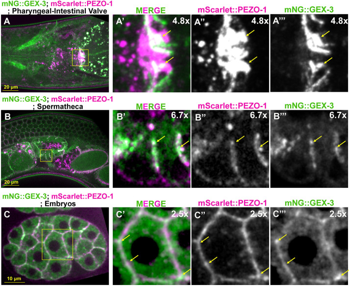
The mechanosensitive PIEZO channel family has been linked to over 26 disorders and diseases. Although progress has been made in understanding these channels at the structural and functional levels, the underlying mechanisms of PIEZO-associated diseases remain elusive. In this study, we engineered four PIEZO-based disease models using CRISPR/Cas9 gene editing. We performed an unbiased chemical mutagen-based genetic suppressor screen to identify putative suppressors of a conserved gain-of-function variant pezo-1[R2405P] that in human PIEZO2 causes distal arthrogryposis type 5 (DA5; p. R2718P). Electrophysiological analyses indicate that pezo-1(R2405P) is a gain-of-function allele. Using genomic mapping and whole-genome sequencing approaches, we identified a candidate suppressor allele in the C. elegans gene gex-3. This gene is an ortholog of human NCKAP1 (NCK-associated protein 1), a subunit of the Wiskott-Aldrich syndrome protein (WASP)-verprolin homologous protein (WAVE/SCAR) complex, which regulates F-actin polymerization. Depletion of gex-3 by RNAi, or with the suppressor allele gex-3(av259[L353F]), significantly increased brood size and ovulation rate, as well as alleviating the crushed oocyte phenotype of the pezo-1(R2405P) mutant. Expression of GEX-3 in the soma is required to rescue the brood size defects in pezo-1(R2405P) animals. Actin organization and orientation were disrupted and distorted in the pezo-1 mutants. Mutation of gex-3(L353F) partially alleviated these defects. The identification of gex-3 as a suppressor of the pathogenic variant pezo-1(R2405P) suggests that the PIEZO coordinates with the cytoskeleton regulator to maintain the F-actin network and provides insight into the molecular mechanisms of DA5 and other PIEZO-associated diseases.

Keywords: Disease modeling in C. elegans; Distal arthrogryposis type 5; Forward genetic screening; Genetic suppressor; PIEZO channel; WAVE complex GEX-3.


Conflict of interest statement

Competing interests The authors declare no competing or financial interests.

Figures

Fig. 1.
Fig. 1.
PIEZO pathogenic variants caused reproductive deficiency in C. elegans. (A) Diagram of the location of the PEZO-1 pathogenic residues in the PEZO-1 channel used in this study. (B) Brood size was reduced in all pezo-1 pathogenic mutants tested when compared with wild type. n indicates the number of animals tested. (C-E) Quantification of the oocyte ovulation rate and percentage of crushed oocytes in wild type and pezo-1 mutants during ovulation at different ages. n indicates the number of gonads tested. The oocyte ovulation rate was significantly reduced in the pezo-1 mutant adults. One-way ANOVA test (B-D). *P=0.0114; ****P<0.0001 (unpaired one-way ANOVA t-test). Data are Data are median±s.d.
Fig. 2.
Fig. 2.
Sperm guidance and navigation is disrupted in PIEZO pathogenic variants. (A) To quantify sperm migration, sperm distribution was counted in three zones, including zone 3, which is the spermathecal region and the space containing the +1 fertilized embryo. Zone 1 is the area closest to the vulva; zone 2 is the area between zone 1 and zone 3. Sperm distribution is measured 1 h after the Mitotracker-labeled males were removed from the mating plate. (B-G′) The distribution of fluorescent male sperm (yellow dots) labeled with MitoTracker in the three zones in both wild type and pezo-1 mutants. Yellow asterisks indicate the vulva (B′,C′,D′,E′,F′,G′). (H) Quantification of sperm distribution values for wild type and each pezo-1 mutant. One-way ANOVA test. n indicates the number of animals tested. ****P<0.0001 (unpaired t-test). Data are median±s.d.
Fig. 3.
Fig. 3.
Electrophysiological characterization of PEZO-1 gain-of function mutation pezo-1(R2450P). (A) Diagram of the location of the PEZO-1(R2405P) pathogenic residue in the PEZO-1 channel. (B) Schematic representation of the mechanical stimulation poked by a blunt pipette applied to Sf9 cells infected with baculovirus containing pezo-1 (wild-type or R2405P constructs) recorded in the whole-cell configuration. Created with BioRender.com. (C) Representative whole-cell patch-clamp recordings (at −60 mV) of currents elicited by mechanical stimulation of Sf9 cells, uninfected (control), expressing pezo-1 wild type or expressing pezo-1(R2405P). Sf9 cells were poked with a heat-polished blunt glass pipette (3-4 µm) driven by a piezo servo controller. Displacement measurements were obtained with a square-pulse protocol consisting of 1 µm incremental indentation steps. Recordings with leak currents>200 pA and with access resistance>10 MΩ, as well as cells with giga seals that did not withstand at least five consecutive steps of mechanical stimulation were excluded from analyses. (D) Current density elicited by maximum displacement (−60 mV) of Sf9 cells expressing pezo-1 wild type or pezo-1(R2405P). Data are mean±s.d. Kruskal–Wallis (H=18.35; ****P=0.0001) and Dunn's multiple comparisons test. (E) Time constants of inactivation elicited by maximum displacement (−60 mV) of Sf9 cells expressing pezo-1 wild type or pezo-1(R2405P). Data are mean±s.d. Two-tailed unpaired t-test with Welch correction (t=−4.29). **P=0.0049. (F) Boxplots show the displacement thresholds required to elicit mechanocurrents of Sf9 cells expressing pezo-1 wild type or pezo-1(R2405P). Boxplots show mean (square), median (bisecting line), bounds of box (75th to 25th percentiles), outlier range with 1.5 coefficient (whiskers), and minimum and maximum data points. Two-tailed Mann–Whitney test (U=13.5). Filled circles come from the representative traces shown in C. n values are indicated under each column. Post-hoc P-values are indicated in the corresponding panels.
Fig. 4.
Fig. 4.
The WAVE regulatory complex NCKAP1 suppresses the reproductive defects in the pezo-1(R2405P) mutant. (A) gex-3(RNAi) treatment reduced brood size in wild-type and pezo-1(T1997M) mutant animals, while significantly restoring the brood size in pezo-1(R2405P) mutants. (B) Depletion of gex-3 by RNAi led to various levels of embryonic lethality in all tested animals; however, the pezo-1(R2405P) allele partially alleviated the lethality when compared with wild-type control. n values indicate the number of animals tested in A and B. (C-E) Quantification of the oocyte ovulation rate and percentage of crushed oocytes of wild type and pezo-1 mutants without or without gex-3(RNAi) treatment. n values indicate the number of gonads tested. One-way ANOVA (A-D) or Chi-squared-test (E). *P=0.0329 (C); *P=0.0247 (D), *P=0.0281 (E); ****P<0.0004 (R2405P gex-3 versus wt gex-3 RNAi in B); ***P=0.0007 (R2405Q ctrl versus R2405Q gex-3 in B); ****P<0.0001 (A and B).
Fig. 5.
Fig. 5.
mScarlet::PEZO-1 colocalizes with mNG::GEX-3 in multiple tissues and cells. (A-A‴) mScarlet::PEZO-1 (magenta in A,A′) colocalizes with mNG::GEX-3 (green in A,A′) at the pharyngeal-intestinal valve. The enlarged pictures of the yellow rectangular area indicates the colocalization of mScarlet::PEZO-1 (magenta in A′) and mNG::GEX-3 (green in A′). (B-B‴) Colocalization of mScarlet::PEZO-1 (magenta in B,B′) and mNG::GEX-3 (green in B,B′) on the spermathecal membrane (yellow square in B). (B′-B‴) Higher magnification views of the area outlined in B showing colocalization of mScarlet::PEZO (magenta in B′) and mNG::GEX-3 (green in B′) on the membrane (yellow arrows). (C-C‴) mNG::GEX-3 and mScarlet::PEZO-1 was observed at the plasma membrane of the early embryos. Both mNG::GEX-3 (green in C′) and mScarlet::PEZO-1 (magenta in C′) were expressed on the embryonic plasma membrane (yellow arrows). Colocalization of mNG::GEX-3 and mScarlet::PEZO-1 appears white.
Fig. 6.
Fig. 6.
gex-3(L353F) suppresses the reproductive defects in the pezo-1(R2405P) mutant. (A) The structure of GEX-3 from Alphafold indicated the Leu353 residue was on the N terminus of a helix (highlighted by red arrow). Different colors represent model confidence: dark blue, very high (pLDDT>90); cyan, confident (90>pLDDT>70); yellow, low (70>pLDDT>50); orange, very low (pLDDT<50). (B) gex-3(L353F) reduced brood size in wild-type animals but suppressed the smaller brood size in the pezo-1(R2405P) mutant animals. (C) The embryonic lethality in pezo-1(R2405P), gex-3(L353F) and double mutants. n values in C indicate the number of animals tested in B and C. (D-F) Quantification of the oocyte ovulation rate and percentage of crushed oocytes of wild type, pezo-1(R2405P), gex-3(L353F) and double mutants at different ages. n values indicate the number of the gonads tested. One-way ANOVA test (B-E) or Chi-squared-test (F). *P=0.0360 (F); **P=0.0015 (D); ***P=0.0008 (B); ****P<0.0001 (B and C).
Fig. 7.
Fig. 7.
Somatic tissue-specific degradation of GEX-3 suppresses the small brood size in the pezo-1(R2405P) mutant. A degron and GFP cassette was inserted at the 5′ end of the gex-3-coding sequence using CRISPR/Cas9-mediated editing. The transgenic TIR-1::BFP::AID was driven by the eft-3 promoter to be expressed in most or all somatic tissues, including the spermatheca and the somatic sheath cells. TIR-1::BFP::AID was driven by the germline-specific promoter mex-5, which drives expression in the germline and oocytes. (A) Brood size was partially restored in the pezo-1(R2405P) degron driven by the eft-3 promoter when animals were treated with 2 mM auxin. (B) Embryonic viabilities were reduced to nearly zero in all degron strains when treated with 2 mM auxin. n values indicate the number of animals tested. ****P<0.0001 (unpaired, one-way ANOVA t-test). The blue circles represent those animals treated with the ethanol-only control; the orange circles represent those treated with auxin.
Fig. 8.
Fig. 8.
Actin organization and orientation were disrupted in pezo-1 mutants. (A,A′) Representative images of the contracted spermatheca labelled by the actin marker GFP::ACT-1. (B,B′) Representative images of wild-type spermathecal cells with parallel actin bundles. (C-D′) Representative images of defective actin bundles, including perpendicular actin and bunching actin in the pezo-1(R2405P) mutant. Bunching actin is indicated by green arrows. (E,E′) Representative images of defective actin bundles in pezo-1Δ mutant. Bunching actin is indicated by green arrows. (F,F′) Actin organization in gex-3(L353F) animals. (G,G′) Double mutant pezo-1(R2405P) gex-3(L353F) suppressed the actin defects when compared with the pezo-1(R2405P) single mutant. The areas outlined in B-G′ are shown at higher magnification in the insets. (H,I) Quantification of actin defects in each strain. Insets in B,C,D,E,F,G are color coded according to z-depth to indicate the bundle organization and orientation. n values indicate the number of the occupied spermatheca tested in H and I. *P=0.0264 (H); **P=0.0088 (H); ****P<0.0001 (H and I) (Chi-squared test).
Fig. 9.
Fig. 9.
Working model for genetic interaction between pezo-1(R2405P) and gex-3(L353F). The proposed model suggests how gex-3(L353F) or partial depletion of gex-3 by RNAi might suppress the reproductive deficiency in the pezo-1(R2405P) mutant. At the cellular level, the pezo-1(R2405P) allele disrupts the actin organization and orientation, such as perpendicular actin and bunching actin in the spermathecal cells. The gex-3(L353F) allele partially alleviates the actin defects in the pezo-1(R2405P) mutant, which may explain its suppression of the low ovulation rate and number of crushed oocytes during ovulation, all leading to suppression of the reduced brood size caused by the pezo-1(R2405P) mutant. The proteins labeled in grey were not explored in this work.

Update of

Similar articles

Cited by

References

    1. Albuisson, J., Murthy, S. E., Bandell, M., Coste, B., Louis-Dit-Picard, H., Mathur, J., Fénéant-Thibault, M., Tertian, G., de Jaureguiberry, J.-P., Syfuss, P.-Y.et al. (2013). Dehydrated hereditary stomatocytosis linked to gain-of-function mutations in mechanically activated PIEZO1 ion channels. Nat. Commun. 4, 1884. 10.1038/ncomms2899 - DOI - PMC - PubMed
    1. Alper, S. L. (2017). Genetic diseases of PIEZO1 and PIEZO2 dysfunction. Curr. Top. Membr. 79, 97-134. 10.1016/bs.ctm.2017.01.001 - DOI - PubMed
    1. Andolfo, I., Alper, S. L., De Franceschi, L., Auriemma, C., Russo, R., De Falco, L., Vallefuoco, F., Esposito, M. R., Vandorpe, D. H., Shmukler, B. E.et al. (2013). Multiple clinical forms of dehydrated hereditary stomatocytosis arise from mutations in PIEZO1. Blood 121, 3925-3935, S3921–3912. 10.1182/blood-2013-02-482489 - DOI - PubMed
    1. Ashley, G. E., Duong, T., Levenson, M. T., Martinez, M. A. Q., Johnson, L. C., Hibshman, J. D., Saeger, H. N., Palmisano, N. J., Doonan, R., Martinez-Mendez, R.et al. (2021). An expanded auxin-inducible degron toolkit for Caenorhabditis elegans. Genetics 217, iyab006. 10.1093/genetics/iyab006 - DOI - PMC - PubMed
    1. Atcha, H., Jairaman, A., Holt, J. R., Meli, V. S., Nagalla, R. R., Veerasubramanian, P. K., Brumm, K. T., Lim, H. E., Othy, S., Cahalan, M. D.et al. (2021). Mechanically activated ion channel Piezo1 modulates macrophage polarization and stiffness sensing. Nat. Commun. 12, 3256. 10.1038/s41467-021-23482-5 - DOI - PMC - PubMed

Supplementary concepts