Review
. 2024 Feb 27;18(8):6038-6094.
doi: 10.1021/acsnano.3c09699. Epub 2024 Feb 13.

Environmental and Health Impacts of Graphene and Other Two-Dimensional Materials: A Graphene Flagship Perspective

Affiliations
Review

Environmental and Health Impacts of Graphene and Other Two-Dimensional Materials: A Graphene Flagship Perspective

Hazel Lin et al. ACS Nano. .

Abstract

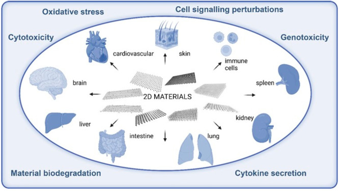
Two-dimensional (2D) materials have attracted tremendous interest ever since the isolation of atomically thin sheets of graphene in 2004 due to the specific and versatile properties of these materials. However, the increasing production and use of 2D materials necessitate a thorough evaluation of the potential impact on human health and the environment. Furthermore, harmonized test protocols are needed with which to assess the safety of 2D materials. The Graphene Flagship project (2013-2023), funded by the European Commission, addressed the identification of the possible hazard of graphene-based materials as well as emerging 2D materials including transition metal dichalcogenides, hexagonal boron nitride, and others. Additionally, so-called green chemistry approaches were explored to achieve the goal of a safe and sustainable production and use of this fascinating family of nanomaterials. The present review provides a compact survey of the findings and the lessons learned in the Graphene Flagship.

Keywords: 2D nanomaterials; biodegradability; carbon materials; environment; exposure; hazard; safe-by-design; test guidelines; toxicity.


Conflict of interest statement

The authors declare no competing financial interest.

Figures

Figure 1
Figure 1
Synthesis of highly enriched 13C-graphene materials for biological and safety applications. Top row panels: schematic representation of the synthesis of carbon nanofibers. Top right panel: typical TEM image of the produced fibers. Middle and bottom panels: (a–f) TEM images and size distribution of graphene obtained by exfoliation of the 13C graphitized (G) carbon nanofibers: (a,d) 13C-GFLG-1, (b,e) 13C-GFLG-2, and (c,f) 13C-GGO. Reproduced with permission from ref (13). Copyright 2023, the American Chemical Society.
Figure 2
Figure 2
Illustration of 2D materials that can potentially come in contact with living organisms within the diverse range of ecosystems and their possible effects.
Figure 3
Figure 3
Images of D. magna individuals after 48 h of exposure to different concentrations of GO and after feeding on fluorescent microbeads visualized by fluorescence microscopy. The beginning of the digestive tract is marked by red arrows, while the end is marked by a green arrow. Reproduced in part with permission under a Creative Commons CC BY 4.0 License from Fekete-Kertesz, I.; Laszlo, K.; Terebesi, C.; Gyarmati, B. S.; Farah, S.; Marton, R.; Molnar, M. Ecotoxicity Assessment of Graphene Oxide by Daphnia magna through a Multimarker Approach from the Molecular to the Physiological Level including Behavioral Changes. Nanomaterials (Basel) 2020, 10, 2048. Copyright 2020, MDPI, Basel.
Figure 4
Figure 4
Ecotoxicology of 2D materials: evaluating the effects of GO on Xenopus laevis tadpoles. Normalized growth rate determined after 2 days (A) or 12 days (B) of exposure to increasing GO concentrations. (C) Pictures of GO intestinal accumulation in tadpole larvae after 2 days or 12 days of exposure. Reproduced with permission from Evariste, L.; Mouchet, F.; Pinelli, E.; Flahaut, E.; Gauthier, L.; Barret, M. Gut Microbiota Impairment following Graphene Oxide Exposure is Associated to Physiological Alterations in Xenopus laevis Tadpoles. Sci. Total Environ. 2022, 857, 159515. Copyright 2022, Elsevier.
Figure 5
Figure 5
Light micrographs of gill tissue samples from (A) control and chitosan functionalized CS-MoS2 at (B) 2 mg/L (C) 10 mg/L and (D) 20 mg/L) Scale bar = 400 μm. Reproduced with permission from Yu, Y.; Yi, Y.; Li, Y.; Peng, T.; Lao, S.; Zhang, J.; Liang, S.; Xiong, Y.; Shao, S.; Wu, N.; Zhao, Y.; Huang, H. Dispersible MoS2 Micro-Sheets Induced a Proinflammatory Response and Apoptosis in the Gills and Liver of Adult Zebrafish. RSC Adv. 2018, 8, 17826–17836. Copyright 2018, the Royal Society of Chemistry.
Figure 6
Figure 6
SEM images of GO-exposed E. gracilis cultivated under phototrophic (A–C) or heterotrophic (D–F) conditions. (A) and (D) are control cells (without GO), (B) and (E) are nanosize GO-exposed cells, and (C) and (F) are microsize GO-exposed cells. Red arrows indicate GO, and gray arrows indicate the damage to the pellicle structure. Reproduced with permission from Kim, K. Y.; Kim, S. M.; Kim, J. Y.; Choi, Y. E. Elucidating the Mechanisms Underlying the Cytotoxic Effects of Nano-/Micro-Sized Graphene Oxide on the Microalgae by Comparing the Physiological and Morphological Changes in Different Trophic Modes. Chemosphere 2022, 309, 136539. Copyright 2022, Elsevier.
Figure 7
Figure 7
Ecotoxicology of 2D materials: interactions of GO with the sexual reproduction of a model plant (summer squash). Experimental design (left) and SEM micrographs (right) of stigmas of Cucurbita pepo flowers treated with dry depositions of 0 (CTRL) (A,B) or 22 μg/mm2 of GO (C) or purified GO (pGO) (D) and pollinated after 3 h. Stigmatic papillae, GO flakes/nanoparticles, and pollen grain are indicated with arrows, arrowheads, and asterisk, respectively. Scale bars = 100 μm. Reproduced with permission from Zanelli, D.; Candotto Carniel, F.; Fortuna, L.; Pavoni, E.; Jehová González, V.; Vázquez, E.; Prato, M.; Tretiach, M, Interactions of Airborne Graphene Oxides with the Sexual Reproduction of a Model Plant: When Production Impurities Matter. Chemosphere 2023, 312, 137138. Copyright 2023, Elsevier.
Figure 8
Figure 8
Illustration of 2D materials that can potentially come in contact with the different barriers and organs of living organisms and their possible effects.
Figure 9
Figure 9
Skin irritation test using the SkinEthicTM reconstructed human epidermis, following the Organisation for Economic Co-operation and Development (OECD) Test Guideline (TG) 439. Presence of GBMs above the epidermis surface and within the stratum corneum (shown by arrows) in reconstructed human epidermis (RhE) exposed to vehicle (A), sodium dodecyl sulfate (SDS) (B) and sodium dodecylbenzenesulfonate (SDBS) (C) positive controls, FLG (D), FLG-SDS (E), FLG-SDBS (F), GO (G), rGO (H), or chemical vapor deposition (CVD) (I). Scale bar: 20 μm. Reproduced in part with permission under a Creative Commons 3.0 Unported License from Fusco, L.; Garrido, M.; Martin, C.; Sosa, S.; Ponti, C.; Centeno, A.; Alonso, B.; Zurutuza, A.; Vazquez, E.; Tubaro, A.; Prato, M.; Pelin, M. Skin Irritation Potential of Graphene-Based Materials using a Non-Animal Test. Nanoscale 2020, 12, 610–622. Copyright 2020, the Royal Society of Chemistry.
Figure 10
Figure 10
Illustration of the various organs and immune cells of the human body.
Figure 11
Figure 11
Gene expression profiling of human macrophages exposed to GO versus GNP. Venn diagrams of differently expressed genes (DEG) indicating material-specific responses in THP-1 macrophages and monocyte-derived macrophage (MDM). Comparison of DEG after 6 and 24 h of exposure to 5 or 20 μg/mL GO or GNP or 100 μg/mL DQ (crystalline quartz) in THP-1 macrophages (A) or MDM (B). Reproduced in part with permission under a Creative Commons BY 4.0 License from Korejwo, D.; Chortarea, S.; Louka, C.; Buljan, M.; Rothen-Rutishauser, B.; Wick, P.; Buerki-Thurnherr, T. Gene Expression Profiling of Human Macrophages After Graphene Oxide and Graphene Nanoplatelets Treatment Reveals Particle-Specific Regulation of Pathways. NanoImpact 2023, 29, 100452. Copyright 2023, Elsevier.
Figure 12
Figure 12
Neutrophil degradation of GO sheets with varying lateral dimensions. (A,B) Freshly isolated human neutrophils were treated with fMLP (10 nM) and cytochalasin B (5 μg/mL) to trigger degranulation and incubated with GO-S (A) or GO-L (B) for the indicated time-points. Raman confocal measurements showed biodegradation of GO-S and GO-L as determined by a reduction in the intensity of both the D and G bands. (C,D) Neutrophils were treated with 25 nM PMA for 3 h to trigger production of neutrophil extracellular traps (NETs). Then, NETs were purified and incubated with GO-L in the presence of NaCl and H2O2 for the indicated time-points and biodegradation was determined by Raman confocal microspectroscopy. Degradation of GO was evidenced in the absence (C), but not in the presence (D) of MPO inhibitor-l (0.6 μM), indicating that the acellular degradation in NETs was MPO-dependent. The data represent an average of the whole scan (10 000 spectra per sample). Reproduced with permission from Mukherjee, S. P.; Gliga, A. R.; Lazzaretto, B.; Brandner, B.; Fielden, M.; Vogt, C.; Newman, L.; Rodrigues, A. F.; Shao, W.; Fournier, P. M.; Toprak, M. S.; Star, A.; Kostarelos, K.; Bhattacharya, K.; Fadeel, B. Graphene Oxide is Degraded by Neutrophils and the Degradation Products are Non-Genotoxic. Nanoscale 2018, 10, 1180–1188. Copyright 2018, the Royal Society of Chemistry.
Figure 13
Figure 13
Comparative study of hexagonal boron nitride (hBN), graphene oxide (GO), and MoS2 using primary human monocyte-derived dendritic cells (DCs). Transmission electron microscopy (TEM) micrographs of DCs maintained in medium along (Control) or exposed to 50 μg/mL hBN, GO, or MoS2 for 24 h. The stars indicate the localization of the materials. Reproduced with permission from Lin, H.; Peng, S.; Guo, S.; Ma, B.; Lucherelli, M. A.; Royer, C.; Ippolito, S.; Samori, P.; Bianco, A. 2D Materials and Primary Human Dendritic Cells: A Comparative Cytotoxicity Study. Small 2022, 18, e2107652. Copyright 2022, Wiley-VCH Verlag GmbH & Co, KGaA, Weinheim.
Figure 14
Figure 14
Pulmonary exposure to GO induces size-dependent granulomatous inflammation. Lung sections from mice intranasally instilled with GO were extracted at 1, 7, 28, and 90 days. (A) Representative images of sections stained with H&E and Masson’s trichrome at days 28 and 90 were acquired. Arrows indicate areas of significant immune cell infiltration in response to the presence of GO, with alveolar wall thickening and granuloma formation. Scale bars = 100 μm. Reproduced in part with permission under a Creative Commons BY License from Rodrigues, A. F.; Newman, L.; Jasim, D.; Mukherjee, S. P.; Wang, J.; Vacchi, I. A.; Menard-Moyon, C.; Bianco, A.; Fadeel, B.; Kostarelos, K.; Bussy, C. Size-Dependent Pulmonary Impact of Thin Graphene Oxide Sheets in Mice: Toward Safe-by-Design. Adv. Sci. (Weinh.) 2020, 7, 1903200. Copyright 2020, Wiley-VCH Verlag GmbH & Co, KGaA, Weinheim.
Figure 15
Figure 15
Graphene dispersed using biocompatible riboflavin causes no liver damage in mice upon intravenous administration. (A) Transmission electron microscopy (TEM), high-resolution (HR)-TEM, and Raman spectroscopy of graphene-riboflavin sheets. (B) Hematoxylin and eosin (H&E) staining of liver sections from control or graphene-riboflavin-exposed mice (low dose: 5 mg/kg body weight and high dose: 15 mg/kg body weight) at different times postadministration. Dotted circles indicate possible nanomaterial accumulation in the tissue. The white arrow indicates the recruitment of Kupffer cells in the liver 24 h after injection. Scale bars: 20 μm. Reproduced with permission from Ruiz, A.; Lucherelli, M. A.; Murera, D.; Lamon, D.; Menard-Moyon, C.; Bianco, A. Toxicological Evaluation of Highly Water Dispersible Few-Layer Graphene in vivo. Carbon 2020, 170, 347–360. Copyright 2020, Elsevier.
Figure 16
Figure 16
Graphene oxide elicits microbiome-dependent type 2 immune responses in zebrafish. Germ-free (GF) wild-type (WT) zebrafish embryos were unexposed or exposed to GO plus butyric acid (BA) (a short-chain fatty acid produced by bacteria in the gastrointestinal tract), and single-cell RNA sequencing was performed on whole zebrafish embryos. (a) The 2D projection of the t-distributed stochastic neighbor embedding (tSNE) analysis showing the lck+ lymphocytes (cluster 5) in control fish. (b) The 2D projection of the tSNE analysis showing the emergence of two separate lck+ clusters in fish exposed to GO+BA, i.e., lck+ (innate lymphoid cell) ILC-like cells (defined as nitr+rag1) (cluster 5) and lck+ T cells (defined as nitrrag1+) (cluster 15). Reproduced in part with permission under a Creative Commons BY 4.0 License from Peng, G.; Sinkko, H. M.; Alenius, H.; Lozano, N.; Kostarelos, K.; Brautigam, L.; Fadeel, B. Graphene Oxide Elicits Microbiome-Dependent Type 2 Immune Responses via the Aryl Hydrocarbon Receptor. Nat. Nanotechnol. 2023, 18, 42–48. Copyright 2023, Nature Publishing Group.
Figure 17
Figure 17
GBM interactions with a 3D model of the human blood–brain barrier (BBB). (A) SEM micrographs of the human multicellular assembloid model showing their spherical morphology. (B) Confocal imaging and 3D reconstruction of the assembloid model. Prestained primary human astrocytes and human pericytes are shown in purple and yellow, respectively; zonula occludens-1 (ZO-1) stained hCMEC/D3 (human brain endothelial cell) tight junctions are shown in red. Representative confocal XY planes, Z projections, and 3D reconstructions from a 20 μm slice of the multicellular assembloid model incubated with 10 μg/mL of GO (C) or FLG (D) for 24 h. Nuclei (Hoechst staining) are visualized in cyan, GO and FLG observed through light reflection mode are reported in yellow, and ZO-1 immunoreactivity is shown in red. Reproduced with permission from ref (367). Copyright 2023, the American Chemical Society.
Figure 18
Figure 18
Evidence of splenic capture and intracellular biodegradation of graphene oxide in mice. (A) Schematic figure showing intravenous injection of GO in a C57BL/6 mouse. (B) Splenic biodegradation of GO over nine months (B) following i.v. administration at a dose of 7.5 mg/kg. (i) Splenic sections of mice that had been stained with hematoxylin and eosin (H&E); scale bars represent 50 μm. Inset images show the presence of GO material in the vicinity of cells of the marginal zone; scale bars represent 10 μm. (ii) Average Raman spectra of GO present in physically homogenized spleen tissue at different time points, n = 10 region of interest (ROI) × 3 mice. (iii) TEM micrographs of GO sequestered within the vesicular compartments of marginal zone splenocytes over time; scale bars represent 1 μm. The inset shows a magnification of the GO material at the respective time points; scale bars represent 500 nm. Reproduced with permission from ref (298). Copyright 2020, the American Chemical Society.
Figure 19
Figure 19
Biodistribution of 14C-graphene oxide following intravenous administration in mice. Comparison between hematoxylin and eosin (H&E), radioimaging and mass spectrometry imaging (MSI) of lung sections from mice exposed to 50 μg and 75 μg of 14/12C-GO. (A) H&E staining. (B) β-Imager acquisition of 50 μg and 75 μg injection dose with a spatial resolution of 150 μm. (C) MSI analysis of the same lung section from mice exposed to 50 μg and 75 μg of 14/12C-GO with a spatial resolution of 25 (inset C.1 and C.2) and 100 μm. Molecular images of GO were represented using the overlay of maps (purple) obtained for m/z 72 (blue) and 74 (red) ions. Reproduced in part with permission under a Creative Commons 3.0 Unported License from Cazier, H.; Malgorn, C.; Georgin, D.; Fresneau, N.; Beau, F.; Kostarelos, K.; Bussy, C.; Campidelli, S.; Pinault, M.; Mayne-L’Hermite, M.; Taran, F.; Junot, C.; Fenaille, F.; Sallustrau, A.; Colsch, B. Correlative Radioimaging and Mass Spectrometry Imaging: A Powerful Combination to Study 14C-Graphene Oxide In Vivo Biodistribution. Nanoscale 2023, 15, 5510–5518. Copyright 2023, the Royal Society of Chemistry.
Figure 20
Figure 20
Evidence for dynamic nanoscrolling of MoS2 nanosheets. (a) STEM image sequence from in situ liquid phase recording of MoS2 sheets in 10 mm H2O2-DPBS. The white and yellow arrows point to sheets that fold and those that scrolled, respectively. (b) STEM image sequence from in situ liquid phase recording of free-standing MoS2 patch scrolling in 5 mm H2O2-DPBS. Time is indicated in min. The last two panels on the right side show the intermediate stages between a free-standing sheet and a fully scrolled needle. These are extracts of a movie showing MoS2 nanosheets forming dynamic nanoscrolls. (c) STEM sequence from in situ liquid etching of MoS2 sheets in DPBS-H2O2 solution. (d) Sequence from in situ liquid STEM displaying internal etching from edge defects of a single MoS2 sheet. Reproduced with permission from Ortiz Pena, N.; Cherukula, K.; Even, B.; Ji, D. K.; Razafindrakoto, S.; Peng, S.; Silva, A. K. A.; Menard-Moyon, C.; Hillaireau, H.; Bianco, A.; Fattal, E.; Alloyeau, D.; Gazeau, F., Resolution of MoS2 Nanosheets-Induced Pulmonary Inflammation Driven by Nanoscale Intracellular Transformation and Extracellular-Vesicle Shuttles. Adv. Mater. 2023, 35, e2209615. Copyright 2023, Wiley-VCH Verlag GmbH & Co, KGaA, Weinheim.
Figure 21
Figure 21
Hazard assessment of thermoplastic composites reinforced with reduced graphene oxide. (A) Characterization of rGO and abraded particles from PA6-rGO composites. SEM images of rGO, abraded particles from PA6-rGO composite and abraded particles from neat PA6. Animals (n = 3) were exposed by oropharyngeal aspiration to abraded polymer (PA6, 15 μg), abraded composite (PA6-rGO, 15 μg; with 2.5% rGO, hence 0.375 μg of rGO in 15 μg of PA6-rGO), reduced graphene oxide (rGO, 0.3 μg or 15 μg; 2.5% of 15 μg equals to about 0.3 μg), or negative control (BSA 0.1% in water). (B) Representative images of hematoxylin and eosin (H&E)-stained lung sections from mice exposed to rGO and abraded composites, following 1, 7, and 28 days after oropharyngeal aspiration. Arrows indicate the formation of granulomas after treatment with rGO. Reproduced with permission from Chortarea, S.; Kuru, O. C.; Netkueakul, W.; Pelin, M.; Keshavan, S.; Song, Z.; Ma, B.; Gomes, J.; Abalos, E. V.; Luna, L. A. V.; Loret, T.; Fordham, A.; Drummond, M.; Kontis, N.; Anagnostopoulos, G.; Paterakis, G.; Cataldi, P.; Tubaro, A.; Galiotis, C.; Kinloch, I.; et al. Hazard Assessment of Abraded Thermoplastic Composites Reinforced with Reduced Graphene Oxide. J. Hazard. Mater. 2022, 435, 129053. Copyright 2022, Elsevier.

Similar articles

  • Safety Assessment of Graphene-Based Materials.
    Fadeel B, Baker J, Ballerini L, Bussy C, Candotto Carniel F, Tretiach M, Pelin M, Buerki-Thurnherr T, Kanerva T, Navas JM, Vázquez E, Rodriguez Unamuno V, Lehtonen P, González M, Rauscher H, Riego Sintes J, Kostarelos K, Bianco A, Prato M. Fadeel B, et al. Small. 2025 Feb;21(7):e2404570. doi: 10.1002/smll.202404570. Epub 2025 Jan 15. Small. 2025. PMID: 39811884 Free PMC article.
  • Assessing and Mitigating the Hazard Potential of Two-Dimensional Materials.
    Guiney LM, Wang X, Xia T, Nel AE, Hersam MC. Guiney LM, et al. ACS Nano. 2018 Jul 24;12(7):6360-6377. doi: 10.1021/acsnano.8b02491. Epub 2018 Jun 18. ACS Nano. 2018. PMID: 29889491 Free PMC article. Review.
  • Two-dimensional nanomaterials: fascinating materials in biomedical field.
    Hu T, Mei X, Wang Y, Weng X, Liang R, Wei M. Hu T, et al. Sci Bull (Beijing). 2019 Nov 30;64(22):1707-1727. doi: 10.1016/j.scib.2019.09.021. Epub 2019 Sep 20. Sci Bull (Beijing). 2019. PMID: 36659785 Review.
  • Solution-Based Processing of Monodisperse Two-Dimensional Nanomaterials.
    Kang J, Sangwan VK, Wood JD, Hersam MC. Kang J, et al. Acc Chem Res. 2017 Apr 18;50(4):943-951. doi: 10.1021/acs.accounts.6b00643. Epub 2017 Feb 27. Acc Chem Res. 2017. PMID: 28240855
  • The Minderoo-Monaco Commission on Plastics and Human Health.
    Landrigan PJ, Raps H, Cropper M, Bald C, Brunner M, Canonizado EM, Charles D, Chiles TC, Donohue MJ, Enck J, Fenichel P, Fleming LE, Ferrier-Pages C, Fordham R, Gozt A, Griffin C, Hahn ME, Haryanto B, Hixson R, Ianelli H, James BD, Kumar P, Laborde A, Law KL, Martin K, Mu J, Mulders Y, Mustapha A, Niu J, Pahl S, Park Y, Pedrotti ML, Pitt JA, Ruchirawat M, Seewoo BJ, Spring M, Stegeman JJ, Suk W, Symeonides C, Takada H, Thompson RC, Vicini A, Wang Z, Whitman E, Wirth D, Wolff M, Yousuf AK, Dunlop S. Landrigan PJ, et al. Ann Glob Health. 2023 Mar 21;89(1):23. doi: 10.5334/aogh.4056. eCollection 2023. Ann Glob Health. 2023. PMID: 36969097 Free PMC article. Review.

Cited by

References

    1. Bhimanapati G. R.; Lin Z.; Meunier V.; Jung Y.; Cha J.; Das S.; Xiao D.; Son Y.; Strano M. S.; Cooper V. R.; Liang L.; Louie S. G.; Ringe E.; Zhou W.; Kim S. S.; Naik R. R.; Sumpter B. G.; Terrones H.; Xia F.; Wang Y.; et al. Recent Advances in Two-Dimensional Materials beyond Graphene. ACS Nano 2015, 9, 11509–39. 10.1021/acsnano.5b05556. - DOI - PubMed
    1. ECHA . Assessment of the Potential Impact of Graphene, Graphene Oxide and other 2D Materials on Health, and the Environment; ECHA-22-R-10-EN; European Chemicals Agency, 2022.
    1. Fadeel B.; Bussy C.; Merino S.; Vazquez E.; Flahaut E.; Mouchet F.; Evariste L.; Gauthier L.; Koivisto A. J.; Vogel U.; Martin C.; Delogu L. G.; Buerki-Thurnherr T.; Wick P.; Beloin-Saint-Pierre D.; Hischier R.; Pelin M.; Candotto Carniel F.; Tretiach M.; Cesca F.; et al. Safety Assessment of Graphene-Based Materials: Focus on Human Health and the Environment. ACS Nano 2018, 12, 10582–10620. 10.1021/acsnano.8b04758. - DOI - PubMed
    1. Novoselov K. S.; Geim A. K.; Morozov S. V.; Jiang D.; Zhang Y.; Dubonos S. V.; Grigorieva I. V.; Firsov A. A. Electric Field Effect in Atomically Thin Carbon Films. Science 2004, 306, 666–9. 10.1126/science.1102896. - DOI - PubMed
    1. Backes C.; Abdelkader A. M.; Alonso C.; Andrieux-Ledier A.; Arenal R.; Azpeitia J.; Balakrishnan N.; Banszerus L.; Barjon J.; Bartali R.; et al. Production and Processing of Graphene and Related Materials. 2D Mater. 2020, 7, 022001.10.1088/2053-1583/ab1e0a. - DOI