Chronic Active Lesions and Larger Choroid Plexus Explain Cognition and Fatigue in Multiple Sclerosis
- PMID: 38350048
- PMCID: PMC11073888
- DOI: 10.1212/NXI.0000000000200205
Chronic Active Lesions and Larger Choroid Plexus Explain Cognition and Fatigue in Multiple Sclerosis
Abstract
Background and objectives: Chronic inflammation may contribute to cognitive dysfunction and fatigue in patients with multiple sclerosis (MS). Paramagnetic rim lesions (PRLs) and choroid plexus (CP) enlargement have been proposed as markers of chronic inflammation in MS being associated with a more severe disease course. However, their relation with cognitive impairment and fatigue has not been fully explored yet. Here, we investigated the contribution of PRL number and volume and CP enlargement to cognitive impairment and fatigue in patients with MS.
Methods: Brain 3T MRI, neurologic evaluation, and neuropsychological assessment, including the Brief Repeatable Battery of Neuropsychological Tests and Modified Fatigue Impact Scale, were obtained from 129 patients with MS and 73 age-matched and sex-matched healthy controls (HC). PRLs were identified on phase images of susceptibility-weighted imaging, whereas CP volume was quantified using a fully automatic method on brain three-dimensional T1-weighted and fluid-attenuated inversion recovery MRI sequences. Predictors of cognitive impairment and fatigue were identified using random forest.
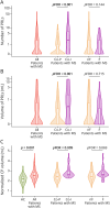
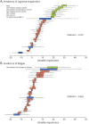
Results: Thirty-six (27.9%) patients with MS were cognitively impaired, and 31/113 (27.4%) patients had fatigue. Fifty-nine (45.7%) patients with MS had ≥1 PRLs (median = 0, interquartile range = 0;2). Compared with HC, patients with MS showed significantly higher T2-hyperintense white matter lesion (WM) volume; lower normalized brain, thalamic, hippocampal, caudate, cortical, and WM volumes; and higher normalized CP volume (p from <0.001 to 0.040). The predictors of cognitive impairment (relative importance) (out-of-bag area under the curve [OOB-AUC] = 0.707) were normalized brain volume (100%), normalized caudate volume (89.1%), normalized CP volume (80.3%), normalized cortical volume (70.3%), number (67.3%) and volume (66.7%) of PRLs, and T2-hyperintense WM lesion volume (64.0%). Normalized CP volume was the only predictor of the presence of fatigue (OOB-AUC = 0.563).
Discussion: Chronic inflammation, with higher number and volume of PRLs and enlarged CP, may contribute to cognitive impairment in MS in addition to gray matter atrophy. The contribution of enlarged CP in explaining fatigue supports the relevance of immune-related processes in determining this manifestation independently of disease severity. PRLs and CP enlargement may contribute to the pathophysiology of cognitive impairment and fatigue in MS, and they may represent clinically relevant therapeutic targets to limit the impact of these clinical manifestations in MS.
Conflict of interest statement
The authors declare that they have no competing interests in relation to this work. Potential conflicts of interest outside the submitted work are as follows: P. Preziosa received speaker honoraria from Roche, Biogen, Novartis, Merck Serono, Bristol-Myers Squibb, Genzyme, Horizon and Sanofi, he has received research support from Italian Ministry of Health and Fondazione Italiana Sclerosi Multipla; E. Pagani has nothing to disclose; A. Meani has nothing to disclose; L. Storelli has nothing to disclose; M. Margoni reports grants and personal fees from Sanofi Genzyme, Merck Serono, Novartis and Almiral; Y. Yudin has nothing to disclose; N. Tedone has nothing to disclose; D. Biondi has nothing to disclose; M. Rubin has nothing to disclose; M.A. Rocca received consulting fees from Biogen, Bristol-Myers Squibb, Eli Lilly, Janssen, Roche, and speaker honoraria from AstraZaneca, Biogen, Bristol-Myers Squibb, Bromatech, Celgene, Genzyme, Horizon Therapeutics Italy, Merck Serono SpA, Novartis, Roche, Sanofi and Teva, she receives research support from the MS Society of Canada, the Italian Ministry of Health, the Italian Ministry of University and Research, and Fondazione Italiana Sclerosi Multipla, she is Associate Editor for Multiple Sclerosis and Related Disorders; M. Filippi is Editor-in-Chief of the Journal of Neurology, Associate Editor of Human Brain Mapping, Neurological Sciences, and Radiology, received compensation for consulting services from Alexion, Almirall, Biogen, Merck, Novartis, Roche, Sanofi, speaking activities from Bayer, Biogen, Celgene, Chiesi Italia SpA, Eli Lilly, Genzyme, Janssen, Merck Serono, Neopharmed Gentili, Novartis, Novo Nordisk, Roche, Sanofi, Takeda, and TEVA, participation in Advisory Boards for Alexion, Biogen, Bristol-Myers Squibb, Merck, Novartis, Roche, Sanofi, Sanofi-Aventis, Sanofi Genzyme, Takeda, scientific direction of educational events for Biogen, Merck, Roche, Celgene, Bristol-Myers Squibb, Lilly, Novartis, Sanofi Genzyme, he receives research support from Biogen Idec, Merck Serono, Novartis, Roche, the Italian Ministry of Health, the Italian Ministry of University and Research, and Fondazione Italiana Sclerosi Multipla. Go to
Figures


References
MeSH terms
LinkOut - more resources
Full Text Sources
Medical
Miscellaneous
