

Isolated laryngeal tuberculosis: A diagnostic dilemma
- PMID: 38350377
- PMCID: PMC10943656
- DOI: 10.1016/j.ijscr.2024.109376
Isolated laryngeal tuberculosis: A diagnostic dilemma
Abstract
Introduction and importance: Primary laryngeal tuberculosis (PLTB) is a rare condition. The symptoms and findings are not specific in most of the cases. Patients are diagnosed essentially based on histopathological examination and mycobacterial culture.
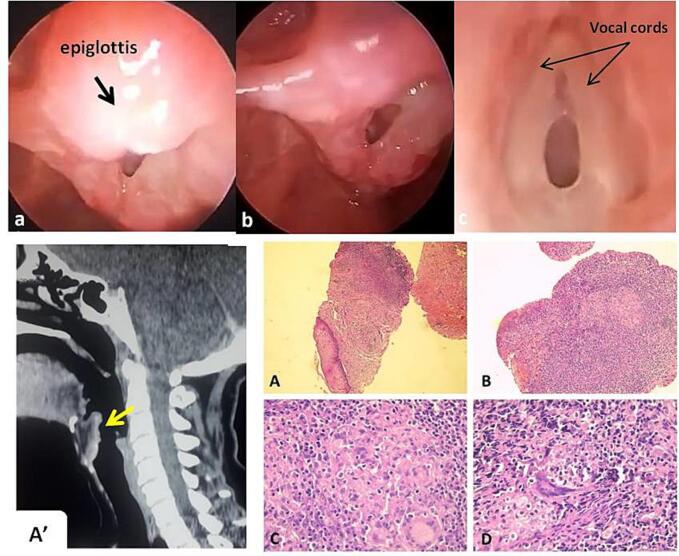
Case report: We report the case of a 42-year-old woman who presented to our hospital with dysphonia and dysphagia. Direct laryngoscopy revealed a lesion of the supraglottis. CT scan found a diffuse thickening of the entire surface of the larynx. A biopsy confirmed the diagnosis of tuberculosis.
Clinical discussion: Primary laryngeal tuberculosis is a rare clinical entity despite its close anatomical and physiological proximity to the lungs.
Conclusion: Clinician should keep in mind the existence of primary laryngeal tuberculosis to avoid delayed diagnosis and treatment, which can lead to high morbidity and mortality.
Keywords: Anti tubercular drugs; Case report; Larynx; Primary; Tuberculosis.
Copyright © 2024 The Authors. Published by Elsevier Ltd.. All rights reserved.
Conflict of interest statement
Conflict of interest statement The authors declare no conflicts of interest.
Figures
Similar articles
-
Primary laryngeal tuberculosis mimicking laryngeal carcinoma: CT scan features.Indian J Radiol Imaging. 2010 Feb;20(1):11-2. doi: 10.4103/0971-3026.59745. Indian J Radiol Imaging. 2010. PMID: 20351985 Free PMC article.
-
Isolated Laryngeal Tuberculosis: a Diagnostic Dilemma.Indian J Otolaryngol Head Neck Surg. 2022 Oct;74(Suppl 2):2308-2310. doi: 10.1007/s12070-020-02139-7. Epub 2020 Sep 10. Indian J Otolaryngol Head Neck Surg. 2022. PMID: 36452854 Free PMC article.
-
Primary Laryngeal Tuberculosis Manifesting as Irregular Vocal Fold Lesion.Turk Arch Otorhinolaryngol. 2022 Mar;60(1):47-52. doi: 10.4274/tao.2021.2021-7-1. Epub 2022 May 12. Turk Arch Otorhinolaryngol. 2022. PMID: 35634235 Free PMC article.
-
Laryngeal manifestations of paracoccidioidomycosis (South American blastomycosis).Arch Otolaryngol Head Neck Surg. 1999 Dec;125(12):1375-8. doi: 10.1001/archotol.125.12.1375. Arch Otolaryngol Head Neck Surg. 1999. PMID: 10604418 Review.
-
Laryngeal tubercolosis: a case report with focus on voice assessment and review of the literature.Acta Otorhinolaryngol Ital. 2022 Oct;42(5):407-414. doi: 10.14639/0392-100X-N2091. Acta Otorhinolaryngol Ital. 2022. PMID: 36541378 Free PMC article. Review.
References
-
- El Ayoubi F., Chariba I., El Ayoubi A., Chariba S., Essakalli L. Primary tuberculosis of the larynx. Eur. Ann. Otorhinolaryngol. Head Neck Dis. 2014;131:361–364. - PubMed
-
- Ponni S., Venkatesan S.K., Saxena S.K., Suryanarayanan G. Primary laryngeal tuberculosis-changing trends and masquerading presentations: a retrospective study. Int J Otorhinolaryngol Head Neck Surg. 2019;5:634.
-
- Ling L., Zhou S.H., Wang S.Q. Changing trends in the clinical features of laryngeal tuberculosis: a report of 19 cases. Int. J. Infect. Dis. 2010;14:e230–e235. - PubMed
Publication types
LinkOut - more resources
Full Text Sources
