. 2024 Feb 13;10(1):e003705.
doi: 10.1136/rmdopen-2023-003705.

Intensive training programme for ultrasound-guided minimally invasive synovial tissue biopsy on knees and wrists in different phases of inflammation

Affiliations

Intensive training programme for ultrasound-guided minimally invasive synovial tissue biopsy on knees and wrists in different phases of inflammation

Lavinia Agra Coletto et al. RMD Open. .

Abstract

Objectives: To develop an intensive training programme for ultrasound (US)-guided synovial tissue (ST) biopsy on knees and wrists in inflammatory arthritis and to assess the learning curve, patient tolerability, sample quality and trainees' expectations.

Methods: Active or remission rheumatoid arthritis patients were enrolled. Nine trainees joined the 4-month programme in a centre experienced in performing US-guided ST biopsies consisting of four sequential phases: (1) observation, (2) performance of guided step-by-step phases, (3) execution of the whole procedure on paired joints (knees or wrists) of the same patient in parallel with the trainer and (4) performance of the procedure autonomously. Sample representativity was assessed by histology, and procedure-related adverse events were recorded. Before and after the programme, trainees' expectations and perceptions were collected.

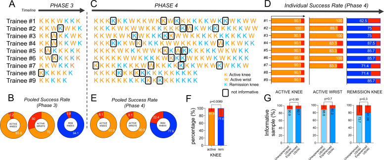
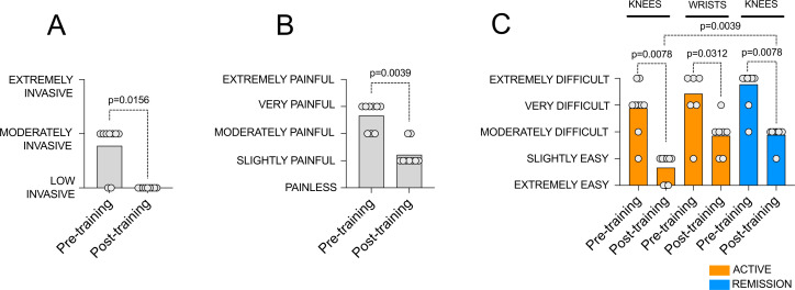
Results: 328 ST biopsy procedures were included. The rate of trainees' informative samples was: (1) comparable to the trainers in active and remission knees, but lower in active wrists (70% for trainees vs 100% for trainers, p=0.06) in phase 3; (2) excellent on active knees and wrists (91.9% and 90.9% respectively) but lower (77.6%, p=0.0089) on remission knees in phase 4. Procedures performed by trainees did not affect patient tolerability. Trainees' expectations about procedure-related invasiveness and pain infliction decreased while the difficulty of procedure execution on active wrists and remission knees remained perceived as moderately difficult.

Conclusions: This intensive training programme develops advanced skills in the performance of US-guided ST biopsy on knees and wrists, yielding high-quality specimens available for basic and translational studies on inflammatory joint diseases.

Keywords: Arthritis, Rheumatoid; Synovitis; Ultrasonography.


Conflict of interest statement

Competing interests: None declared.

Figures

Figure 1
Figure 1
Intensive training programme for minimally invasive ultrasound-guided synovial tissue biopsy of active and remission joints. (A) Schematic of the different phases of minimally invasive ultrasound-guided synovial tissue (ST) biopsy on active knee; (B) Schematic of the different phases of minimally invasive ultrasound-guided ST biopsy on active wrist; (C) Schematic of the different phases of minimally invasive ultrasound-guided ST biopsy on remission knee; (D) Schematic of the steps of the intensive training programme for minimally invasive ultrasound-guided ST biopsy: (1) visualisation of the procedure performed by the trainer, (2) step-by-step performance of the procedure phases by the trainee guided by the trainer, (3) performance of the entire procedure by the trainee compared with the procedure performed by the trainer on the contralateral joint and (4) autonomous performance of the entire procedure by the trainee.
Figure 2
Figure 2
Comparative success rate of synovial tissue retrieval through minimally invasive ultrasound guided biopsy between trainee and trainer. Representative Ultrasound (Grey scale and power Doppler) images and histological (H&E staining) microphotographs and comparative success rate of informative sample retrieval between trainees and trainers on (A) paired active knees, (B) paired active wrists and (C) paired remission knees.
Figure 3
Figure 3
Trainee’s individual path and pooled/individual success rate of the intensive training programme for minimally invasive ultrasound-guided synovial tissue (ST) biopsy of active and remission joints. Temporal schematic representation of each trainee’s individual path during phase 3 (A, B) and phase 4 (C–G) of the intensive training programme for minimally invasive ultrasound-guided ST biopsy of active and remission joints. (A) Phase 3: the trainee performed the procedure immediately after the trainer on the contralateral joint with comparable imaging features of the same patient. Each letter (red K, red W, blue K) represents a procedure (on active knee, active wrist and remission knee, respectively). Black rectangles highlight non-representative procedures. (B) Donut graphs represent pooled success and unsuccess rate of biopsy retrieval for each joint condition of phase 3. (C) Phase 4: the trainee performed the entire procedure autonomously. (D) Phase 4 individual success rate per trainee for each joint condition. (E) Donut graphs represent pooled success and unsuccess rate of biopsy retrieval for each joint condition of phase 4. (F) Pooled success rate of representative ST specimens’ retrieval by minimally invasive ultrasound guided ST biopsy autonomously performed by trainees on active and remission knees in phase 4. (G) Pooled success rate of informative samples autonomously collected by trainees based on their pre-training ultrasound and intra-articular injections’ experience.
Figure 4
Figure 4
Pre-training and post-training perception of trainees on procedure invasiveness and performance difficulty on wrists and knees across disease activity. (A) Pre-training and post-training perception of trainees on procedure invasiveness; (B) Pre-training and post-training perception of trainees on procedure-related pain; (C) Pre-training and post-training perception of trainees on procedure performance difficulty on wrists and knees across diseases activity.

References

    1. Alivernini S, Firestein GS, McInnes IB. The pathogenesis of rheumatoid arthritis. Immunity 2022;55:2255–70. 10.1016/j.immuni.2022.11.009 - DOI - PubMed
    1. Orr C, Vieira-Sousa E, Boyle DL, et al. . Synovial tissue research: a state-of-the-art review. Nat Rev Rheumatol 2017;13:463–75. 10.1038/nrrheum.2017.115 - DOI - PubMed
    1. Humby F, Durez P, Buch MH, et al. . Rituximab versus tocilizumab in anti-TNF inadequate responder patients with rheumatoid arthritis (R4RA): 16-week outcomes of a stratified, biopsy-driven, multicentre, open-label, phase 4 randomised controlled trial. The Lancet 2021;397:305–17. 10.1016/S0140-6736(20)32341-2 - DOI - PMC - PubMed
    1. Alivernini S, Tolusso B, Petricca L, et al. . Synovial features of patients with rheumatoid arthritis and psoriatic arthritis in clinical and ultrasound remission differ under anti-TNF therapy: a clue to interpret different chances of relapse after clinical remission? Ann Rheum Dis 2017;76:1228–36. 10.1136/annrheumdis-2016-210424 - DOI - PMC - PubMed
    1. Alivernini S, Peluso G, Fedele AL, et al. . Tapering and discontinuation of TNF-α blockers without disease relapse using ultrasonography as a tool to identify patients with rheumatoid arthritis in clinical and histological remission. Arthritis Res Ther 2016;18:39. 10.1186/s13075-016-0927-z - DOI - PMC - PubMed

LinkOut - more resources