. 2024 Mar;43(5):868-885.
doi: 10.1038/s44318-024-00045-0. Epub 2024 Feb 14.

The development of early human lymphatic vessels as characterized by lymphatic endothelial markers

Affiliations

The development of early human lymphatic vessels as characterized by lymphatic endothelial markers

Shoichiro Yamaguchi et al. EMBO J. 2024 Mar.

Abstract

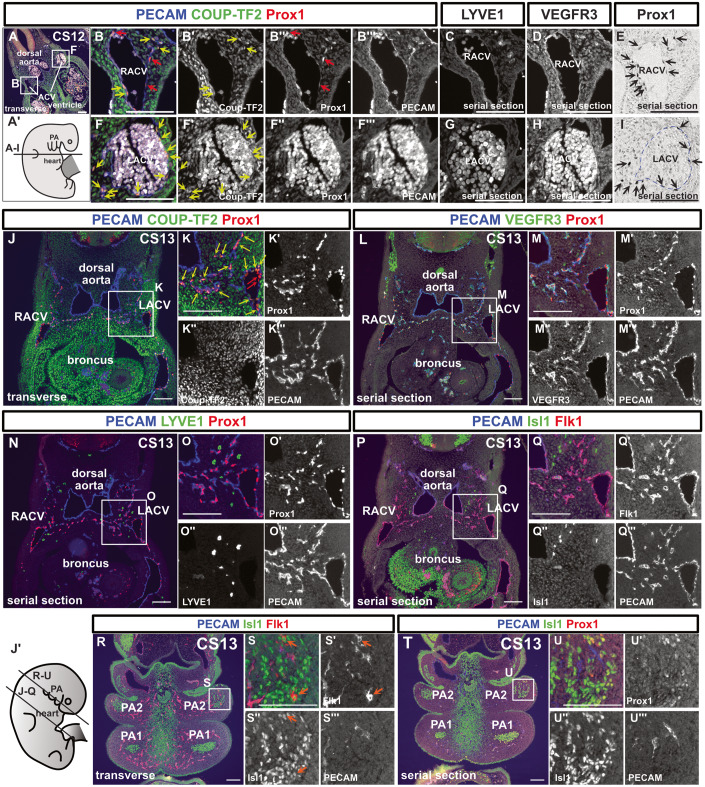
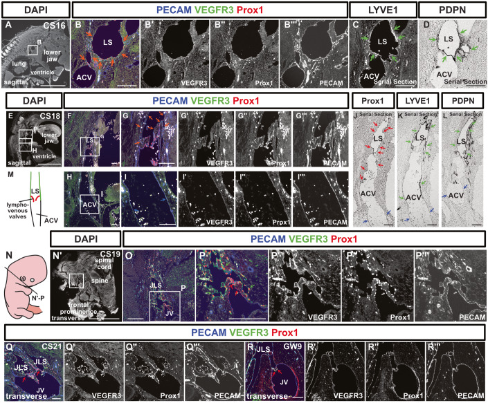
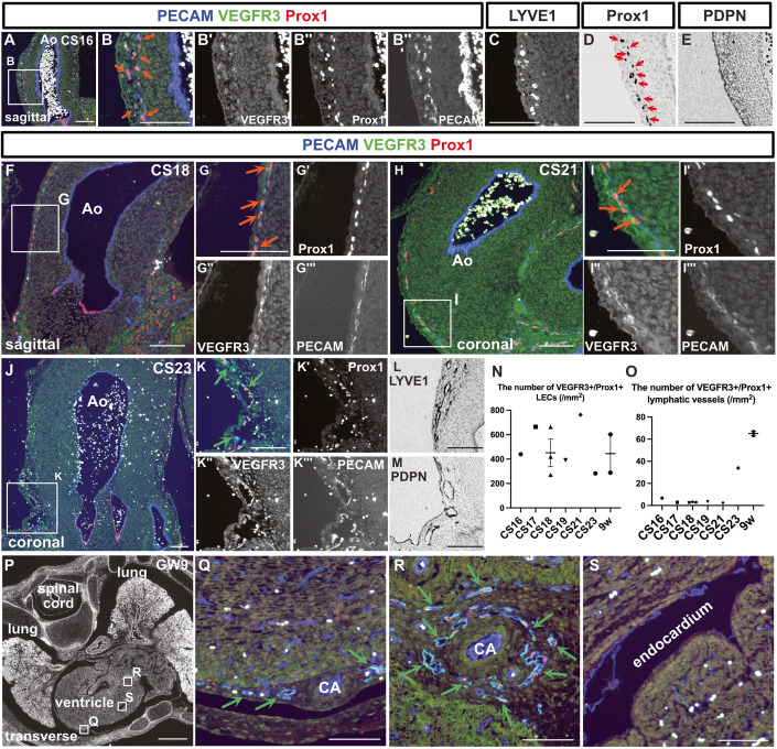
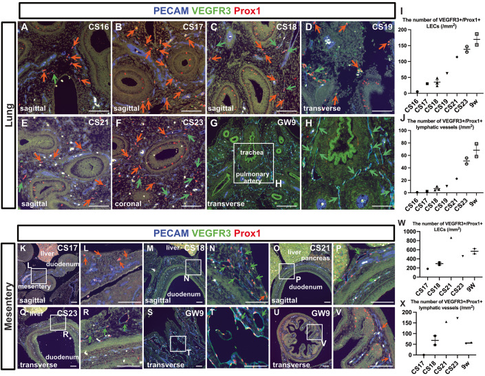
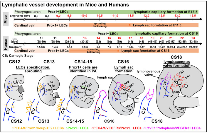
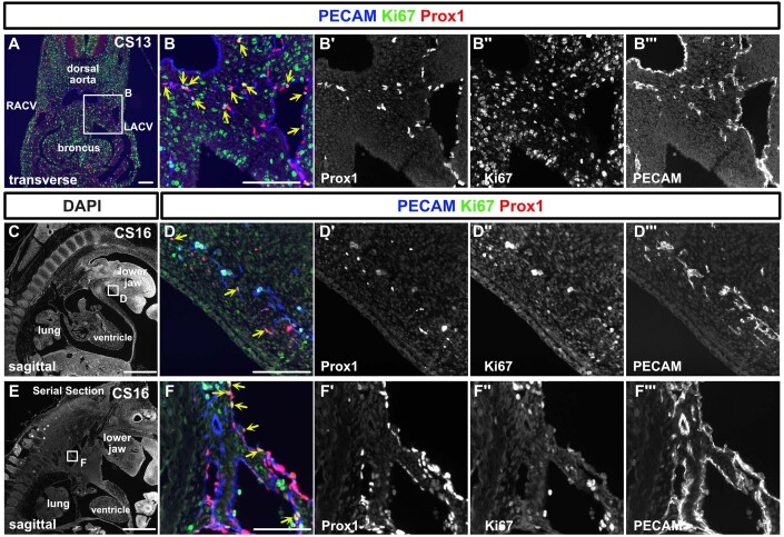
Lymphatic vessel development studies in mice and zebrafish models have demonstrated that lymphatic endothelial cells (LECs) predominantly differentiate from venous endothelial cells via the expression of the transcription factor Prox1. However, LECs can also be generated from undifferentiated mesoderm, suggesting potential diversity in their precursor cell origins depending on the organ or anatomical location. Despite these advances, recapitulating human lymphatic malformations in animal models has been difficult, and considering lymphatic vasculature function varies widely between species, analysis of development directly in humans is needed. Here, we examined early lymphatic development in humans by analyzing the histology of 31 embryos and three 9-week-old fetuses. We found that human embryonic cardinal veins, which converged to form initial lymph sacs, produce Prox1-expressing LECs. Furthermore, we describe the lymphatic vessel development in various organs and observe organ-specific differences. These characterizations of the early development of human lymphatic vessels should help to better understand the evolution and phylogenetic relationships of lymphatic systems, and their roles in human disease.

Keywords: Cellular Origin of Lymphatic Endothelial Cells; Human Embryos; Lymphatic Vessel Development.


Conflict of interest statement

The authors declare no competing interests.

Figures

Figure 1
Figure 1. Lymphatic endothelial cells originate from the cardinal veins in human embryos.
(AI) Immunostaining of transverse sections using the indicated antibodies in a CS12 embryo and schematic representation of the locations of the sections; We detected Prox1+/PECAM+/Coup-TF2 cells (red arrows) and Prox1+/PECAM+/Coup-TF2+ cells (yellow arrows: 78.9% of PECAM+/Prox1+ cells were Coup-TF2+, n = 1) in and around the ACVs. On average, there were 18.25 nuclei per cross-section of the CV; of these, 4.5 were Prox1/PECAM+ blood endothelial cells (BECs), and 13.75 were Prox1+/PECAM+ LECs. Therefore, BECs constituted 24.7%, and LECs constituted 75.3%. There were an average of 9.75 Prox1+/PECAM+ cells located externally to the CV. (E, I) Enzyme-antibody method; Prox1+ cells were present around the ACVs (black arrows). These Prox1+/PECAM+ cells were localized around the cardinal veins in the CS12 embryos. (JU”’) Fluorescent immunostaining of transverse sections using the indicated antibodies in a CS13 embryo and a schematic representation of the positions of the sections; The PECAM+/Prox1+ LECs budding from the ACVs consisted of Coup-TF2-expressing LECs (yellow arrows: 82.8 ± 2.18% of the PECAM+/Prox1+ cells were Coup-TF2+, n = 2) and Coup-TF2 LECs (red arrows). PECAM/Isl1+/Flk1+ cardiovascular progenitor cells were observed in the pharyngeal arch mesoderm (orange arrow). CV cardinal vein, RACV right anterior cardinal vein, LACV left anterior cardinal vein, PA1 first pharyngeal arch, PA2 second pharyngeal arch. Scale bar, 100 μm (AU). Source data are available online for this figure.
Figure 2
Figure 2. Lymph sacs express LYVE1 and PDPN and establish connections with the anterior cardinal veins to form lymphovenous valves.
(AD) Immunostaining of sagittal sections from a CS16 embryo using the indicated antibodies. (B, C) Fluorescent immunostaining. (D) Immunostaining using the enzyme-antibody method. At CS16, lymph sacs emerged, which exhibited Prox1, VEGFR3, and PECAM expression (orange arrows). (C, D) The expression of LYVE1 and PDPN began within these lymph sacs at this stage (green arrows). (EL) Immunostaining of sagittal sections from a CS18 embryo using the indicated antibodies. (EI”’) Fluorescent immunostaining. (JL) The enzyme-antibody method. (EI”’) Lymph sacs expressing PECAM, VEGFR3, and Prox1 (orange arrows) formed connections with the ACVs, which expressed PECAM (blue arrows). (JL) While Prox1 (red arrows), LYVE1, and PDPN (green arrows) were expressed in the lymph sacs, these markers were absent from the ACVs (blue arrows). There were four CS18-stage embryos, but only one included lymph sacs. (M) Schematic representation of the relationship between the ACVs and lymph sacs in a CS18 embryo. The lymph sacs and the ACVs were continuous, featuring a valve structure on the lymph sac side. (NP”’) Transverse sections and schematic representation of the positions of the sections in a CS19 embryo; Fluorescent immunostaining of PECAM, Prox1, and VEGFR3 was conducted. VEGFR3 was expressed in lymph sacs at CS19 (green arrows). The LVVs were overlaid with Prox1+ cells (red arrows) (LVVs were identifiable in 1 of 5 embryos). (QR”’) Transverse sections of a CS21 embryo and a GW9 fetus, highlighting the areas containing LVVs; as was the case at CS19, LVVs were visible (red arrows). (At CS21, LVVs were identified in 1 out of 2 embryos; at GW9, LVVs were identified in 1 out of 4 fetuses). LS lymph sacs, ACV anterior cardinal vein, JLS jugular lymph sacs, JV jugular vein. Scale bars, 1 mm (A, E, N’) or 100 μm (BD, FL, OR). Source data are available online for this figure.
Figure 3
Figure 3. Development of the cardiac lymphatic vessels.
(AE) Immunostaining of sagittal sections from a CS16 embryo using the indicated antibodies. (AC) Fluorescent immunostaining. (D, E) Enzyme-antibody method, followed by DAB color development; PECAM+/Prox1+/VEGFR3+ LECs were present in the aortic wall (orange arrows). (D) Prox1+ cells were also identified by the enzyme-antibody method (red arrows). (FI”’) Immunostaining of sections from CS18 and CS21 embryos using the indicated antibodies. Consistent with the CS16 embryo, PECAM+/Prox1+/VEGFR3+ LECs could be identified in the aortic wall (orange arrows). (JM) Immunostaining of coronal sections from a CS23 embryo using the indicated antibodies. (JK”’) Fluorescent immunostaining. (L, M) Enzyme-antibody method, followed by DAB color development; LYVE1+ and PDPN+ lymphatic vessels were identified around the aorta (green arrows). (N, O) Changes in the number of PECAM+/Prox1+/VEGFR3+ LECs in the aortic wall as development advanced (N). Changes in the number of PECAM+/Prox1+/VEGFR3+ luminal lymphatic vessels as development advanced (O). (N, O) Data are presented as the mean ± standard error of the mean (SEM). Each dot represents a single individual. (PS) Fluorescent immunostaining of PECAM, Prox1, and VEGFR3 in transverse sections of a GW9 fetus. By GW9, lymphatic vessels could be seen around the coronary arteries in the epicardium (green arrows). Ao aorta, CA coronary artery. Scale bars, 1 mm (P) or 100 μm (AM, QS). Source data are available online for this figure.
Figure 4
Figure 4. Development of the lung lymphatic vessels and mesenteric lymphatic vessels.
(AH) Immunostaining of PECAM, Prox1, and VEGFR3 in the lungs at each stage. The orange arrows indicate isolated or small clusters of LECs. The green arrows indicate lymphatic vessels that had formed luminal structures. (I, J) Changes in the number of PECAM+/Prox1+/VEGFR3+ LECs in the aortic wall as development advanced (I). Changes in the number of PECAM+/Prox1+/VEGFR3+ luminal lymphatic vessels as development advanced (J). Each dot represents a single individual. (I, J) Data are presented as the mean ± standard error of the mean (SEM). (KV) Immunostaining of PECAM, Prox1, and VEGFR3 in the intestine and mesentery at each indicated stage. The orange arrows indicate isolated or small clusters of LECs that had not formed luminal structures, while the green arrows indicate lymphatic vessels that had formed luminal structures. (W, X) Changes in the number of PECAM+/Prox1+/VEGFR3+ LECs in the aortic wall as development advanced (W). Changes in the number of PECAM+/Prox1+/VEGFR3+ luminal lymphatic vessels as development advanced (X). (W, X) Data are presented as the mean ± standard error of the mean (SEM). Each dot represents a single individual. Scale bars, 100 μm (AH, KV). Source data are available online for this figure.
Figure 5
Figure 5. Comparative overview of early lymphatic vessel development in mouse and human embryos.
In mouse embryos, Prox1 expression is initiated in the cardiac pharyngeal mesoderm from E9.0 to E9.5, leading to the distribution of LECs in the head and neck, mediastinal, and cardiac outflow regions. By E13.5, these LECs start forming capillary lymphatics, which further develop into larger lymphatic vessels. Prox1 expression in the cardinal vein starts at E9.5, with the lymph sac forming post-sprouting by E11.5. In contrast, for human embryos, Prox1 expression in the pharyngeal arch area is first noted at CS15. Following this, LECs in the mandibular and cardiac outflow tract regions slowly establish a capillary lymphatic network, which by CS23 progresses to clearly defined lymphatic vessels with luminal structures. Prox1 expression in the cardinal vein begins at CS12, with the lymph sac being discernible by CS16. By CS18, the lymphovenous valve is identifiable between the lymph sac and the ACV. Across both embryos, early expression of transcription factors such as Prox1 and Coup-TF2 is observed, with the subsequent expression of VEGFR3, LYVE1, and Podoplanin. CS Carnegie stage, ACV the anterior cardinal veins.
Figure EV1
Figure EV1. Multiple lymphatic markers are expressed in fetal lymph sacs.
(AH) Schema showing the positions of the sections in a GW9 fetus (A), and immunostaining of transverse sections (B–H); (CC”’) Fluorescent immunostaining of PECAM, Prox1, and VEGFR3; These markers were expressed in lymph sacs (green arrows). (DH) Immunostaining of Prox1, LYVE1, VEGFR3, PDPN, and PECAM using the enzyme-antibody method, with color development by DAB. (IL) Imaging of the secondary antibody-only staining. (M, N) Immunostaining of Flk1, and Isl1 using the enzyme-antibody method, with color development by DAB. Scale bars, 1 mm (B) or 100 μm (C, D, I, J, M).
Figure EV2
Figure EV2. Prox1 expression is not observed in the precardial vein of the CS11 embryo.
(AC’) Immunostaining of transverse sections of a CS11 embryo with the indicated antibodies and schema showing a CS11 embryo (n = 1). Scale bars, 1 mm (B) or 100 μm (C, C’).
Figure EV3
Figure EV3. LECs bud from the cardinal veins and form luminal structures.
(AJ) Immunostaining of sagittal sections of a CS14 embryo with the indicated antibodies and schema showing the CS14 embryo. (BC”’) Cardiovascular progenitor cells, which were composed of FLK1+/Isl1+/PECAM cells, were observed in the second pharyngeal arch (white arrows). (FG”’) PECAM+/Prox1+/Coup-TF2+ cells (yellow arrows: Frequency of Coup-TF2+ cells among PECAM+/Prox1+ cells=44.1%, n = 1 [the ACVs could not be identified in another embryo]), and PECAM+/Prox1+/Coup-TF2- cells (red arrows) were observed in and around the ACVs. (KP’) HE staining of a sagittal section of a CS15 embryo (K) and immunostaining of sagittal sections of the same CS15 embryo with the indicated antibodies (LP’); At this stage, scattered Prox1+ cells were observed in the pharyngeal arch (red arrows). (QS”’) Immunostaining of sagittal sections of a CS16 embryo with the indicated antibodies. PA1 first pharyngeal arch, PA2 second pharyngeal arch, ACV anterior cardinal vein. scale bars, 1 mm (K) or 100 μm (BG, L).
Figure EV4
Figure EV4. Development of lymphatic vessels in the lower jaw, kidneys, and thoracic duct.
(AO) Immunostaining of sagittal sections of embryos collected at the described stages with the indicated antibodies; The orange arrows indicate isolated or small clusters of LECs. The green arrows indicate lymphatic vessels that had formed luminal structures. (P, Q) Changes in the number of PECAM+/Prox1+/VEGFR3+ LECs in the aortic wall (P). Changes in the number of PECAM+/Prox1+/VEGFR3+ luminal lymphatic vessels (Q). Each dot represents a single individual. (RU) Fluorescent immunostaining of PECAM, Prox1, and VEGFR3 in sections from embryos collected at the described stages. The orange arrows indicate isolated or small clusters of LECs. The green arrows indicate lymphatic vessels that had formed luminal structures. (V, W) The green arrows indicate lymphatic vessels surrounding the aorta at GW9. (X, Y) Immunostaining for PDPN and LYVE1 in the lung, heart, kidney, mesentery, intestine, and lower jaw. Scale bars, 1 mm (A, V) or 100 μm (B, C, FO, RU, W, X).
Figure EV5
Figure EV5. Proliferation activity of lymphatic endothelial cells at CS13 and CS16.
(AF”’) Immunostaining of transverse and sagittal sections using the indicated antibodies in a CS13 and 16 embryos. We detected Prox1+/PECAM+/Ki67+ LECs (indicated by yellow arrows) as well as Prox1+/PECAM+/Ki67- LECs. At CS13, within CV, 19.6% of LECs were Ki67 positive, while outside the CV, the percentage was 43.6%. At CS16, 22.7% of LECs in the lymph sacs were Ki67 positive, and in the lower jaw, the positivity rate was 31% (based on an average of two sections at n = 1). CV cardinal vein, RACV right anterior cardinal vein, LACV left anterior cardinal vein. scale bar, 1 mm (C, E), 100 μm (A, B, D, F).

Similar articles

Cited by

References

    1. Alitalo K. The lymphatic vasculature in disease. Nat Med. 2011;17:1371–1380. doi: 10.1038/nm.2545. - DOI - PubMed
    1. Antila S, Karaman S, Nurmi H, Airavaara M, Voutilainen MH, Mathivet T, Chilov D, Li Z, Koppinen T, Park J-H, et al. Development and plasticity of meningeal lymphatic vessels. J Exp Med. 2017;214:3645–3667. doi: 10.1084/jem.20170391. - DOI - PMC - PubMed
    1. Banda CH, Shiraishi M, Mitsui K, Okada Y, Danno K, Ishiura R, Maemura K, Chiba C, Mizoguchi A, Imanaka-Yoshida K, et al. Structural and functional analysis of the newt lymphatic system. Sci Rep. 2023;13:6902. doi: 10.1038/s41598-023-34169-w. - DOI - PMC - PubMed
    1. Budras K‐D, Hullinger RL, Rautenfeld DBV. Lymph heart musculature in birds. J Morphol. 1987;191:77–87. doi: 10.1002/jmor.1051910108. - DOI - PubMed
    1. François M, Caprini A, Hosking B, Orsenigo F, Wilhelm D, Browne C, Paavonen K, Karnezis T, Shayan R, Downes M, et al. Sox18 induces development of the lymphatic vasculature in mice. Nature. 2008;456:643–647. doi: 10.1038/nature07391. - DOI - PubMed

Substances

Supplementary concepts