Review
. 2024 Oct;96(1):150-169.
doi: 10.1111/prd.12548. Epub 2024 Feb 13.

The role of programmed death receptor (PD-)1/PD-ligand (L)1 in periodontitis and cancer

Affiliations
Review

The role of programmed death receptor (PD-)1/PD-ligand (L)1 in periodontitis and cancer

Sabine Groeger et al. Periodontol 2000. 2024 Oct.

Abstract

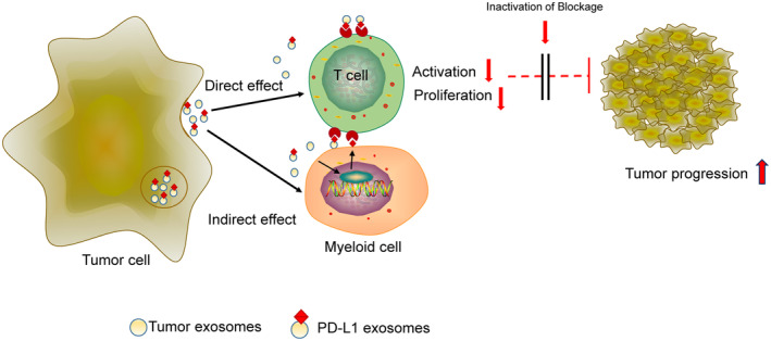
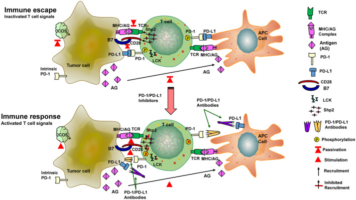
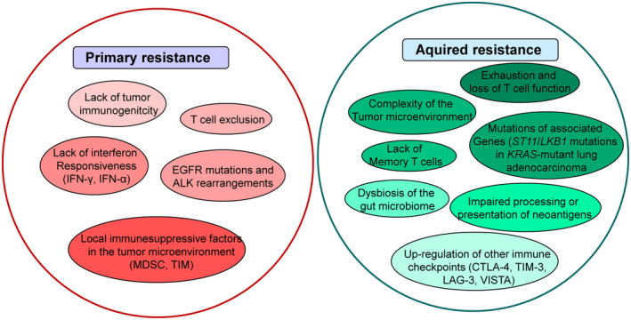
The programmed-death-ligand-1 (PD-L1) is an immune-modulating molecule that is constitutively expressed on various immune cells, different epithelial cells and a multitude of cancer cells. It is a costimulatory molecule that may impair T-cell mediated immune response. Ligation to the programmed-death-receptor (PD)-1, on activated T-cells and further triggering of the related signaling pathways can induce T-cells apoptosis or anergy. The upregulation of PD-L1 in various cancer types, including oral squamous cell carcinomas, was demonstrated and has been linked to immune escape of tumors and poor prognosis. A bidirectional relationship exists between the increased PD-L1 expression and periodontitis as well as the epithelial-mesenchymal transition (EMT), a process of interconversion of epithelial cells to mesenchymal cells that may induce immune escape of tumors. Interaction between exosomal PD-L1 and PD-1 on T-cells may cause immunosuppression by blocking the activation and proliferation of T-cells. The efficacy and importance of treatment with PD-1/PD-L1 checkpoint inhibitors and their prognostic influence on human cancers was demonstrated. Regarding PD-1/PD-L1 checkpoint inhibitors, resistances exist or may develop, basing on various factors. Further investigations of the underlying mechanisms will help to overcome the therapeutic limitations that result from resistances and to develop new strategies for the treatment of cancer.

Keywords: EMT; PD‐L1; cancer; exosomes; immune checkpoint; immune escape; periodontitis.


Conflict of interest statement

The authors declare that they have no conflicts of interest.

Figures

FIGURE 1
FIGURE 1
PD‐L1 as mechanistic link between periodontitis and cancer. The PD‐1/PD‐L1 checkpoint in periodontitis: Bacterial peptidoglycans (i.e. P. gingivalis, F. nucleatum) may induce the upregulation of PD‐L1 and PD‐1 expressed on gingival keratinocytes and lymphocytes T‐cells. The PD‐1/PD‐L1 interaction causes immune suppressive effects that contribute to the process of periodontitis. PD‐L1‐ expression inhibiting drugs or PD‐L1 functional blockage could be useful to revers immune escape and inflammation linked with periodontitis. Modified after Bailly et al.
FIGURE 2
FIGURE 2
PD‐L1 as mechanistic link between periodontitis and cancer. Adaptive resistance to tumor immunity mediated by PD‐1/PD‐L1: After activation in lymphoid organs, tumorspecific T effector cells (Teffs) migrate into the tumor site and then develop tumor‐infiltrating lymphocytes (TILs). Following recognition of tumor antigens, TILs release cytokines including IFN‐γ, which induces the expression of PD‐L1 in the tumor microenvironment. Binding to PD1, PD‐L1 provides a suppressive signal to T‐cells and an anti‐apoptotic signal to tumor cells, causing dysfunction of T‐cells and tumor survival. Modified after Ghosh et al.
FIGURE 3
FIGURE 3
Epithelial–mesenchymal transition (EMT) as factor in tumor progression and metastatic expansion. Outline of a typical EMT programme: Epithelial cells showing apical–basal polarity are connected by tight junctions, adherens junctions and desmosomes and are attached to the underlying basement membrane by hemidesmosomes. Molecules associated with epithelial state and cell polarity: E‐cadherin, epithelial cell adhesion molecules, occludins, claudins, Α6β4 inegrins, cytokeratins. The induction of EMT results in the expression of the EMT‐maintaining transcription factors (EMT‐TFs) ZEB, SNAIL and TWIST that inhibit the expression of genes associated with the epithelial state, coming along with activation of the expression of genes associated with the mesenchymal state: N‐cadherin, Vimentin, Fibronectin, β1 and β3 integrins, MMPs. Changed gene expression lead to cellular modifications including disassembly of epithelial cell–cell junctions and the dissolution of apical–basal cell polarity by the downregulation of crumbs, PALS1‐associated tight junction protein (PATJ) and lethal giant larvae (LGL), all proteins that specifically maintain tight junction formation and apical–basal polarity. The process of EMT leads to enhanced cellular motility and the cells derive invasive capabilities. The process of EMT is reversible, and mesenchymal cells can go back to the epithelial morphology by undergoing mesenchymal–epithelial transition (MET). Both, EMT and MET, take place during normal development as well as cancer progression. E‐cadherin, epithelial cadherin; MMP, matrix metalloproteinase; N‐cadherin, neural cadherin. Modified after Dongre et al.
FIGURE 4
FIGURE 4
Epithelial–mesenchymal transition (EMT) as factor in tumor progression and metastatic expansion. Schematic overview of the crosstalk between EMT and IFNγ‐induced PD‐L1 expression: IFNγ‐induced PD‐L1 upregulation supports the processes of EMT. EMT can also be induced by, i.e.TGF‐β, enhanced PD‐L1 expression levels, promoting tumor immunoevasion. Modified after Burger et al.
FIGURE 5
FIGURE 5
Mechanisms of exosomal PD‐L1 induced immunosuppression. Interaction between exosomal PD‐L1 and PD‐1 on T‐cells may directly cause immunosuppression. Additionally, exosomal PD‐L1 upregulates the expression of PD‐L1 in myeloid cells via the NF‐κB pathway, induces their transformation into myeloid immune cells, and indirectly suppresses the activation and proliferation of T‐cells. The final result of these processes is T‐cell inactivation and tumor progression. Modfied after Ye et al.
FIGURE 6
FIGURE 6
PD‐L1 as therapeutic target in cancer. Mechanisms of PD‐1/PD‐L1 inhibitor treatment response: PD‐1 mainly is expressed in T‐cells and PD‐L1 in tumor cells and antigen presenting (APC) cells. Some tumor cells can also express intrinsic PD‐1. After PD‐1 – PD‐L1 ligation immune escape is enabled. PD‐1 can be phosphorylated by LCK to further recruit tyrosine phosphatase Src homologous phosphatase 2 (Shp2), resulting in inactivation of CD28 and TCR function and signaling pathway. This leads to attenuation of the T‐cell activating signal that causes immune escape. Lymphocyte‐specific protein tyrosine kinase (LCK) kinase activity is needed to mediate PD‐1/SHP‐2‐maintained inhibiting signaling. The interaction of PD‐1/PD‐L1 immunocheckpoint inhibitors with its targets can effectively block the binding between PD‐L1 and PD‐1, which in turn inhibits the recruitment of SHP‐2 and can reactivate T‐cells signaling and their immunologic function. Modified after Tang et al.
FIGURE 7
FIGURE 7
PD‐L1 as therapeutic target in cancer. Mechanisms of PD‐1/PD‐L1 inhibitors resistance: PD‐1/PD‐L1 inhibitor resistance is classified as primary resistance and acquired resistance. Mechanisms of primary resistance include lack of tumor immunogenicity; T‐cell rejection; lack of interferon responsiveness, such as IFNg (interferon‐γ) and IFN‐α (interferon‐α); EGFR (epidermal growth factor receptor) mutations and ALK rearrangements; local immunosuppressive factors within the tumor microenvironment, such as MDSC (myeloid‐derived suppressor cell) and TIM (tumorinfiltrating myeloid cell). The mechanisms of acquired resistance are attributed to exhaustion and loss of T‐cell function; impaired processing or presentation of neoantigens; complexity of the tumor microenvironment; mutations in associated genes, such as STK11/LKB1; dysbiosis of the gut microbiome; lack of Memory T‐cells and upregulation of other Immune Checkpoints, such as CTLA‐4 (cytotoxic T‐lymphocyte antigen‐4), TIM‐3 (T‐cell immunoglobulin and mucin domain‐containing molecule‐3), LAG‐3 (lymphocyte activation gene‐3) and VISTA (V‐domain Ig suppressor of T‐cell activation). Modified after Tang et al.

Similar articles

Cited by

References

    1. Darveau RP. Periodontitis: a polymicrobial disruption of host homeostasis. Nat Rev Microbiol. 2010;8:481‐490. - PubMed
    1. Larsen T, Fiehn NE. Dental biofilm infections—an update. APMIS. 2017;125:376‐384. - PubMed
    1. Picolos DK, Lerche‐Sehm J, Abron A, Fine JB, Papapanou PN. Infection patterns in chronic and aggressive periodontitis. J Clin Periodontol. 2005;32:1055‐1061. - PubMed
    1. Hajishengallis G, Darveau RP, Curtis MA. The keystone‐pathogen hypothesis. Nat Rev Microbiol. 2012;10:717‐725. - PMC - PubMed
    1. Dong H, Strome SE, Salomao DR, et al. Tumor‐associated B7‐H1 promotes T‐cell apoptosis: a potential mechanism of immune evasion. Nat Med. 2002;8:793‐800. - PubMed

MeSH terms

LinkOut - more resources