. 2024 Apr;41(2):61-73.
doi: 10.12701/jyms.2023.01347. Epub 2024 Feb 14.

Comprehensive overview of the role of mitochondrial dysfunction in the pathogenesis of acute kidney ischemia-reperfusion injury: a narrative review

Affiliations

Comprehensive overview of the role of mitochondrial dysfunction in the pathogenesis of acute kidney ischemia-reperfusion injury: a narrative review

Min-Ji Kim et al. J Yeungnam Med Sci. 2024 Apr.

Abstract

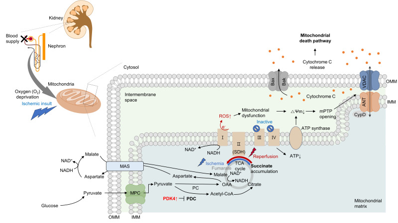
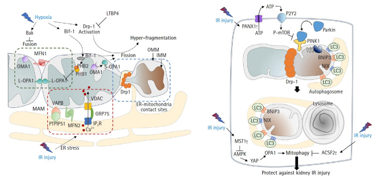
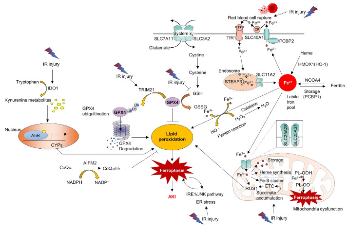
Acute kidney ischemia-reperfusion (IR) injury is a life-threatening condition that predisposes individuals to chronic kidney disease. Since the kidney is one of the most energy-demanding organs in the human body and mitochondria are the powerhouse of cells, mitochondrial dysfunction plays a central role in the pathogenesis of IR-induced acute kidney injury. Mitochondrial dysfunction causes a reduction in adenosine triphosphate production, loss of mitochondrial dynamics (represented by persistent fragmentation), and impaired mitophagy. Furthermore, the pathological accumulation of succinate resulting from fumarate reduction under oxygen deprivation (ischemia) in the reverse flux of the Krebs cycle can eventually lead to a burst of reactive oxygen species driven by reverse electron transfer during the reperfusion phase. Accumulating evidence indicates that improving mitochondrial function, biogenesis, and dynamics, and normalizing metabolic reprogramming within the mitochondria have the potential to preserve kidney function during IR injury and prevent progression to chronic kidney disease. In this review, we summarize recent advances in understanding the detrimental role of metabolic reprogramming and mitochondrial dysfunction in IR injury and explore potential therapeutic strategies for treating kidney IR injury.

Keywords: Acute kidney injury; Ferroptosis; Mitochondria; Mitochondrial dynamics; Mitophagy; Reactive oxygen species.


Conflict of interest statement

Conflicts of interest

No potential conflict of interest relevant to this article was reported.

Figures

Fig. 1.
Fig. 1.
Ischemia induces mitochondrial death pathway and aberrant succinate accumulation. Ischemia significantly impacts mitochondrial functions, notably causing a decline in adenosine triphosphate (ATP) production within kidney proximal tubule cells. During reperfusion, a compromised state becomes evident with a significant increase in reactive oxygen species (ROS), which leads to mitochondrial dysfunction. This dysfunction causes a decrease in mitochondrial membrane potential (ΔΨm), subsequently triggering the opening of the mitochondrial permeability transition pore (mPTP). A crucial event in this process is the opening of the mPTP, which allows the release of cytochrome c and initiates the mitochondrial death pathway. Concurrently, hypoxia induces a reversal in the activity of complex II, leading to an excessive accumulation of succinate. Upon reperfusion, the accumulated succinate is rapidly oxidized back by succinate dehydrogenase (SDH). This phenomenon amplifies ROS generation, ultimately culminating in cellular death. Pyruvate dehydrogenase kinase 4 (PDK4) inhibition enhances pyruvate dehydrogenase complex (PDC) activity. Inhibition of the malate/aspartate shuttle (MAS) by activating the pyruvate dehydrogenase (PDH) flux can mitigate this succinate accumulation. VDAC, voltage-dependent anion channel; ANT, adenine nucleotide translocase; OMM, outer mitochondrial membrane; IMM, inner mitochondrial membrane; CypD, cyclophilin D; NAD+, oxidized nicotinamide adenine dinucleotide; NADH, reduced nicotinamide adenine dinucleotide; TCA, tricarboxylic acid cycle; MPC, mitochondrial pyruvate carrier; PC, pyruvate carboxylase.
Fig. 2.
Fig. 2.
Mitochondrial dynamics and quality control in kidney IR injury. (A) Mitochondrial fusion-fission: mitochondria undergo fusion or division in response to stimuli like hypoxia. Dynamin-related protein 1 (Drp-1) predominantly causes fission. Importantly, OMA1-mediated proteolysis of optic atrophy 1 (OPA1) present in two main isoforms, a long form and a short form, plays a differential role in these processes. The long form of OPA1 (L-OPA1) promotes mitochondrial fusion, while the short form of OPA1 (S-OPA1) is more associated with fission. Stress can lead to inner membrane cleavage through OMA1’s action on OPA1, converting L-OPA1 to its S-OPA1 counterpart. In kidney injury, such processes are linked to tubular cell apoptosis. Under stressful conditions including IR injury, the endoplasmic reticulum (ER) also undergoes stress. Persistent ER stress causes a large amount of Ca2+ release from the ER. This released Ca2+ results in mitochondrial Ca2+ overload through the inositol 1,4,5-trisphosphate receptor (IP3R)-voltage-dependent anion channel (VDAC) 1, which forms a bridge between the two organelles at mitochondria-associated membranes. Furthermore, under such stressful conditions, Bax-interacting factor 1 (Bif-1) regulates mitochondrial membranes by interacting with prohibitin-2. This interaction leads to disruption of prohibitin (PHB) complexes, culminating in the inactivation of OPA1, particularly the L-OPA1 isoform, thereby favoring mitochondrial fission. (B) Mitochondrial quality control: mitophagy is the process of clearing damaged mitochondria from cells essential in kidney ischemia-reperfusion (IR) injury. PTEN-induced kinase 1 (PINK1) and parkin can detect mitochondrial quality, leading to removal of damaged mitochondria. Activation of mitophagy is critical for reducing IR injury. Disruption of mitophagy worsens the injury. Several proteins and pathways, such as pannexin 1 (PANX1), mammalian sterile 20-like kinase 1 (MST1), and acyl-CoA synthetase family member 2 (ACSF2), have roles in mitophagy and kidney IR injury protection. LTBP4, latent transforming growth factor-beta binding protein 4; MFN, mitofusin; OMM, outer mitochondrial membrane; IMM, inner mitochondrial membrane; VAPB, vesicle-associated membrane protein-associated protein B; GRP75, glucose-regulated protein 75; PTPIP51, protein tyrosine phosphatase interacting protein 51; ATP, adenosine triphosphate; P-mTOR, phosphorylated mammalian target of rapamycin; LC3, microtubule-associated proteins 1A/1B light chain 3; BNIP3, Bcl-2 interacting protein 3; NIX, NIP3-like protein X; AMPK, AMP-activated protein kinase; YAP, yes-associated protein.
Fig. 3.
Fig. 3.
Ferroptosis as a central mechanism in kidney ischemia-reperfusion (IR) injury. IR injury can induce lipid peroxidation through various pathways, ultimately triggering ferroptosis and leading to acute kidney injury (AKI). Cells have various antioxidant enzymes (e.g., Catalase) and coenzymes (e.g., coenzyme Q10 [CoQ10]) to eliminate reactive oxygen species (ROS), thus maintaining cellular homeostasis. However, excessive ROS generated due to prolonged injury can eventually lead to lipid peroxidation. IR injury causes the release of Fe3+ from ruptured red blood cells, which binds to transferrin (TF) and then associates with TF receptor 1 (TfR1) on the cell membrane, forming endosomes. Within the endosome, six-transmembrane epithelial antigen of the prostate 3 (STEAP3) converts Fe3++ to Fe2+, which is subsequently released into the cytoplasm through solute carrier family 11 member 2 (SLC11A2), establishing a labile iron pool. Fe2++ is extruded from the cytoplasm to the extracellular space through cell membrane proteins such as SLC40A1, solute carrier family 40 member 1 (SCL40A1) and adaptor protein (poly(rC)-binding protein [PCBP] 2). Extracellular Fe2+ is converted back to Fe3+ by ceruloplasmin (CP). It can bind to TF, re-entering the cytoplasm by binding to TfR1, leading to endosome formation. Intracellularly, Fe2+ within the labile iron pool is regulated by PCBP1, which can store it as ferritin or convert it back to Fe2+ through nuclear receptor coactivator 4 (NCOA4). Additionally, heme oxygenase 1 (HMOX1) contributes to intracellular Fe2+ accumulation. Prolonged IR injury can lead to excessive cytoplasmic Fe2+, promoting the Fenton reaction, converting Fe2+ to Fe3+, and generating H2O2 and hydroxyl radicals (•OH) with subsequent lipid peroxidation. Intracellular Fe2+ also accumulates within mitochondria through membrane proteins such as solute carrier family 25 member 28 (SLC25A28) and solute carrier family 25 member 37 (SLC25A37), contributing to heme synthesis and Fe-S cluster formation. Fe2+ within mitochondria can accumulate ROS including peroxides (polyunsaturated fatty acid-phospholipid ethanolamine peroxyl radical, PL-OO•). Additionally, excessive succinate accumulation during IR injury can lead to ROS generation through the electron transport chain (ETC), causing lipid peroxidation both inside and outside the mitochondria. The antioxidant enzyme glutathione peroxidase 4 (GPX4) plays a crucial role in reducing lipid peroxidation by using reduced glutathione (GSH) to convert oxidized glutathione (GSSG) while countering ROS. GSH is produced intracellularly through the Xc (solute carrier family 7 member 11 [SCL7A11], solute carrier family 3 member 2 [SCL3A2]) transport system, converting cystine to cysteine. However, reduced GSH during IR injury can compromise GPX4’s activity. Increased tripartite motif-containing 21 (TRIM21) activity during IR injury can lead to GPX4 ubiquitination and degradation, intensifying ROS and lipid peroxidation. Activated inositol-requiring enzyme 1 (IRE1)/jun N-terminal kinase (JNK) pathways during IR injury can induce endoplasmic reticulum (ER) stress, promoting ferroptosis. Ferroptosis can exacerbate ER stress. IR-induced activation of indoleamine 2,3‑dioxygenase 1 (IDO1) can lead to increased conversion of intracellular tryptophan to kynurenine metabolites. These metabolites can bind to the aryl hydrocarbon receptor (AhR) in the nucleus, enhancing the expression of cytochrome P450 superfamily of enzymes (CYPs). CYPs can further increase ROS production, contributing to lipid peroxidation. AIFM2, apoptosis-inducing factor mitochondria-associated 2; CoQ10H2, reduced coenzyme Q10; NADPH, reduced nicotinamide adenine dinucleotide phosphate; NADP+, oxidized nicotinamide adenine dinucleotide phosphate; DMT1, divalent metal transporter 1; PL-OOH, polyunsaturated fatty acid containing phospholipid hydroperoxides.

Similar articles

Cited by

References

    1. Khwaja A. KDIGO clinical practice guidelines for acute kidney injury. Nephron Clin Pract. 2012;120:c179–84. - PubMed
    1. Rewa O, Bagshaw SM. Acute kidney injury-epidemiology, outcomes and economics. Nat Rev Nephrol. 2014;10:193–207. - PubMed
    1. Eltzschig HK, Eckle T. Ischemia and reperfusion--from mechanism to translation. Nat Med. 2011;17:1391–401. - PMC - PubMed
    1. Sabouny R, Shutt TE. The role of mitochondrial dynamics in mtDNA maintenance. J Cell Sci. 2021;134:jcs258944. - PubMed
    1. Javadov S, Kozlov AV, Camara AK. Mitochondria in health and diseases. Cells. 2020;9:1177. - PMC - PubMed

LinkOut - more resources