. 2024 Apr;11(15):e2305541.
doi: 10.1002/advs.202305541. Epub 2024 Feb 13.

Phosphorylation of AGO2 by TBK1 Promotes the Formation of Oncogenic miRISC in NSCLC

Affiliations

Phosphorylation of AGO2 by TBK1 Promotes the Formation of Oncogenic miRISC in NSCLC

Xian Zhao et al. Adv Sci (Weinh). 2024 Apr.

Abstract

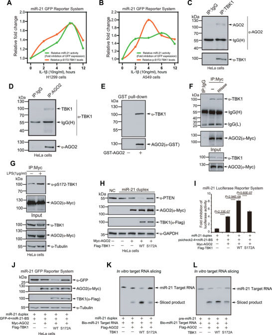
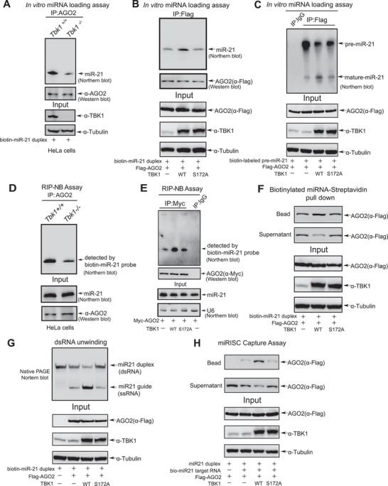
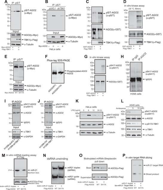
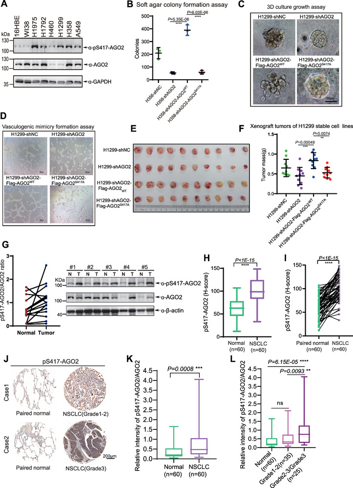
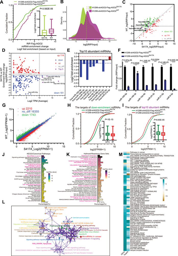
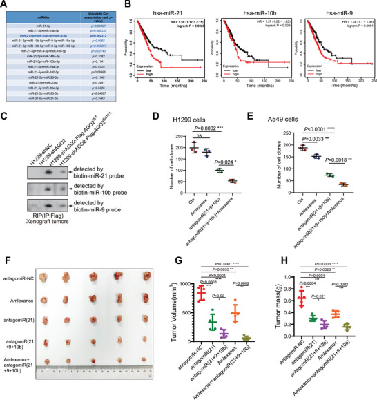
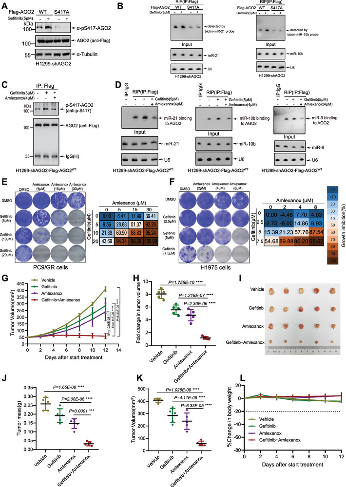
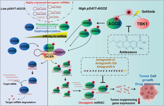
Non-small-cell lung cancer (NSCLC) is a highly lethal tumor that often develops resistance to targeted therapy. It is shown that Tank-binding kinase 1 (TBK1) phosphorylates AGO2 at S417 (pS417-AGO2), which promotes NSCLC progression by increasing the formation of microRNA-induced silencing complex (miRISC). High levels of pS417-AGO2 in clinical NSCLC specimens are positively associated with poor prognosis. Interestingly, the treatment with EGFR inhibitor Gefitinib can significantly induce pS417-AGO2, thereby increasing the formation and activity of oncogenic miRISC, which may contribute to NSCLC resistance to Gefitinib. Based on these, two therapeutic strategies is developed. One is jointly to antagonize multiple oncogenic miRNAs highly expressed in NSCLC and use TBK1 inhibitor Amlexanox reducing the formation of oncogenic miRISC. Another approach is to combine Gefitinib with Amlexanox to inhibit the progression of Gefitinib-resistant NSCLC. This findings reveal a novel mechanism of oncogenic miRISC regulation by TBK1-mediated pS417-AGO2 and suggest potential therapeutic approaches for NSCLC.

Keywords: AGO2; Gefitinib; TBK1; miRISC; non‐small‐cell lung cancer (NSCLC).


Conflict of interest statement

The authors declare no conflict of interest.

Figures

Figure 1
Figure 1
TBK1 directly interacts with AGO2 and promotes miRNA‐guided gene silencing. A,B) H1299 A) or A549 B) cells transfected with GFP‐4×miR‐21‐BS were treated with IL‐1β (10 ng ml−1) for the indicated time, and then harvested for the miR‐21 miRISC GFP reporter assay. C) HeLa cell lysates were used for Co‐IP with anti‐TBK1 antibody, and followed by Western blotting analysis with anti‐AGO2 antibody. D) HeLa cell lysates were used for Co‐IP with anti‐AGO2 antibody, and followed by Western blotting analysis with anti‐TBK1 antibody. E) Purified GST‐AGO2 was incubated with HeLa cell lysates for pull‐down assay. F) Lysates form 293T cells transfected with Myc‐AGO2 were treated with RNase (100 ng ml−1) for 30 min at 37 °C, and then used for Co‐IP with anti‐Myc antibody, and followed by Western blotting analysis with anti‐TBK1 antibody. G) HeLa cells transfected with Myc‐AGO2 were treated with LPS (1 µg ml−1) for 12 h before harvested. Lysates were used for IP with anti‐Myc antibody, and followed by Western blotting analysis with pS172‐TBK1 antibody. H) Lysates form HeLa cells co‐transfected miR‐21 duplex, Myc‐AGO2 with Flag‐TBK1WT or TBK1S172A were used for Western blotting analysis with anti‐PTEN antibody. I) 293T cells co‐transfected miR‐21 duplex mimics, psiCHECK2‐4×miR21‐BS, Myc‐AGO2 with Flag‐TBK1WT or TBK1S172A were harvested for the dual‐luciferase activity assay. The Renilla luciferase values were normalized to the Firefly luciferase activity and plotted as relative luciferase activity. Data were presented as mean ± SD, n = 3. Statistical analysis was performed using one‐way ANOVA. ****p < 0.0001. J) HeLa cells co‐transfected miR‐21 duplex mimics, GFP‐4×miR‐21‐BS, Myc‐AGO2 with Flag‐TBK1WT or TBK1S172A were harvested for Western blotting analysis with anti‐GFP antibody. K‐L) Lysates from 293T cells co‐transfected Flag‐AGO2 with TBK1WT or TBK1S172A were immunoprecipitated by Flag‐AGO2, and then this purified AGO2 protein were co‐incubated with miR‐21 duplex K) or pre‐miR‐21 L) and biotin‐tagged miR‐21 target for in vitro target RNA slicing assay. The cleavage products were detected on 20% urea PAGE by Northern blotting analysis.
Figure 2
Figure 2
TBK1 promotes miRNAs loading and miRISC formation. A) Lysates from HeLa‐Tbk1+/+ and HeLa‐Tbk1−/‐ cells were immunoprecipitated by AGO2, and then the beads coupled with AGO2 were co‐incubated with biotin‐miR‐21 duplex for in vitro miRNA loading assay. The miR‐21 associated with AGO2 was detected by Northern blotting analysis. B‐C) Lysates from 293T cells co‐transfected Flag‐AGO2 with TBK1WT or TBK1S172A were immunoprecipitated by Flag‐AGO2, then the beads were co‐incubated with purified biotin‐miR‐21 duplex B) or pre‐miR‐21 C) for in vitro miRNA loading assay. The miR‐21 associated with AGO2 was detected by Northern blotting analysis. D) HeLa‐Tbk1+/+ and HeLa‐Tbk1−/− cells were lysed by for the RIP‐NB assay with anti‐AGO2 antibody, and then endogenous miR‐21 associated with AGO2 was detected by Northern blotting analysis with biotin‐miR‐21 probe. E) 293T cells co‐transfected Myc‐AGO2 with Flag‐TBK1WT or TBK1S172A were lysed by for the RIP‐NB assay with anti‐Myc antibody, and then endogenous miR‐21 associated with AGO2 was detected by Northern blotting analysis with biotin‐miR‐21 probe. F) Lysates from 293T cells co‐transfected Flag‐AGO2 with TBK1WT or TBK1S172A were incubated with streptavidin‐Dynabeads‐coupled‐biotinylated miR‐21 duplex for biotinylated RNA‐streptavidin pull down assay. AGO2 pulled down by miR‐21 on the beads and not pulled down by miR‐21 in the supernatant were examined by Western blotting analysis with anti‐Flag antibody. G) Flag‐AGO2 purified from 293T cells co‐transfected Flag‐AGO2 with TBK1WT or TBK1S172A using 3×Flag peptide was incubated with miR‐21 duplex mimics for miR‐21 unwinding assays. The single‐stranded (ss) RNA molecules unwinding from double‐stranded (ds) miR‐21 substrates was detected on native polyacrylamide gels. H) Lysates from 293T cells co‐transfected Flag‐AGO2 with TBK1WT or TBK1S172A were co‐incubated with miR‐21 duplex and biotin‐tagged miR‐21 target RNA. Complex of AGO2, miR‐21, and miR‐21 target RNA was pulled down by streptavidin beads, and then detected by Western blotting analysis with anti‐Flag antibody.
Figure 3
Figure 3
TBK1 phosphorylates AGO2 at S417 to enhance the formation and activity of miRISC. A) 293T cells transfected with Myc‐AGO2 with or without Flag‐TBK1 were lysed for IP with anti‐phospho‐Ser/Thr antibody, and followed by Western blotting analysis with anti‐Myc antibody. B) Lysates from HeLa‐shTBK1 or ‐pLKO.1 stable cells were used for IP with anti‐phospho‐Ser/Thr antibody, and followed by Western blotting analysis with anti‐Myc antibody. C) Purified GST‐AGO2 protein was incubated in lysates from 293T cells expressing Flag‐TBK1 or the control vector. The phosphorylation of GST‐AGO2 was analyzed by GST pull‐down and followed by Western blotting analysis with anti‐phospho‐Ser/Thr antibody. D) Purified GST‐AGO2 and Flag‐TBK1 were co‐incubated in the kinase reaction buffer containing ATP, subsequently the phosphorylation of GST‐AGO2 was analyzed by GST pull‐down and followed by Western blotting analysis with anti‐phospho‐S/T antibody. E) 293T cells transfected with Myc‐AGO2WT or Myc‐AGO2S417A were lysed for IP with anti‐phospho‐S/T antibody, and followed by Western blotting analysis with anti‐Myc antibody. F) Phos‐tag SDS‐PAGE showing the phosphorylation level of Myc‐AGO2WT or Myc‐AGO2S417A in 293T cells co‐transfected with TBK1. G) Purified GST‐AGO2 and Flag‐TBK1 were co‐incubated in the kinase reaction buffer containing ATP, subsequently the phosphorylation of GST‐AGO2 was analyzed by GST pull‐down and followed by Western blotting analysis with specific pS417‐AGO2 antibody. H–J) Lysates from H358 cells (H), stable cells H1299‐shTBK1 or ‐pLKO.1 I), and H1299‐TBK1 or ‐Ctrl () were used for IP with anti‐AGO2 antibody, and then immunoblotted by specific pS417‐AGO2 antibody. K) HeLa cells treated with LPS (1 µg ml−1) for the indicated times were lysed for Western blotting analysis by using the specific pS417‐AGO2 antibody. L) A549 cells treated with Amlexanox for 12 h before harvested. Lysates were used for Western blotting analysis with the indicated antibodies. M) Lysates from 293T cells transfected with Flag‐AGO2WT or Flag‐AGO2S417A were immunoprecipitated by Flag‐AGO2, then the beads were co‐incubated with biotin‐tagged miR‐21 duplex for in vitro miRNA loading assay. The miR‐21 recruited to AGO2 was detected by Northern blotting analysis. N) Flag‐AGO2 or Flag‐AGO2S417A purified from 293T cells using 3×Flag peptide was incubated with miR‐21 duplex mimics for miR‐21 unwinding assays. The single‐stranded (ss) RNA molecules unwinding from double‐stranded (ds) miR‐21 substrates was detected on native polyacrylamide gels. O) Lysates from 293T cells transfected with Flag‐AGO2WT or Flag‐AGO2S417A were incubated with streptavidin‐Dynabeads‐coupled‐biotinylated miR‐21 duplex for biotinylated RNA‐streptavidin pull down assay. AGO2 pulled down by miR‐21 on the beads and not pulled down by miR‐21 were examined by Western blotting analysis with anti‐Flag antibody. P) Lysates from 293T cells transfected with Flag‐AGO2WT or Flag‐AGO2S417A were immunoprecipitated by Flag‐AGO2, and then were co‐incubated with miR‐21 duplex and biotin‐tagged miR‐21 target for in vitro target RNA slicing assay. The cleavage products were detected by Northern blotting analysis.
Figure 4
Figure 4
Phosphorylation of AGO2 at S417 promotes NSCLC progression. A) The phosphorylation levels of AGO2 in normal human bronchial epithelial cell line 16HBE, normal human lung fibroblasts cell line WI38, and NSCLC cell lines H1975, H1972, H460, H1299, H358 and A549 were analyzed by Western blotting analysis with specific anti‐pS417‐AGO2 and anti‐AGO2 antibodies. B) Soft agar colony formation assay for H358 stable cell lines was performed according to Methods. Data were presented as mean ± SD, n = 3. Statistical analysis was performed using one‐way ANOVA. ****p < 0.0001. C,D) 3D culture growth and vasculogenic mimicry (VM) for H1299 stable cell lines. Representative images of cell morphology C) and vasculogenic networks D) in extracellular matrix were taken. E,F) H1299 stable cell lines were subcutaneously injected into 6‐week‐old BALB/c nude mice individually. Mice were killed after 5 weeks of injection. Tumors were dissected E), and weight was assessed F). Data were presented as mean ± SD, n = 10. Statistical analysis was performed using one‐way ANOVA. **p < 0.01 and ***p < 0.001. (G) The phosphorylation levels of AGO2 in lung adenocarcinoma specimens and paired adjacent normal tissues were analyzed by Western blotting analysis with the specific pS417‐AGO2 antibody. Quantitative analysis for the ratio of pS417‐AGO2/AGO2 (left panel) (n = 17) and representative Western blotting analysis for pS417‐AGO2 levels (right panel). H‐J) IHC detection for pS417‐AGO2 in lung cancer tissue arrays. (H‐I) IHC staining scores for pS417‐AGO2 levels in paired normal tissues (n = 60) and NSCLC tissues (n = 60) were analyzed. Statistical analysis was performed using paired two‐tailed t‐test. ****p < 0.0001. J) Representative images of IHC staining for pS417‐AGO2 levels in normal tissues and pathological sub‐stages of NSCLC tissues. K) pS417‐AGO2/AGO2 ratio in normal tissues (n = 60) and NSCLC tissues (n = 60) were analyzed. Statistical analysis was performed using paired two‐tailed t‐test. ***p < 0.001. (L) pS417‐AGO2/AGO2 ratio in normal tissues (n = 60) and pathological sub‐stages of NSCLC tissues (Grade1‐2, n = 35; Grade2‐3/Grade2, n = 25) were analyzed. In box plots, the lines represent the median, first and third quartiles, the whiskers denote the minima and maxima. Statistical analysis was performed using one‐way ANOVA. **p < 0.01 and ****p < 0.0001.
Figure 5
Figure 5
pS417‐AGO2 promotes the loadings of high‐abundance oncogenic miRNAs into AGO2 in NSCLC. A–C) Cumulative fraction analysis A), density distribution map B) and scatter plot C) of RIP‐Seq for miRNAs bound to AGO2 in stable cells H1299‐shAGO2‐Flag‐AGO2WT and H1299‐shAGO2‐Flag‐AGO2S417A. P‐value was calculated using a two‐sided Mann‐Whitney U test for cumulative fraction analysis, n = 172; In box plots, the lines represent the median, first and third quartiles, the whiskers denote the minima and maxima. D) Scatterplot showing the fold change enrichment and abundance of miRNAs bound to AGO2. Significantly up‐enrichment (red), significantly down‐enrichment (blue) and top 5 most abundant (purple) miRNAs were indicated. E) Histograms showing the log2 fold change in the enrichment of top 10 most abundant miRNAs. F) Most abundant miRNAs including miR‐100‐5p, miR‐148‐3p, miR‐10a‐5p, miR‐24‐3p, miR‐21‐5p, miR‐9‐5p bound to AGO2 were examined by RIP‐qPCR. The enrichment of miRNAs associated with AGO2 was normalized by Input abundance of miRNAs. Data were presented as mean ± SD, n = 3. Statistical analysis was performed using unpaired two‐sided t‐test. **p < 0.01, ***p < 0.001 and ****p < 0.0001. G) Scatter plot showing the differentially expressed genes (DEGs) with log2 [fold change] ≥ 0.5 (up‐regulated) or log2 [fold change] < 0.5 (down‐regulated). H‐I) Cumulative fraction analyses for the abundance of targets of down‐enrichment miRNAs in RNA‐Seq, n = 2719 H), and the abundance of targets(of top 10 most abundant miRNAs in RNA‐Seq, n = 332 I). P‐values were calculated using a two‐sided Mann–Whitney U test; In box plots, the lines represent the median, first and third quartiles, the whiskers denote the minima and maxima. J) KEGG/Canonical Pathways enrichment analysis for targets of the top 10 most abundant miRNAs. K) Hallmark/Reactome Gene sets analysis for targets of the top 10 most abundant miRNAs. L) Representation of the most enriched pathways within targets of the top 10 most abundant miRNAs. M) Heatmap showing the top enrichment pathways by targets of the top 10 most abundant miRNAs and DEGs together, one row per cluster, using a discrete color scale to represent statistical significance.
Figure 6
Figure 6
Combining antagonism of oncogenic miRNAs and reduction of oncogenic miRISC formation for the treatment of NSCLC. A) The optimal miRNA combination of miRNA‐based therapeutic strategies for NSCLC patients was developed by univariate cox analysis. B) The correlations between the expression levels of miRNAs (miR‐21, miR‐10b, miR‐9) and the survival of lung adenocarcinoma(LUAD) patients were analyzed by the Kaplan‐Meier analysis and and compared by the log‐rank test. C) The miR‐21, miR‐9, and miR‐10b loading in xenografted tumors were detected by RIP‐Northern blotting analysis. D,E) Colony formation assays of H1299 D) and A549 E) cells. Cells were grown in the absence or presence of the indicated only antagomiRs, Amlexanox, or combinations for 10–12 days, stained and the number of colonies was scored. Data were presented as mean ± SD, n = 3. Statistical analysis was performed using one‐way ANOVA. *p < 0.05, **p < 0.01, ***p < 0.001 and ****p < 0.0001. F–H) Mice were subcutaneously injected with 1 × 10⁶ H1299 cells. Once tumors reached an average of 5 mm × 5 mm (14 days), the mice were treated with antagomiRs (5 nmol) by intratumoral injection and TBK1 inhibitor Amlexanox (25 mg k−1g) by intraperitoneal injection for twice per week. 28 days after inoculation, xenograft tumors were dissected F), tumor volume G) and weight H) were assessed. Data were presented as mean ± SD, n = 5 in G) and H). Statistical analysis was performed using unpaired two‐sided t‐test. *p < 0.05, **p < 0.01, ***p < 0.001 and ****p < 0.0001.
Figure 7
Figure 7
Gefitinib combined with Amlexanox for the treatment of drug‐resistant NSCLC. A) H1299‐shAGO2‐Flag‐AGO2WT or S417A cells were treated with Gefitinib (5 µM) and together with or without Amlexanox (4 µM) simultaneously for 12 h before harvested. Cells were lysed for IP with anti‐Flag antibody, and then immunoblotted by specific anti‐pS417‐AGO2 antibody. B) H1299‐shAGO2‐Flag‐AGO2WT or S417A cells treated with or without Gefitinib (5 µM) were lysed for the RIP assay with anti‐Flag antibody, and then miR‐21, miR‐10b associated with AGO2 were detected by Northern blotting analysis. C) H1299‐shAGO2‐Flag‐AGO2WT cells were treated with Gefitinib (5 µM) together with or without Amlexanox (4 µM) simultaneously for 12 h before harvested. Cells were lysed for IP with anti‐Flag antibody, and then immunoblotted by specific anti‐pS417‐AGO2 antibody. D) H1299‐shAGO2‐Flag‐AGO2WT cells treated with Gefitinib (5 µM) together with or without Amlexanox (4 µM) simultaneously for 12 h before harvested. Cells were lysed for the RIP assay with anti‐Flag antibody, and then miR‐21, miR‐9, and miR‐10b associated with AGO2 were detected by Northern blotting analysis. E,F) PC9/GR E) and H1975 F) cells were treated with Gefitinib, Amlexanox or their combination at the indicated concentrations. Cells were fixed and stained after 10–12 days. Representative data from three independent experiments. G–L) Mice were subcutaneously injected with 4 × 10⁶ PC9/GR cells. Once tumors reached an average of 5 mm × 5 mm, the mice were treated with Gefitinb (20 mg k−1 g) by oral gavage, Amlexanox (20 mg k−1 g) by intraperitoneal injection, or combination therapy for every 2 days. The tumor growth curve during treatment period G) and fold change in tumor volume pre or post‐treatment was analysed H). Xenograft tumors were dissected I). Tumor mass J) and Tumor volume K) after treatment were assessed. L) The change of body weight during treatment period were assessed. Data were presented as mean ± SD, n = 5. Statistical analysis was performed using one‐way ANOVA. ***p < 0.001 and ****p < 0.0001.
Figure 8
Figure 8
A model summarizing that TBK1‐mediated pS417‐AGO2 promotes NSCLC progression and resistance to Gefitinib by increasing the formation and activity of oncogenic miRISCs.

Similar articles

Cited by

References

    1. Siegel R. L., Miller K. D., Wagle N. S., Jemal A., CA Cancer J Clin 2023, 73, 17. - PubMed
    1. Gandhi L., Garassino M. C., N. Engl. J. Med. 2018, 379, e18. - PubMed
    1. Sun X., Xu S., Yang Z., Zheng P., Zhu W., Expert Opin. Ther. Pat. 2021, 31, 223. - PubMed
    1. Yang H., Liang S. Q., Schmid R. A., Peng R. W., Front Oncol 2019, 9, 953. - PMC - PubMed
    1. Denis M. G., Bennouna J., Cancer Manag Res 2020, 12, 12593. - PMC - PubMed

MeSH terms