Extramural recurrence of tracheal glomus tumour following resection by rigid bronchoscopy
- PMID: 38351921
- PMCID: PMC10864103
- DOI: 10.1002/rcr2.1302
Extramural recurrence of tracheal glomus tumour following resection by rigid bronchoscopy
Abstract
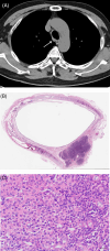
Glomus tumour of the trachea is very rare neoplasm that is generally benign and arises most commonly from the distal portion of the respiratory tree. This report presents the case of a 67-year-old man who was referred to our institute for excision of a tracheal mass that had been found incidentally, and subsequently recurred extramurally. Initial contrast-enhanced computed tomography images of the chest revealed a nodular lesion in the trachea, 2.5 cm above the carina, that demonstrated strong enhancement similar to blood vessels. The tumour was excised by rigid bronchoscopy, but an extramural tracheal lesion was detected 18 months later. Tracheal resection and end-to-end anastomosis were performed, and histopathological examination confirmed the extramural lesion as recurrence of the tracheal glomus tumour. The histologic features and treatment are discussed.
Keywords: rigid bronchoscopy; tracheal glomus tumour; tracheoplasty.
© 2024 The Authors. Respirology Case Reports published by John Wiley & Sons Australia, Ltd on behalf of The Asian Pacific Society of Respirology.
Conflict of interest statement
None declared.
Figures


Similar articles
-
Tracheal Glomus Tumor: A Case Report with CT Imaging Features.Medicina (Kaunas). 2022 Jun 13;58(6):791. doi: 10.3390/medicina58060791. Medicina (Kaunas). 2022. PMID: 35744054 Free PMC article.
-
Glomus tumor of the trachea.Asian Cardiovasc Thorac Ann. 2015 Mar;23(3):325-7. doi: 10.1177/0218492314528184. Epub 2014 Apr 2. Asian Cardiovasc Thorac Ann. 2015. PMID: 24696105
-
Successful resection of a glomus tumor arising from the lower trachea: report of a case.Surg Today. 1998;28(3):332-4. doi: 10.1007/s005950050134. Surg Today. 1998. PMID: 9548322
-
Malignant glomus tumor of trachea: a case report with literature review.Asian Cardiovasc Thorac Ann. 2016 Jan;24(1):104-6. doi: 10.1177/0218492315608546. Epub 2015 Sep 28. Asian Cardiovasc Thorac Ann. 2016. PMID: 26420909 Review.
-
Successful resection of a glomus tumor of the trachea.Gen Thorac Cardiovasc Surg. 2011 Dec;59(12):815-8. doi: 10.1007/s11748-010-0772-y. Epub 2011 Dec 16. Gen Thorac Cardiovasc Surg. 2011. PMID: 22173681 Review.
Cited by
-
Non-Surgical Treatment of Tracheal Glomus Tumour Using Rigid Fiberoptic Bronchoscopy: A Case Report.Respirol Case Rep. 2025 Jun 19;13(6):e70236. doi: 10.1002/rcr2.70236. eCollection 2025 Jun. Respirol Case Rep. 2025. PMID: 40546266 Free PMC article.
References
-
- Specht K, Antonescu CR. Glomus tumour. WHO classification of Tumours of soft tissue and bone. 5th ed. Lyon, France: International Agency for Research on Cancer (IARC); 2020. p. 179–181.
Publication types
LinkOut - more resources
Full Text Sources

