Review
. 2024 Feb 1:6:100223.
doi: 10.1016/j.crmicr.2024.100223. eCollection 2024.

Toxoplasma-host endoplasmic reticulum interaction: How T. gondii activates unfolded protein response and modulates immune response

Affiliations
Review

Toxoplasma-host endoplasmic reticulum interaction: How T. gondii activates unfolded protein response and modulates immune response

Obed Cudjoe et al. Curr Res Microb Sci. .

Abstract

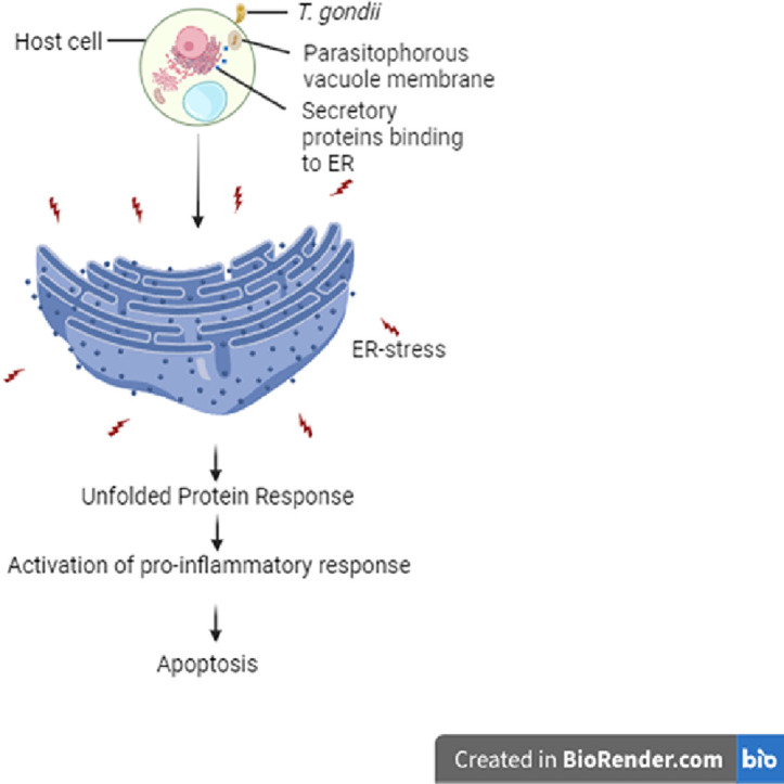
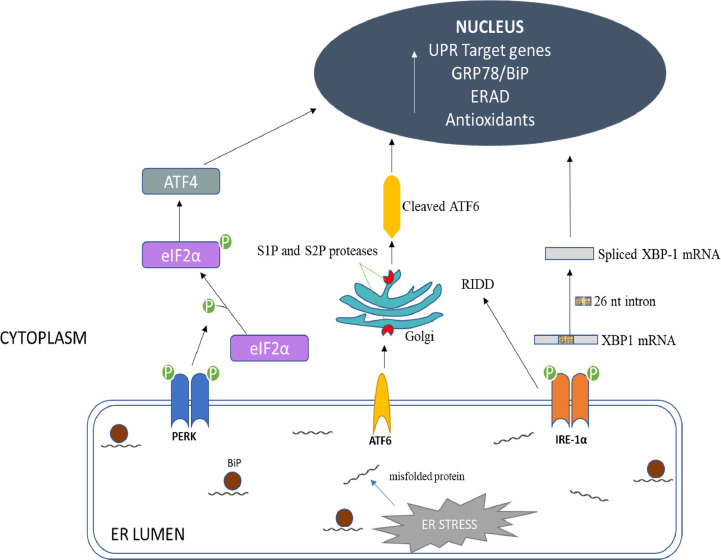
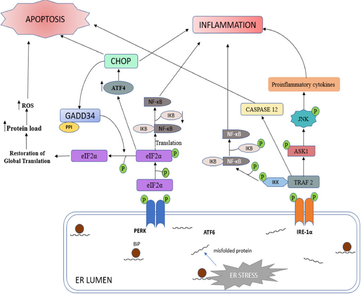
Toxoplasma gondii is a neurotropic single-celled zoonotic parasite that can infect human beings and animals. Infection with T. gondii is usually asymptomatic in immune-competent individual, however, it can cause symptomatic and life-threatening conditions in immunocompromised individuals and in developing foetuses. Although the mechanisms that allow T. gondii to persist in host cells are poorly understood, studies in animal models have greatly improved our understanding of Toxoplasma-host cell interaction and how this interaction modulates parasite proliferation and development, host immune response and virulence of the parasite. T. gondii is capable of recruiting the host endoplasmic reticulum (ER), suggesting it may influence the host ER function. Herein, we provide an overview of T. gondii infection and the role of host ER during stressed conditions. Furthermore, we highlight studies that explore T. gondii's interaction with the host ER. We delve into how this interaction activates the unfolded protein response (UPR) and ER stress-mediated apoptosis. Additionally, we examine how T. gondii exploits these pathways to its advantage.

Keywords: Apoptosis; ER–stress; Endoplasmic reticulum; Immune response; Toxoplasma gondii; UPR.


Conflict of interest statement

The authors declare that they have no known competing financial interests or personal relationships that could have appeared to influence the work reported in this paper.

Figures

Image, graphical abstract
Graphical abstract
Fig 1
Fig. 1
Representation of a T. gondii tachyzoite illustrating the internal structures and secretory organelles. Adapted from Blader et al. (2015).
Fig 2
Fig. 2
Invasion/entry of the tachyzoite of T. gondii into the host cell. T. gondii re–orientates and aligns its apical pole with the membrane of the host cell. The apical end of the microneme secretes microneme proteins (MIC adhesins), which enables attachment to host cell glycosaminoglycans (GAGs). Together with micronemes, RON proteins form a moving junction that progresses along the parasite. This moving junction is crucial for the PV's firm anchorage to the cytoskeleton of the host cell, as well as the acquisition of host proteins that are incorporated into the PVM. Rhoptry proteins (ROP) play a critical role in the PVM formation, while the dense granule proteins (GRA) are released after PV formation and allow the PV to become functional. Adapted from Mercier and Cesbron-Delauw (2012).
Fig 3
Fig. 3
Unfolded Protein Response. The dissociation of BiP/ GRP78 away from the ER transmembrane signalling stress sensors triggers PERK, ATF6 and IRE–1α activation in response to ER stress. PERK, a type I transmembrane protein, handles the immediate ER stress response through its signalling pathway. Dimerization of PERK activates cytosolic kinase and trans-autophosphorylates eukaryotic translation-initiation factor 2α (eIF2α), inhibiting protein syntheses. However, phosphorylation of eIF2α (eIF2α-P) selectively increases ribosome translation downstream, leading to the translation of other mRNA subunits, including ATF4, which initiates an antioxidant response and regulates ER homeostasis. ATF6 is a 90 kDa type II ER transmembrane protein with a carboxyl terminus acting as an intraluminal sensor and an amino terminus acting as a bZIP transcription factor. Recruitment of GRP–78 from the luminal domain allows ATF6 to translocate from the endoplasmic reticulum to the Golgi apparatus, where it is cleaved by specialized enzymes (site–1 and site–2 proteases(SIP/S2P)) to release ATF6 cytosolic fragment, which increases gene production for ER-associated protein degradation (ERAD) and folding chaperones. IRE-1α is a type I transmembrane signal activator protein similar to PERK. It possesses both kinase and site–specific endoribonuclease (RNase) activity. ER stress causes IRE1 to dimerize, activate its kinase domain, and catalyzes the excision of a 26-base intron from the mRNA encoding the X–box binding protein 1 (XBP-1), regulating genes involved in ERAD and protein folding. Adapted from Sprenkle et al. (2017).
Fig 4
Fig. 4
Apoptosis and Inflammatory Signal Pathways Associated with Unremitted ER Stress and UPR. Chronic ERS triggers both inflammatory and apoptotic pathways of the UPR. IRE–1α and PERK both trigger UPR–mediated inflammation and apoptosis. Dimerization of PERK phosphorylates eIF2α which attenuates global mRNA translation. However, downstream signalling increases the expression of inflammatory genes. Furthermore, the activation of CHOP by p–eIF2α can also initiate inflammation and apoptosis. Prolonged IRE–1α activation recruits (TNF–α)–receptor–associated factor 2 (TRAF2) and MAP kinase (MAPKKK) (ASK1), causing downstream signalling activation of NF–κB and JNK which upregulate the transcription of inflammatory genes. PERK upregulates CHOP to promote host cell apoptosis. The formation of IRE–1α and TRAF2 complex induces apoptosis via caspase activation. Adapted from Sprenkle et al. (2017).

Similar articles

References

    1. Adams C.J., Kopp M.C., Larburu N., Nowak P.R., Ali M.M.U. Structure and molecular mechanism of ER stress signaling by the unfolded protein response signal activator IRE1. Front. Mol. Biosci. 2019;6(11) doi: 10.3389/fmolb.2019.00011. - DOI - PMC - PubMed
    1. Ahn H.J., Kim S., Kim H.E., Nam H.W. Interactions between secreted GRA proteins and host cell proteins across the paratitophorous vacuolar membrane in the parasitism of Toxoplasma gondii. Korean J. Parasitol. 2006;44(4):303–312. doi: 10.3347/kjp.2006.44.4.303. - DOI - PMC - PubMed
    1. Al-Malki E.S. Toxoplasmosis: stages of the protozoan life cycle and risk assessment in humans and animals for an enhanced awareness and an improved socio-economic status. Saudi J. Biol. Sci. 2021;28(1):962–969. doi: 10.1016/j.sjbs.2020.11.007. - DOI - PMC - PubMed
    1. Almanza A., Carlesso A., Chintha C., Creedican S., Doultsinos D., Leuzzi B., et al. Endoplasmic reticulum stress signalling - from basic mechanisms to clinical applications. FEBS. J. 2019;286(2):241–278. doi: 10.1111/febs.14608. - DOI - PMC - PubMed
    1. Amen O.M., Sarker S.D., Ghildyal R., Arya A. Endoplasmic reticulum stress activates unfolded protein response signaling and mediates inflammation, obesity, and cardiac dysfunction: therapeutic and molecular approach. Front. Pharmacol. 2019;(977):10. doi: 10.3389/fphar.2019.00977. - DOI - PMC - PubMed

LinkOut - more resources