

Anatomical variations in the circle of Willis on magnetic resonance angiography in a south Trinidad population
- PMID: 38352180
- PMCID: PMC10860579
- DOI: 10.1093/bjro/tzad002
Anatomical variations in the circle of Willis on magnetic resonance angiography in a south Trinidad population
Abstract
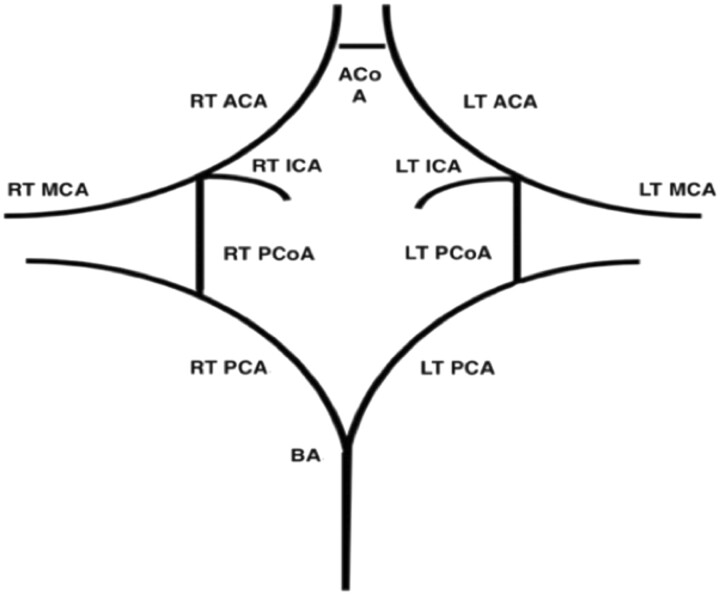
Objectives: This article seeks to determine the prevalence of a complete circle of Willis (CoW) and its common morphological variations in a south Trinidad population, while also investigating the influence of gender, age, and ethnicity on CoW morphology.
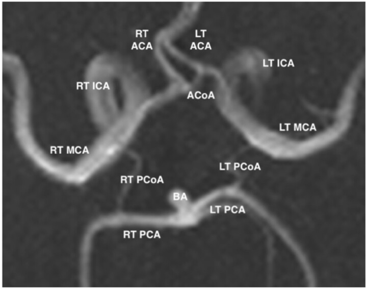
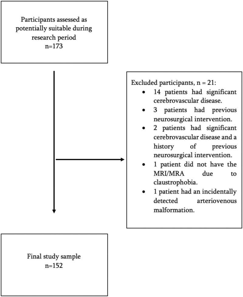
Methods: A prospective, descriptive, cross-sectional study was done on the magnetic resonance images for consecutive patients who had a brain MRI/magnetic resonance angiography at a tertiary health institution in south Trinidad between October 2019 and September 2020. Patients with significant cerebrovascular disease and/or a history of prior neurosurgical intervention were excluded.
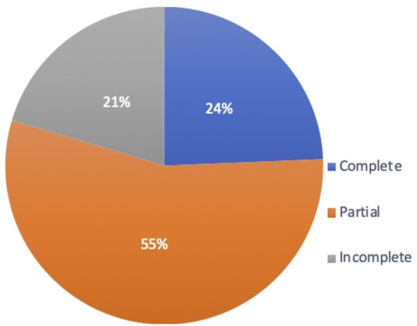
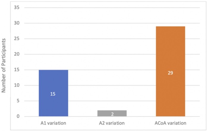
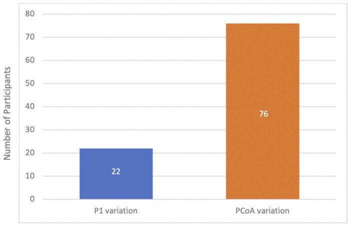
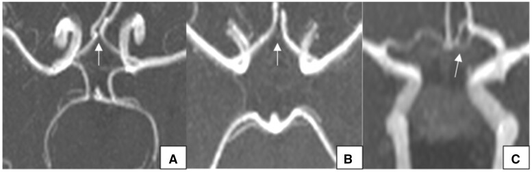
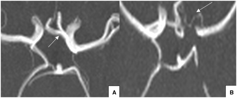
Results: A complete CoW was seen in 24.3%, with more complete circles observed in younger participants (≤45 years) and Afro-Trinidadians. No gender predilection for a complete CoW was demonstrated. The most common variations in the anterior and posterior parts of the circle were a hypoplastic anterior communicating artery (8.6%, n = 13) and bilateral aplastic posterior communicating arteries (18.4%, n = 28), respectively.
Conclusions: Significant variations exist in the CoW of a south Trinidad population with a frequency of complete in 24.3%, and more complete circles in younger patients and Afro-Trinidadians. Gender did not influence CoW morphology.
Advances in knowledge: Structural abnormalities in the CoW may be linked to future incidence of cerebrovascular diseases and should therefore be communicated to the referring physician in the written radiology report. Knowledge of variant anatomy and its frequency for a particular populations is also required by neurosurgeons and neuro-interventional radiologists to help with preprocedural planning and to minimize complications.
Keywords: Trinidad; Trinidad and Tobago; anatomical variations circle of Willis; circle of Willis; magnetic resonance angiography; magnetic resonance imaging; neuroradiology; stroke.
© The Author(s) 2023. Published by Oxford University Press on behalf of the British Institute of Radiology.
Conflict of interest statement
None declared.
Figures
Similar articles
-
Distinct Circle of Willis anatomical configurations in healthy preterm born adults: a 3D time-of-flight magnetic resonance angiography study.BMC Med Imaging. 2025 Jan 30;25(1):33. doi: 10.1186/s12880-025-01562-y. BMC Med Imaging. 2025. PMID: 39885378 Free PMC article.
-
Circle of Willis: anatomical variations of configuration. A magnetic resonance angiography study.Folia Morphol (Warsz). 2023;82(1):24-29. doi: 10.5603/FM.a2021.0134. Epub 2021 Dec 30. Folia Morphol (Warsz). 2023. PMID: 34966998
-
Configuration of the circle of Willis and its two parts among Egyptian: a magnetic resonance angiographic study.Folia Morphol (Warsz). 2019;78(4):703-709. doi: 10.5603/FM.a2019.0015. Epub 2019 Feb 14. Folia Morphol (Warsz). 2019. PMID: 30761512
-
Anatomical variations of the circle of Willis and their prevalence, with a focus on the posterior communicating artery: A literature review and meta-analysis.Clin Anat. 2021 Oct;34(7):978-990. doi: 10.1002/ca.23662. Epub 2020 Aug 6. Clin Anat. 2021. PMID: 32713011 Review.
-
Anatomical variations in the Circle of Willis and the formation and rupture of intracranial aneurysms: A systematic review and meta-analysis.Front Neurol. 2023 Jan 16;13:1098950. doi: 10.3389/fneur.2022.1098950. eCollection 2022. Front Neurol. 2023. PMID: 36726753 Free PMC article.
Cited by
-
Morphological evaluation of completeness of Circle of Willis.Anat Sci Int. 2025 Mar 3. doi: 10.1007/s12565-025-00832-7. Online ahead of print. Anat Sci Int. 2025. PMID: 40032796
-
Anatomical investigation of the morphometry of the cerebral arteries using digital subtraction angiography in the Thai population.Surg Radiol Anat. 2024 Nov;46(11):1775-1781. doi: 10.1007/s00276-024-03484-w. Epub 2024 Sep 18. Surg Radiol Anat. 2024. PMID: 39292256
-
Distinct Circle of Willis anatomical configurations in healthy preterm born adults: a 3D time-of-flight magnetic resonance angiography study.BMC Med Imaging. 2025 Jan 30;25(1):33. doi: 10.1186/s12880-025-01562-y. BMC Med Imaging. 2025. PMID: 39885378 Free PMC article.
References
-
- Siddiqi H, Tahir M, Lone KP.. Variations in cerebral arterial circle of Willis in adult Pakistani population. J Coll Physicians Surg Pak. 2013;23(9):615-619. - PubMed
-
- Saikia B, Handique A, Phukan P, Lynser D, Jamil M.. Study of anomalies in the circle of Willis using magnetic resonance angiography in north eastern India. J Anat Soc India. 2014;63(1):67-73.
LinkOut - more resources
Full Text Sources
