

Body weight-based iodinated contrast immersion timing for human fetal postmortem microfocus computed tomography
- PMID: 38352185
- PMCID: PMC10860501
- DOI: 10.1093/bjro/tzad006
Body weight-based iodinated contrast immersion timing for human fetal postmortem microfocus computed tomography
Abstract
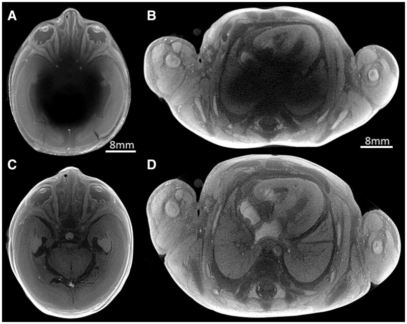
Objectives: The aim of this study was to evaluate the length of time required to achieve full iodination using potassium tri-iodide as a contrast agent, prior to human fetal postmortem microfocus computed tomography (micro-CT) imaging.
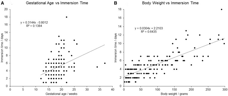
Methods: Prospective assessment of optimal contrast iodination was conducted across 157 human fetuses (postmortem weight range 2-298 g; gestational age range 12-37 weeks), following micro-CT imaging. Simple linear regression was conducted to analyse which fetal demographic factors could produce the most accurate estimate for optimal iodination time.
Results: Postmortem body weight (r2 = 0.6435) was better correlated with iodination time than gestational age (r2 = 0.1384), producing a line of best fit, y = [0.0304 × body weight (g)] - 2.2103. This can be simplified for clinical use whereby immersion time (days) = [0.03 × body weight (g)] - 2.2. Using this formula, for example, a 100-g fetus would take 5.2 days to reach optimal contrast enhancement.
Conclusions: The simplified equation can now be used to provide estimation times for fetal contrast preparation time prior to micro-CT imaging and can be used to manage service throughput and parental expectation for return of their fetus.
Advances in knowledge: A simple equation from empirical data can now be used to estimate preparation time for human fetal postmortem micro-CT imaging.
Keywords: fetal; immersion time; micro-CT; potassium tri-iodide.
© The Author(s) 2023. Published by Oxford University Press on behalf of the British Institute of Radiology.
Conflict of interest statement
The authors declare that they have no competing financial interests.
Figures
Similar articles
-
Postmortem microfocus computed tomography for early gestation fetuses: a validation study against conventional autopsy.Am J Obstet Gynecol. 2018 Apr;218(4):445.e1-445.e12. doi: 10.1016/j.ajog.2018.01.040. Epub 2018 Feb 2. Am J Obstet Gynecol. 2018. PMID: 29410108
-
Comparison of postmortem whole-body contrast-enhanced microfocus computed tomography and high-field magnetic resonance imaging of human fetuses.Ultrasound Obstet Gynecol. 2022 Jul;60(1):109-117. doi: 10.1002/uog.24827. Ultrasound Obstet Gynecol. 2022. PMID: 34826157 Free PMC article.
-
Novel imaging techniques to study postmortem human fetal anatomy: a systematic review on microfocus-CT and ultra-high-field MRI.Eur Radiol. 2020 Apr;30(4):2280-2292. doi: 10.1007/s00330-019-06543-8. Epub 2019 Dec 13. Eur Radiol. 2020. PMID: 31834508 Free PMC article.
-
Postmortem microfocus computed tomography for noninvasive autopsies: experience in >250 human fetuses.Am J Obstet Gynecol. 2021 Jan;224(1):103.e1-103.e15. doi: 10.1016/j.ajog.2020.07.019. Epub 2020 Jul 16. Am J Obstet Gynecol. 2021. PMID: 32682860 Free PMC article.
-
Fetal postmortem imaging: an overview of current techniques and future perspectives.Am J Obstet Gynecol. 2020 Oct;223(4):493-515. doi: 10.1016/j.ajog.2020.04.034. Epub 2020 May 4. Am J Obstet Gynecol. 2020. PMID: 32376319 Review.
References
-
- MBRRACE-UK. MBRRACE-UK Intrapartum Confidential Enquiry Report—Term, Singleton, Intrapartum Stillbirth and Intrapartum-Related Neonatal Death. The Infant Mortality and Morbidity Studies; 2017.
-
- Gordijn SJ, Erwich JJ, Khong TY.. Value of the perinatal autopsy: critique. Pediatr Dev Pathol. 2002;5(5):480-488. - PubMed
-
- Osborn M, Lowe J, Cox PG, Hargitai B, Marton T.. Guidelines on Autopsy Practice. Fetal Autopsy 2nd Trimester Fetal Loss and Termination of Pregnancy for Congenital Anomaly. The Royal College of Pathologists; 2017.
-
- Lewis C, Simcock IC, Arthurs OJ.. Improving uptake of perinatal autopsy. Curr Opin Obstet Gynecol. 2021;33(2):129-134. - PubMed
Grants and funding
LinkOut - more resources
Full Text Sources
