

Preparation and in vivo evaluation of nano sized cubosomal dispersion loaded with Ruta graveolens extracts as a novel approach to reduce asthma-mediated lung inflammation
- PMID: 38352238
- PMCID: PMC10862413
- DOI: 10.1016/j.jsps.2024.101968
Preparation and in vivo evaluation of nano sized cubosomal dispersion loaded with Ruta graveolens extracts as a novel approach to reduce asthma-mediated lung inflammation
Abstract
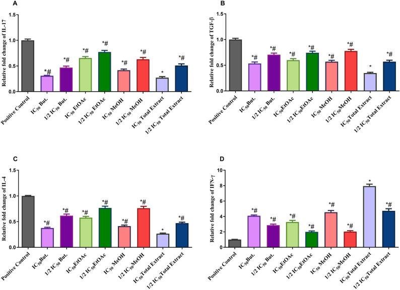
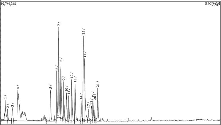
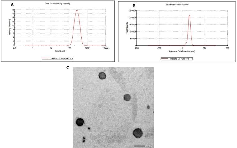
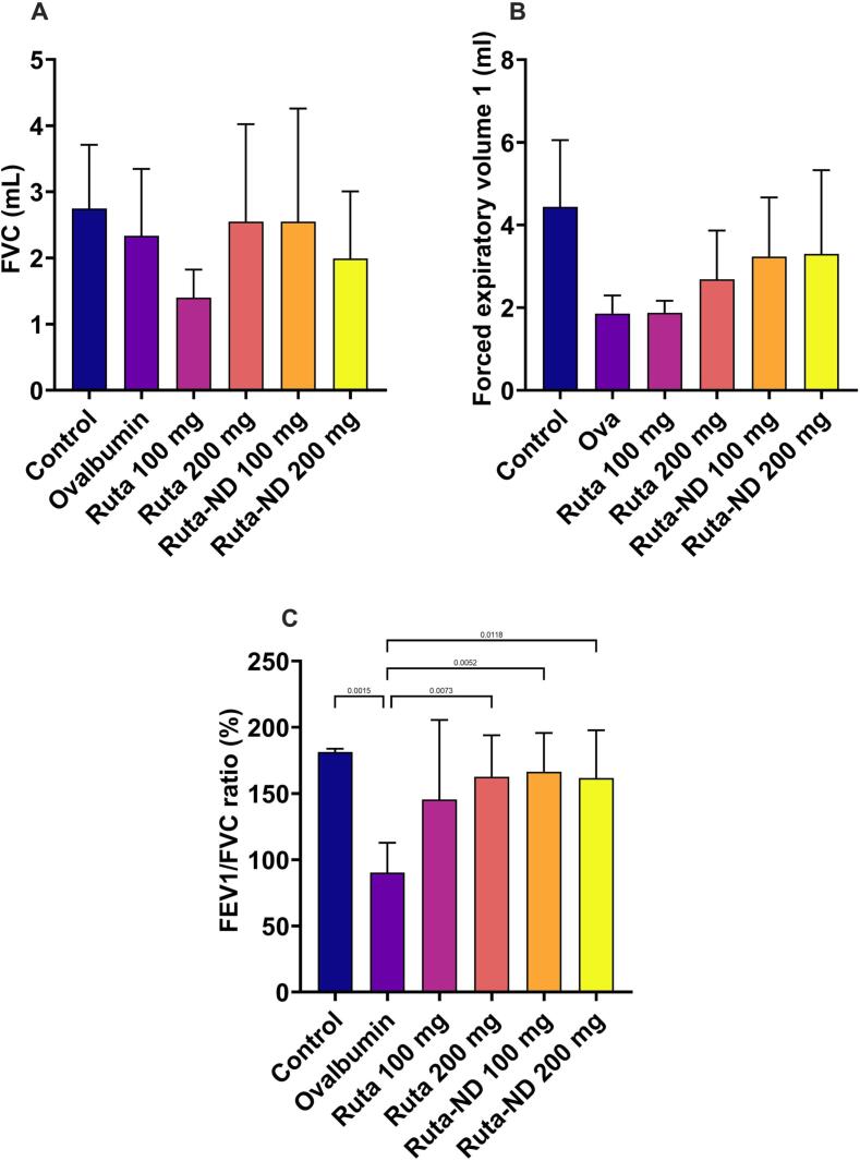
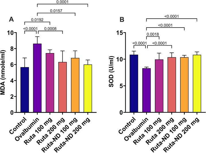
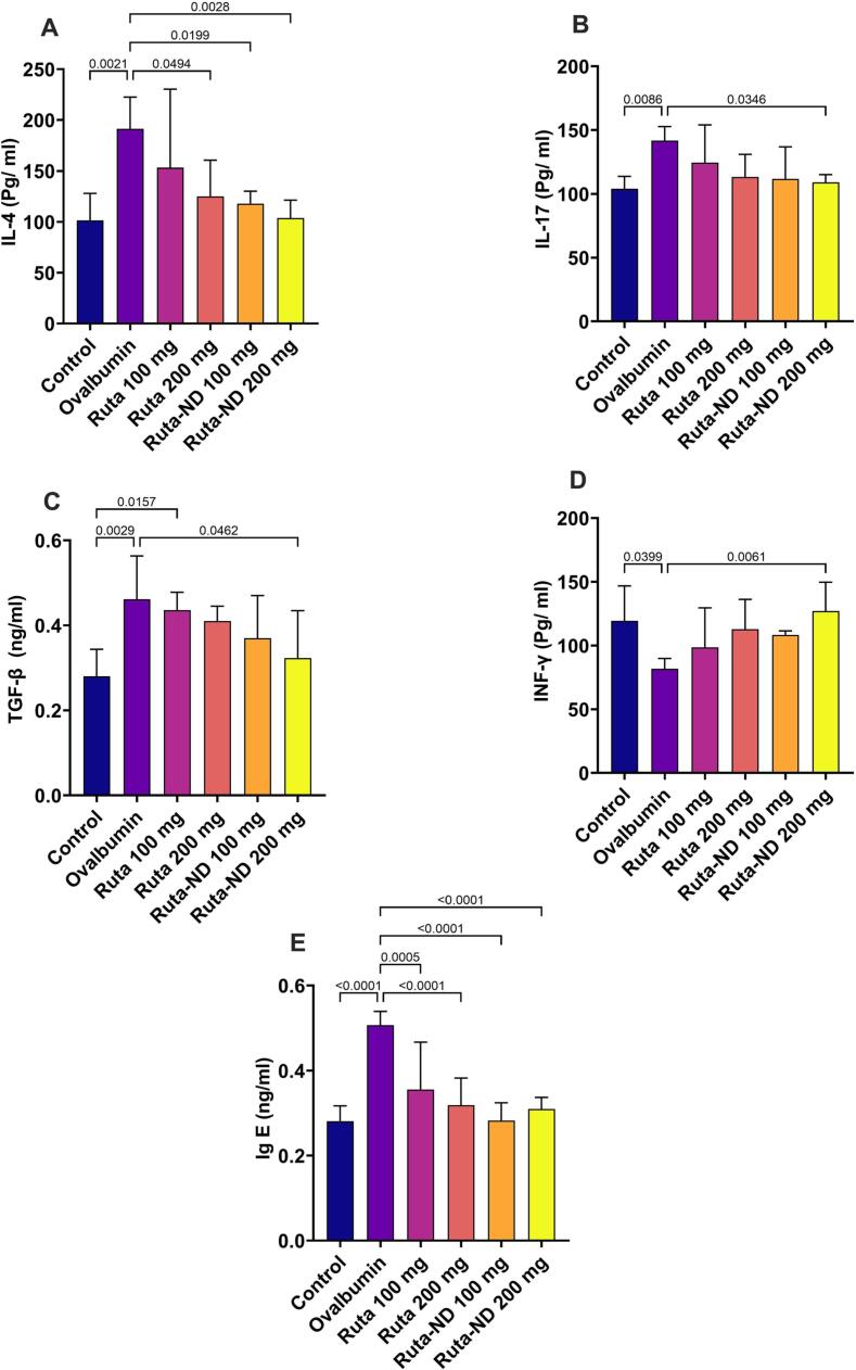
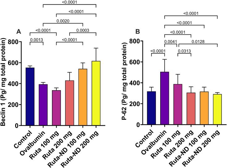
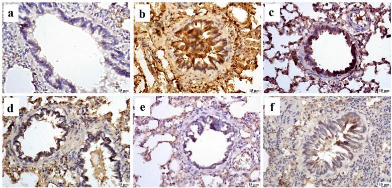
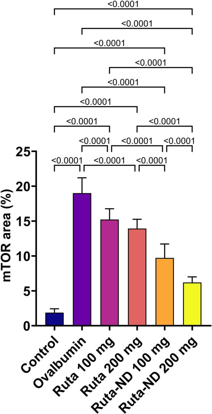
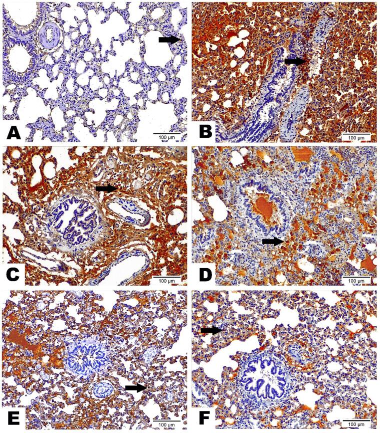
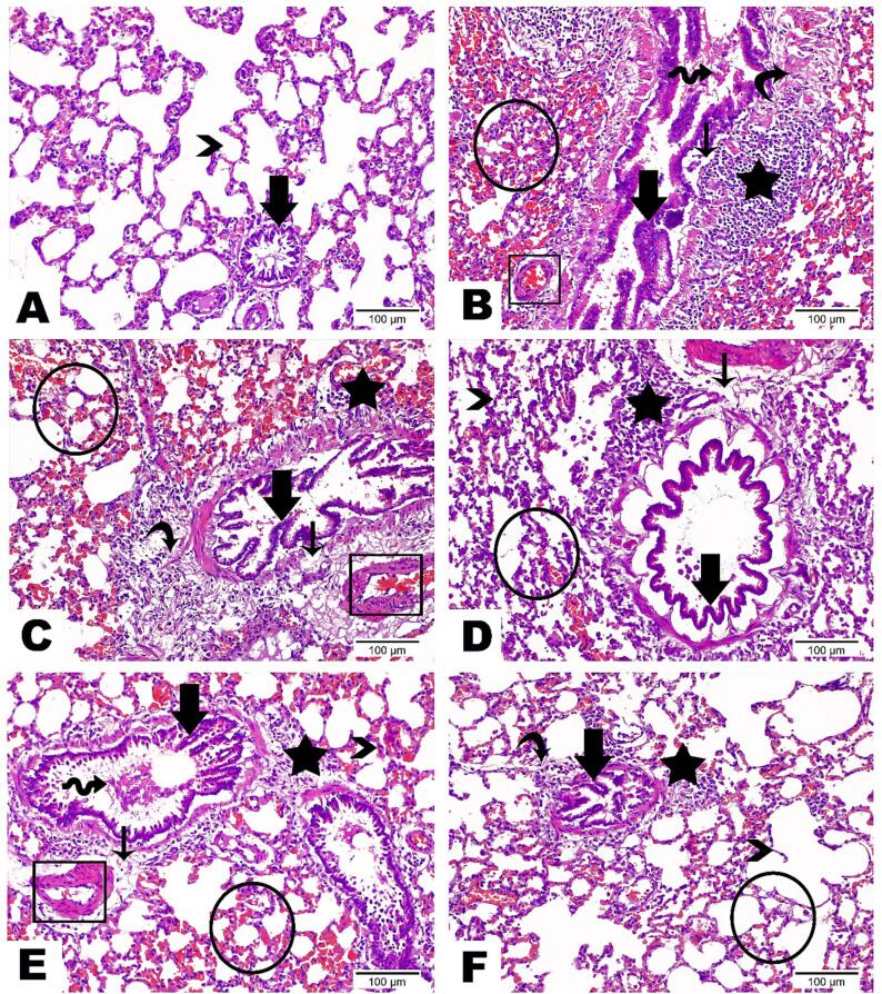
Asthma is a chronic disease affecting people of all ages. Asthma medications are associated with adverse effects restricting their long-term usage, demanding newer alternative therapies. This study aimed to investigate the anti-asthmatic properties of Ruta graveolens extract and its prepared nano-cubosomal dispersion (Ruta-ND). Firstly, the R. graveolens methanolic extract exhibited higher anti-inflammatory activity on Lipopolysaccharide (LPS)-activated BEAS-2B cells. To ensure best bioavailability and hence best cellular uptake, R. graveolens extract was loaded in nano-cubosomal dispersion (ND). Then, the anti-asthmatic effects of Ruta extract and ND were simultaneously evaluated in rats' model with ovalbumin-induced allergic asthma. R. graveolens extract and Ruta-ND subsided asthma score and improved lung function by restoring FEV1/FVC ratio to the expected values in control rats. Also, it showed strong antioxidant and anti-inflammatory activities manifested by lowering levels of malondialdehyde (MDA), IL-4, IL-7, TGF-β, and Ig-E, and increasing levels of superoxide dismutase (SOD) and INF-γ in bronchoalveolar lavage fluid. Our research findings also indicate autophagy induction and apoptosis inhibition by Ruta extract and Ruta-ND. Finally, the HPLC MS/MS phytochemical profiling of R. graveolens extract evident production of various alkaloids, flavonoids, coumarins, and other phenolics with reported pharmacological properties corresponding to/emphasize our study findings. In conclusion, R. graveolens exhibited promise in managing Ova-induced allergic asthma and could be developed as an alternative anti-allergic asthma drug.
Keywords: Apoptosis; Autophagy; Beclin-1; Behavior scoring; Ovalbumin Allergic airway; Respiratory function.
© 2024 The Author(s).
Figures

Similar articles
-
Phenolic Content and in Vitro Antioxidant, Anti-Inflammatory and antimicrobial Evaluation of Algerian Ruta graveolens L.Chem Biodivers. 2022 Sep;19(9):e202200545. doi: 10.1002/cbdv.202200545. Epub 2022 Aug 16. Chem Biodivers. 2022. PMID: 35866461
-
Phytochemical Characterization, and Antioxidant and Antimicrobial Properties of Agitated Cultures of Three Rue Species: Ruta chalepensis, Ruta corsica, and Ruta graveolens.Antioxidants (Basel). 2022 Mar 20;11(3):592. doi: 10.3390/antiox11030592. Antioxidants (Basel). 2022. PMID: 35326242 Free PMC article.
-
Antinociceptive, anti-inflammatory and antipyretic activities of the leaf methanol extract of Ruta graveolens L. (Rutaceae) in mice and rats.Afr J Tradit Complement Altern Med. 2014 Apr 3;11(3):173-81. doi: 10.4314/ajtcam.v11i3.25. eCollection 2014. Afr J Tradit Complement Altern Med. 2014. PMID: 25371580 Free PMC article.
-
Methanolic extract of Ruta graveolens L. inhibits inflammation and oxidative stress in adjuvant induced model of arthritis in rats.Inflammopharmacology. 2009 Apr;17(2):100-5. doi: 10.1007/s10787-009-8044-0. Inflammopharmacology. 2009. PMID: 19205849
-
Genus Ruta: A natural source of high value products with biological and pharmacological properties.J Ethnopharmacol. 2020 Oct 5;260:113076. doi: 10.1016/j.jep.2020.113076. Epub 2020 Jun 10. J Ethnopharmacol. 2020. PMID: 32534112 Review.
Cited by
-
Vesicular Carriers for Phytochemical Delivery: A Comprehensive Review of Techniques and Applications.Pharmaceutics. 2025 Apr 2;17(4):464. doi: 10.3390/pharmaceutics17040464. Pharmaceutics. 2025. PMID: 40284459 Free PMC article. Review.
-
Augmented glycerosomes as a promising approach against fungal ear infection: Optimization and microbiological, ex vivo and in vivo assessments.Int J Pharm X. 2024 Oct 22;8:100295. doi: 10.1016/j.ijpx.2024.100295. eCollection 2024 Dec. Int J Pharm X. 2024. PMID: 39525529 Free PMC article.
References
-
- Abbas, A.K., Lichtman, A.H., Pillai, S., 2019. Basic Immunology: Functions and Disorders of the Immune System, 6e: Sae-E-Book. Elsevier India.
-
- Albash R., Yousry C., Al-Mahallawi A.M., Alaa-Eldin A.A. Utilization of PEGylated cerosomes for effective topical delivery of fenticonazole nitrate: in-vitro characterization, statistical optimization, and in-vivo assessment. Drug Deliv. 2021;28:1–9. doi: 10.1080/10717544.2020.1859000. - DOI - PMC - PubMed
-
- Al-Mahallawi A.M., Abdelbary A.A., El-Zahaby S.A. Norfloxacin loaded nano-cubosomes for enhanced management of otitis externa: In vitro and in vivo evaluation. Int. J. Pharm. 2021;600 - PubMed
LinkOut - more resources
Full Text Sources
