[Preprint]. 2024 Feb 2:2024.01.31.577760.
doi: 10.1101/2024.01.31.577760.

Activation of XBP1s attenuates disease severity in models of proteotoxic Charcot-Marie-Tooth type 1B

Affiliations

Activation of XBP1s attenuates disease severity in models of proteotoxic Charcot-Marie-Tooth type 1B

Thierry Touvier et al. bioRxiv. .

Update in

Abstract

Mutations in myelin protein zero (MPZ) are generally associated with Charcot-Marie-Tooth type 1B (CMT1B) disease, one of the most common forms of demyelinating neuropathy. Pathogenesis of some MPZ mutants, such as S63del and R98C, involves the misfolding and retention of MPZ in the endoplasmic reticulum (ER) of myelinating Schwann cells. To cope with proteotoxic ER-stress, Schwann cells mount an unfolded protein response (UPR) characterized by activation of the PERK, ATF6 and IRE1α/XBP1 pathways. Previous results showed that targeting the PERK UPR pathway mitigates neuropathy in mouse models of CMT1B; however, the contributions of other UPR pathways in disease pathogenesis remains poorly understood. Here, we probe the importance of the IRE1α/XBP1 signalling during normal myelination and in CMT1B. In response to ER stress, IRE1α is activated to stimulate the non-canonical splicing of Xbp1 mRNA to generate spliced Xbp1 (Xbp1s). This results in the increased expression of the adaptive transcription factor XBP1s, which regulates the expression of genes involved in diverse pathways including ER proteostasis. We generated mouse models where Xbp1 is deleted specifically in Schwann cells, preventing XBP1s activation in these cells. We observed that Xbp1 is dispensable for normal developmental myelination, myelin maintenance and remyelination after injury. However, Xbp1 deletion dramatically worsens the hypomyelination and the electrophysiological and locomotor parameters observed in young and adult CMT1B neuropathic animals. RNAseq analysis suggested that XBP1s exerts its adaptive function in CMT1B mouse models in large part via the induction of ER proteostasis genes. Accordingly, the exacerbation of the neuropathy in Xbp1 deficient mice was accompanied by upregulation of ER-stress pathways and of IRE1-mediated RIDD signaling in Schwann cells, suggesting that the activation of XBP1s via IRE1 plays a critical role in limiting mutant protein toxicity and that this toxicity cannot be compensated by other stress responses. Schwann cell specific overexpression of XBP1s partially re-established Schwann cell proteostasis and attenuated CMT1B severity in both the S63del and R98C mouse models. In addition, the selective, pharmacologic activation of IRE1α/XBP1 signaling ameliorated myelination in S63del dorsal root ganglia explants. Collectively, these data show that XBP1 has an essential adaptive role in different models of proteotoxic CMT1B neuropathy and suggest that activation of the IRE1α/XBP1 pathway may represent a therapeutic avenue in CMT1B and possibly for other neuropathies characterized by UPR activation.

Keywords: Charcot-Marie-Tooth; Schwann cell; XBP1; demyelinating neuropathy; proteostasis; unfolded protein response.


Conflict of interest statement

Competing interests JWK and RLW are shareholders and scientific advisory board members for Protego Biopharma who have licensed IRE1/XBP1s activators including IXA4 for therapeutic development.

Figures

Figure 1
Figure 1. Schwann cell specific ablation of Xbp1 worsens pathology in developing S63del mice.
(A) qRT-PCR analysis for Xbp1 exon 2 (which is deleted following CRE mediated recombination) on mRNA extracts from P30 sciatic nerves; n = 5–7 reverse transcription (RT) per genotype from independent nerves. (B) Transverse semithin sections from P30 sciatic nerves. Scale bar 10μm. (C) G-ratio analysis performed on P30 sciatic nerves semithin sections; n = 3–4 mice per genotype (D) Average g-ratio plotted by axon diameter. (E) Percentage of amyelinated axons (axons > 1μm in a 1:1 relationship with a Schwann cell but not myelinated); 8–10 microscopy field from semithin sections per mouse were evaluated from n = 3 mice per genotype. Error bars represent SEM and ****P < 0.0001, *** P < 0.001, ** P <0.01, * P <0.05 by one-way ANOVA followed by Tukey post hoc test.
Figure 2
Figure 2. Schwann cell specific ablation of Xbp1 worsens neurophysiological, behavioral and morphological parameters in adult S63del mice.
Analysis of nerve conduction velocities (NCV) (A) and F-wave latencies (B) in 6-months old mice; S63del/Xbp1SC-KO mice show severe neurophysiological worsening as compared to S63del; n = 12–16 mice per genotype. ****P < 0.0001 and ** P <0.01 by one-way ANOVA followed by Tukey post hoc test. (C) Rotarod analysis performed at 6 months show reduced motor capacity in S63de/Xbp1SC-KO mice. ** P <0.01 by unpaired Student’s t-test. (D) Transverse semithin sections from 6-months old sciatic nerves. Scale bar 10μm. (E) Western blot analysis on sciatic nerve extracts and (F) relative quantification for the myelin proteins MBP and PMP22; β-tubulin was used as loading control. ** P <0.01 by one-way ANOVA followed by Tukey post hoc test; n = 4. (G) G-ratio analysis performed on 6-month-old sciatic nerve semithin sections and (H) average g-ratio plotted by axon diameter from n = 3 mice per genotype. Error bars represent SEM and ****P < 0.0001, *** P < 0.001, ** P <0.01, * P <0.05 by one-way ANOVA followed by Tukey post hoc test.
Figure 3
Figure 3. Schwann cell specific ablation of XBP1 worsens neurophysiological, behavioral and morphological parameters in adult R98C mice.
Analysis of nerve conduction velocities (NCV) (A) and F-wave latencies (B) in 6-months old mice. n = 6–12 mice per genotype. ****P < 0.0001 by one-way ANOVA followed by Tukey post hoc test. (C) Rotarod analysis performed at 6-months show reduced motor capacity in R98C/Xbp1SC-KO mice. * P <0.05 by unpaired Student’s t-test. (D) Transverse semithin sections from 6-months old sciatic nerves. Scale bar 10μm. (E) Electron microscopy on sciatic nerve transverse sections from R98C and R98C/Xbp1SC-KO mice at 6-months show severely reduced myelin thickness in CMT1B mice lacking Xbp1; Scale bar 5μm. (F) G-ratio analysis performed on 6-month-old sciatic nerve semithin sections and (G) average g-ratio plotted by axon diameter from n = 3 mice per genotype. Error bars represent SEM and ****P < 0.0001, ** P <0.01, * P <0.05 by unpaired Student’s t-test.
Figure 4
Figure 4. RNA-sequencing in S63del and S63de/Xbp1SC-KO
(A) Volcano plot showing the genes differentially regulated in P30 S63del nerves in comparison to WT. (B) Volcano plot showing the genes differentially regulated in P30 S63del/Xbp1SC-KO nerves in comparison to WT. (C) Gene ontology (GO) of biological processes upregulated in S63del vs WT. (D) GO of biological processes upregulated in S63del/Xbp1SC-KO vs WT. (E) Venn-diagram of genes upregulated in S63del nerves as compared to WT, and genes down-regulated in S63del/Xbp1SC-KO as compared to S63del. The 362 common genes are likely direct or indirect targets of XBP1. (F) GO of the 362 putative targets of XBP1s.
Figure 5
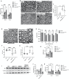
Figure 5. Ablation of Xbp1 limits ER-associated degradation, exacerbates ER-stress and impairs Schwann cell differentiation in S63del mice.
qRT-PCR analysis on mRNAs extracted from P30 sciatic nerves for (A) the XBP1s targets Erdj4 and Erdj6 (B) the PERK/ATF4 pathway targets Chop, Gadd34 and Mthfd2 and (C) the ATF6 pathway targets BiP, Grp94 and Sel1l. (D) qRT-PCR analysis for the negative regulator of myelination Id2. (E-F) qRT-PCR analysis on mRNAs extracted from P180 sciatic nerves for the (E) PERK/ATF4 targets Chop, Gadd34 and Mthfd2 and (F) the ATF6 pathway targets BiP, Grp94 and Sel1l. For all qRT-PCR experiments n = 8–10 RTs from independent nerves for each genotype. Values are expressed as fold change relative to WT (arbitrarily set to 100); error bars represent SEM. (G) Representative Western blots form P180 sciatic nerve extracts for IRE1α, GRP94, P-JNK, BiP and P-eIF2α; b-tubulin was used as loading control. One representative blot of four (quantified in the graphs in H, I and J) is shown per each genotype. Values are expressed as arbitrary units relative to WT that was set to 1. Bars represent SEM. For all panels ****P < 0.0001, *** P < 0.001, ** P <0.01, * P <0.05 by one-way ANOVA followed by Tukey post hoc test.
Figure 6
Figure 6. Schwann cell specific overexpression of spliced XBP1 ameliorates disease parameters in S63del mice.
(A) qRT-PCR analysis on mRNAs extracted from P30 (upper graph) and P180 (lower graph) sciatic nerves for the XBP1s targets Erdj4 and Erdj6. n = 6–8 RTs from independent nerves for each genotype. Values are expressed as fold change relative to WT (arbitrarily set to 100); error bars represent SEM. ****P < 0.0001, * P <0.05 by one-way ANOVA followed by Tukey post hoc test. (B) Transverse semithin sections from P30 sciatic nerves. Scale bar 10μm. (C) G-ratio analysis performed on the P30 semithin sections. ** P <0.01 by one-way ANOVA (D) EM transverse section (scale bar 5μm) from P30 sciatic nerves and (E) relative g-ratio values plotted by axon diameter. Error bars represent SEM; ** P <0.01, * P <0.05 by unpaired Student’s t-test. n = 80–100 fibers per nerve were measured from 5 mice per genotype. (F) NCV and (G) F-wave latency measurements on 6-month-old mice; n = 4–8 mice per genotype. ****P < 0.0001, ** P <0.01, by one-way ANOVA followed by Tukey post hoc test. (H) qRT-PCR analysis on mRNAs extracted from P180 sciatic nerves for the negative regulator of myelination Id2. n = 8–10 RTs from independent nerves for each genotype. Values are expressed as fold change relative to WT (arbitrarily set to 100); error bars represent SEM, *** P < 0.001 by one-way ANOVA followed by Tukey post hoc test. (I) Western blot analysis on P180 sciatic nerve extracts for P-eIF2α. Two representative samples out of four per genotype are shown; (J) quantification of P-eIF2α levels as arbitrary units (a.u.) relative to WT. (K) qRT-PCR analysis on mRNAs extracted from P180 sciatic nerves for Chop and Mthfd2. n = 5 RTs from independent nerves for each genotype. Values are expressed as fold change relative to WT (arbitrarily set to 100); error bars represent SEM, ****P < 0.0001, *** P < 0.001, * P <0.05 by one-way ANOVA followed by Tukey post hoc test.
Figure 7
Figure 7. Schwann cell specific overexpression of spliced XBP1 ameliorates disease parameters in R98C mice.
(A) Transverse semithin sections and (B) relative g-ratio analysis from adult (P160) sciatic nerves. (C) EM transverse sections (scale bar 5μm) and (D) g-ratio analysis from adult R98C and R98C/XBP1SC-OE sciatic nerves. n = 150 fibers per nerve were measured. ****P < 0.0001, ** P < 0.01 by unpaired Student’s t-test. (E) Nerve conduction velocity studies. n = 4–12 mice per genotype. ****P < 0.0001, *** P < 0.001 by one-way ANOVA followed by Tukey post hoc test. (F) Western blot analysis on sciatic nerve extracts for P0 and P-eIF2α; β-tubulin was used as loading control. Two representative samples out of four are shown for each genotype. (G) protein levels quantification for P0 and (H) P-eIF2α expressed as arbitrary units. Error bars represent SEM and ** P < 0.01, * P <0.05 by one-way ANOVA followed by Tukey post hoc test. (I) qRT-PCR analysis for Chop and (J) Mthfd2 on mRNA extracts form P180 nerves. n = 8–12 RTs from independent nerves for each genotype. Values are expressed as fold change relative to WT (arbitrarily set to 100); error bars represent SEM, * P <0.05 by one-way ANOVA followed by Tukey post hoc test.
Figure 8
Figure 8. Pharmacologic activation of IRE1α mediated Xbp1 splicing improves myelination in S63del dorsal root ganglia cultures.
(A) Dorsal root ganglia were dissected from E13.5 WT and S63del embryos and myelination induced with 50μm ascorbic acid in the presence or absence of 1μm IXA4. Myelinating internodes were visualized with an antibody against myelin protein zero (MBP) (B) qRT-PCRs from mRNA extracted from WT or S63del cultures treated with vehicle or 1μm IXA4 for Xbp1s and its target Erdj4. n = 3–4 RT from independent experiments. Error bars represent SEM and ****P < 0.0001, ** P < 0.01, * P <0.05 by one-way ANOVA followed by Tukey post hoc test. (C) Quantification of MBP+ internodes in S63del cultures after IXA4 treatment. * P <0.05 by Student t-test.

Similar articles

References

    1. Taveggia C, Feltri ML. Beyond Wrapping: Canonical and Noncanonical Functions of Schwann Cells. Annu Rev Neurosci. 2022;45:561–580. - PMC - PubMed
    1. Volpi VG, Touvier T, D’Antonio M. Endoplasmic Reticulum Protein Quality Control Failure in Myelin Disorders. Front Mol Neurosci. 2017;9:162. - PMC - PubMed
    1. Bosch-Queralt M, Fledrich R, Stassart RM. Schwann cell functions in peripheral nerve development and repair. Neurobiol Dis. 2023;176:105952. - PubMed
    1. Giese KP, Martini R, Lemke G, Soriano P, Schachner M. Mouse P0 gene disruption leads to hypomyelination, abnormal expression of recognition molecules, and degeneration of myelin and axons. Cell. 1992;71(4):565–576. - PubMed
    1. Scherer SS, Wrabetz L. Molecular mechanisms of inherited demyelinating neuropathies. Glia. 2008;56(14):1578–1589. - PMC - PubMed

Publication types

LinkOut - more resources