[Preprint]. 2024 Jan 30:2024.01.30.577861.
doi: 10.1101/2024.01.30.577861.

Non-CG DNA methylation and MeCP2 stabilize repeated tuning of long genes that distinguish closely related neuron types

Affiliations

Non-CG DNA methylation and MeCP2 stabilize repeated tuning of long genes that distinguish closely related neuron types

J Russell Moore et al. bioRxiv. .

Update in

Abstract

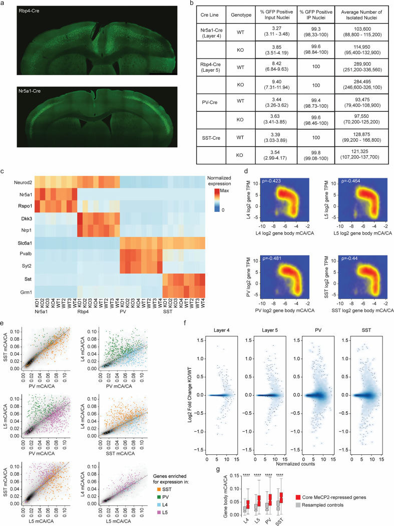
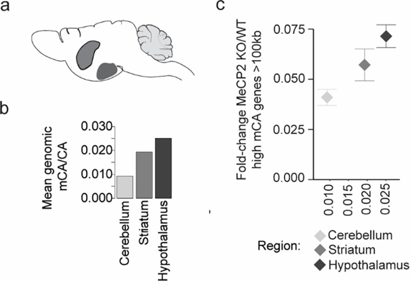
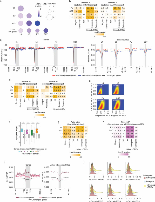
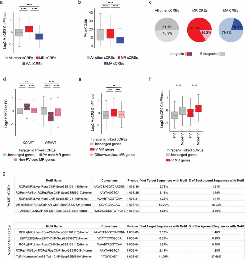
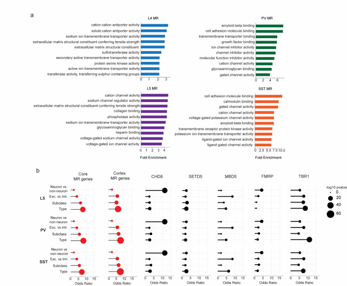
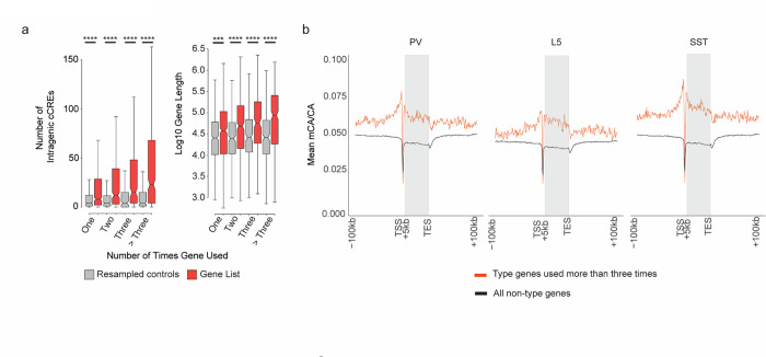
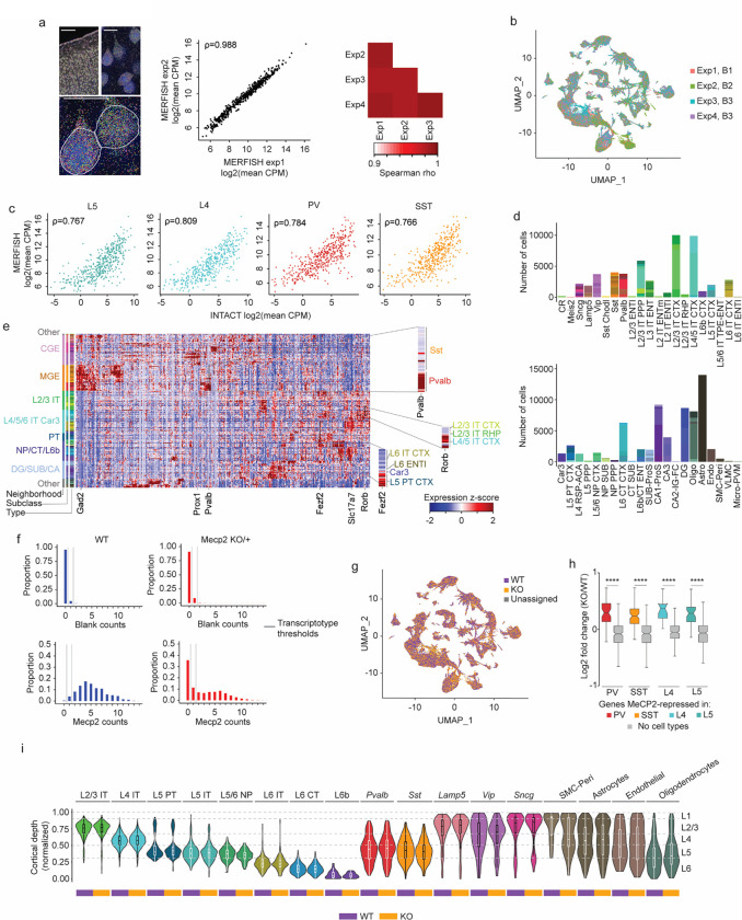
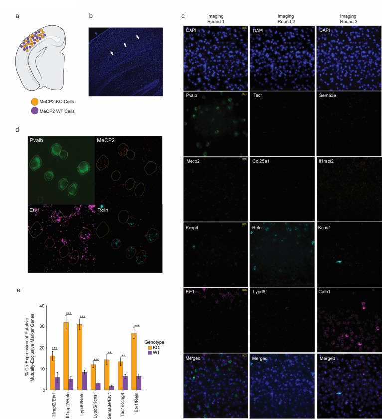
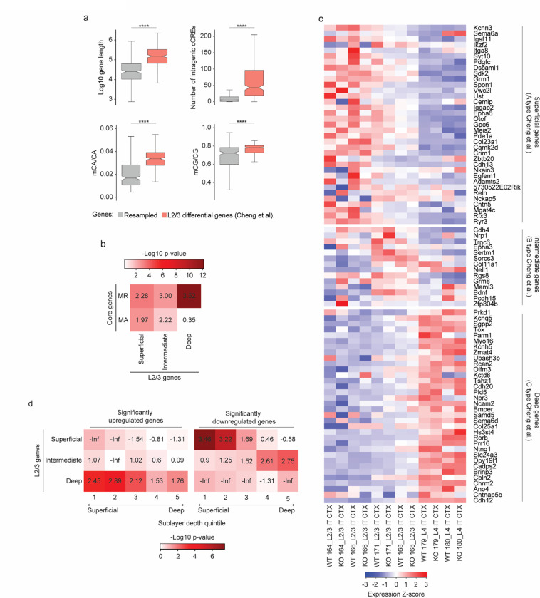
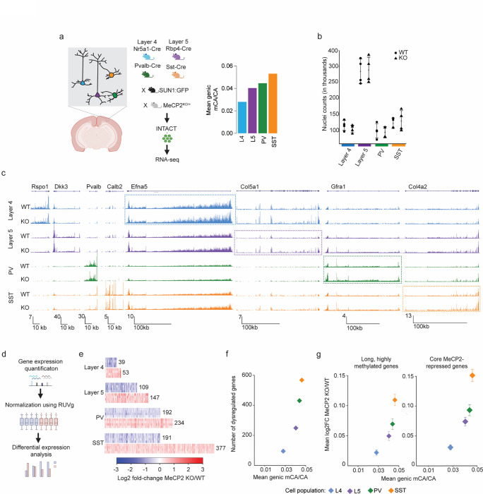
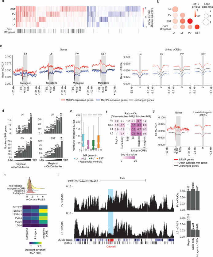
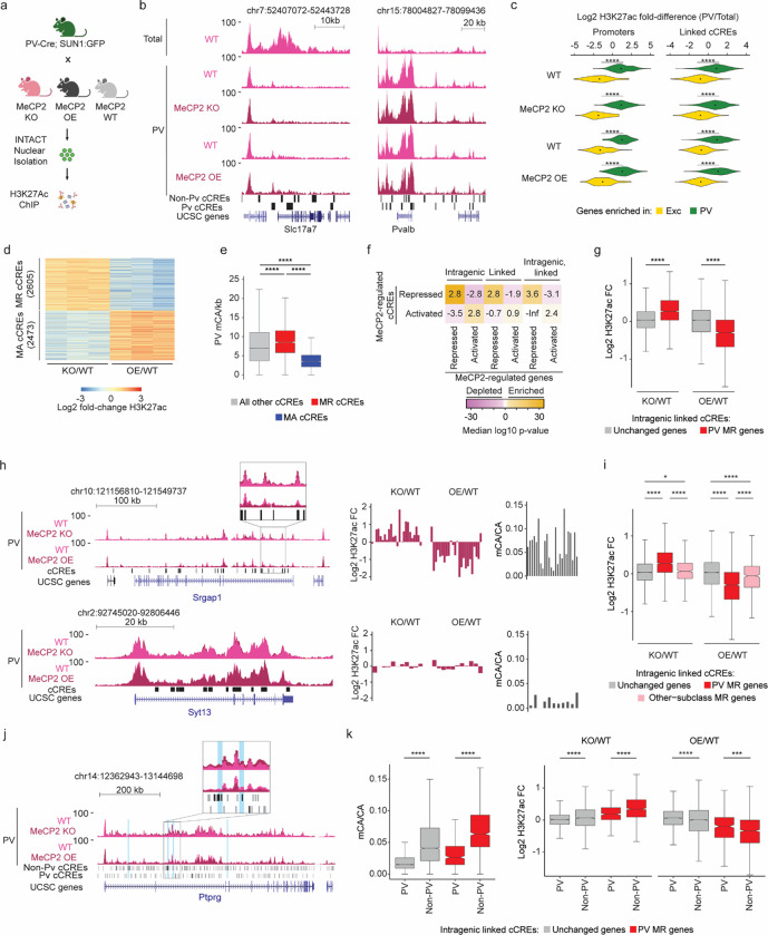
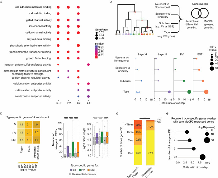
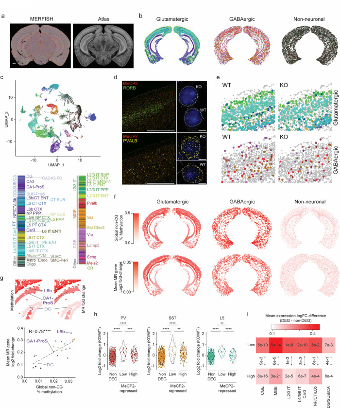
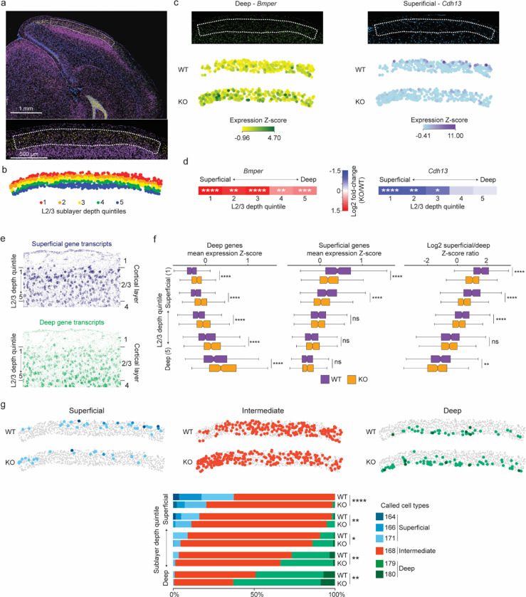
The extraordinary diversity of neuron types in the mammalian brain is delineated at the highest resolution by subtle gene expression differences that may require specialized molecular mechanisms to be maintained. Neurons uniquely express the longest genes in the genome and utilize neuron-enriched non-CG DNA methylation (mCA) together with the Rett syndrome protein, MeCP2, to control gene expression, but the function of these unique gene structures and machinery in regulating finely resolved neuron type-specific gene programs has not been explored. Here, we employ epigenomic and spatial transcriptomic analyses to discover a major role for mCA and MeCP2 in maintaining neuron type-specific gene programs at the finest scale of cellular resolution. We uncover differential susceptibility to MeCP2 loss in neuronal populations depending on global mCA levels and dissect methylation patterns and intragenic enhancer repression that drive overlapping and distinct gene regulation between neuron types. Strikingly, we show that mCA and MeCP2 regulate genes that are repeatedly tuned to differentiate neuron types at the highest cellular resolution, including spatially resolved, vision-dependent gene programs in the visual cortex. These repeatedly tuned genes display genomic characteristics, including long length, numerous intragenic enhancers, and enrichment for mCA, that predispose them to regulation by MeCP2. Thus, long gene regulation by the MeCP2 pathway maintains differential gene expression between closely-related neurons to facilitate the exceptional cellular diversity in the complex mammalian brain.


Figures

Extended Data Fig. 1.
Extended Data Fig. 1.. Gene expression and methylation in INTACT-isolated PV, SST, L4, and L5 neurons.
a, Representative images of Rbp4-Cre;SUN1:GFP labeling of L5 excitatory neurons and Nr5a1-Cre;SUN1:GFP labeling of L4 excitatory neurons. b, Summary statistics of INTACT experiments organized by Cre-line and genotype (no significant differences found between genotypes). c, Heatmap of marker gene expression in RNA sequencing data from each subclass profiled. d, Log2 gene snmc-seq mCA/CA vs log2 gene TPMs for L4, L5, SST, and PV cells. ρ = Spearman’s correlation coefficient. e, Pairwise comparisons of gene body mCA/CA across cell types. Genes enriched for expression >5 fold in one cell type over another are colored according to the cell type where they are highly expressed. f, Log2 fold-change (MeCP2 KO/WT) of gene expression in L4, L5, SST, and PV cells. The y-axis is normalized counts of genes from DESeq2. g, Gene body mCA level of core MeCP2-repressed genes in each cell type compared to expression-matched, resampled control genes. ****p < 0.0001 two-sided Wilcoxon rank-sum test. The center line of each boxplot is the median. Each box encloses the first and third quartile of the data. The whiskers extend to the most extreme values, excluding outliers which are outside 1.5 times the interquartile range. n=4 biological replicates for PV, SST, L4, and L5 RNA-seq.
Extended Data Fig. 2.
Extended Data Fig. 2.. Analysis of global mCA levels and gene dysregulation in different brain regions of MeCP2 KO mice.
a, Depiction of the three brain regions analyzed: cerebellum, striatum, and hypothalamus. b, Global mCA/CA levels across the three brain regions analyzed. c, Scatter plot of global mCA levels in each brain region and average fold-change in mRNA expression for long (greater than 100 kb), high mCA (top decile of gene body mCA) genes in the MeCP2 KO vs. WT. n=1 biological replicate for cerebellum, striatum, and hypothalamus methylation analysis.
Extended Data Fig. 3.
Extended Data Fig. 3.. Methylation patterns at MeCP2-regulated genes in PV, SST, L4, and L5 neurons.
a, Significance of overlap of MeCP2-activated genes from each cell type and core MeCP2-activated genes from multiple datasets. −Log10 p-value is calculated from two-sided Fisher’s exact test. b, Heatmap of mCA/CA enrichment in regions, gene bodies, and linked cCREs of cell-type MR genes or cell-type MA genes over those of unchanged genes, colored by the log10 two-sided Wilcoxon rank-sum p-value. Numbers in the tiles represent the ratio of median methylation level of elements associated with subclass MR or MA genes to the median methylation level of elements associated with unchanged genes. c, Left: Aggregate mCG/CG levels for MeCP2-regulated genes in L4, L5, SST, and PV neurons. Mean mCG/CG is reported for 1 kb bins. “Metagene’’ refers to 50 equally sized bins within gene bodies. Right: aggregate mCG levels centered at cCREs linked to MeCP2-regulated genes in L4, L5, SST, and PV neurons. Mean mCG/CG is reported for 100 bp bins. Gray rectangle = 700 bp, ~ median length of all cCREs. d, Heatmap of mCG/CG enrichment in regions, gene bodies, and linked cCREs of cell-type MR genes or cell-type MA genes over those of unchanged genes, colored by the log10 two-sided Wilcoxon rank-sum p-value. Numbers in the tiles represent the ratio of median methylation level of elements associated with cell-type MR or MA genes to the median methylation level of elements associated with unchanged genes. e, Scatter plot showing regional mCA/CA levels and cCRE mCA/CA levels for all cCREs in the genome in each cell type. This neuron subclass specific analysis shows that cCREs mCA is correlated with the mCA set-point for large-scale genomic regions, as previously shown for whole cortex. Here, regions are defined as topologically associated domains (TADs) identified in the cortex. ρ = Spearman’s correlation coefficient. f, Log 10 gene length of genes MeCP2-repressed in L4, L5, PV, and SST genes. The gray box next to each subclass represents the expression-matched genes resampled from that subclass’s list of unchanged genes. The center line of each boxplot is the median. Each box encloses the first and third quartile of the data. The whiskers extend to the most extreme values, excluding outliers which are outside 1.5 times the interquartile range. g, Heatmap of mCA/CA enrichment in regions, gene bodies, and linked cCREs core MR genes over those of all other genes, colored by the log10 Wilcoxon rank-sum p-value. Numbers in the tiles represent the ratio of median mCA/CA of elements associated with core MR genes to the median mCA/CA of elements associated with all other genes. h, Heatmap of mCA/CA enrichment in regions, gene bodies, and linked cCREs of non-subclass core MR genes over those of subclass core MR genes, colored by the log10 two-sided Wilcoxon rank-sum p-value. Numbers in the tiles represent the ratio of median mCA/CA of elements associated with subclass MR genes to the median mCA/CA of elements associated with other-subclass MR genes. i, Aggregate mCA/CA levels at gene bodies (left) and linked cCREs (right) of L5 core MR genes, non-L5 core MR genes, and unchanged genes. j, Density plots of pairwise mCA/CA ratios between cell types in 1 kb extragenic regions, intragenic regions, and regions centered at intragenic cCREs. DNA methylation data were compiled from previously published single-cell methylomic analysis, see methods. RNA-seq data are from INTACT analysis described in Figure 1, n=4 biological replicates per genotype per cell type.
Extended Data Fig. 4.
Extended Data Fig. 4.. Distribution of epigenomic signals at MeCP2-regulated cCREs in PV interneurons.
a, Log2 input-normalized MeCP2 ChIP signal at MeCP2-repressed (MR) and MeCP2-activated (MA) cCREs in PV cells. ****p < 0.0001 two-sided Wilcoxon rank-sum test. b, Boxplot of PV mCG/kb in MeCP2-regulated cCREs. ****p < 0.0001 two-sided Wilcoxon rank-sum test. c, Genic distributions of MeCP2-regulated cCREs showing enrichment of MR cCREs to be intragenic. d, Log2 H3K27ac ChIP fold-change (MeCP2 mutant/wild-type) in cCREs inside and linked to PV core MR, non-PV core MR, or unchanged genes. **p < 0.01, ****p < 0.0001 two-sided Wilcoxon rank-sum test. e, Log2 input-normalized MeCP2 ChIP-seq signal in cCREs inside and linked to PV MR genes, other-subclass MR genes, or unchanged genes. **p < 0.01, ****p < 0.0001 two-sided Wilcoxon rank-sum test. f, Log2 input-normalized MeCP2 ChIP-seq signal in PV cCREs and non-PV cCREs inside and linked to PV MR genes or unchanged genes. ****p < 0.0001 two-sided Wilcoxon rank-sum test. g, HOMER output showing ROR family motif enrichment in PV and non-PV cCREs. n=3 biological replicates for PV WT, MeCP2 KO, and MeCP2 OE H3K27ac ChIP-seq. In all boxplots, the center line of each boxplot is the median. Each box encloses the first and third quartile of the data. The whiskers extend to the most extreme values, excluding outliers which are outside 1.5 times the interquartile range.
Extended Data Fig. 5.
Extended Data Fig. 5.. Functional annotation of subclass-defined MeCP2-repressed genes and their representation in other datasets.
a, Gene ontology of MeCP2-repressed (MR) genes in L4, L5, PV, and SST neurons. Top 10 terms for Molecular Function shown. b, Overlap of differentially expressed genes across L5, PV, and SST cellular hierarchy with core MeCP2-repressed genes, MeCP2-repressed genes previously identified in the cortex, and dysregulated genes detected in other NDD mouse models. Note that each differential list labeled on the left for L5, PV and SST reflects the same neuron vs. non-neuron and excitatory vs. inhibitory lists, but the lists of differential genes at the subclass and type level reflect the genes that specifically define these differentiations in L5, PV or SST cells. Analysis was performed on differentially expressed genes from INTACT RNA-seq analysis described in Figure 1. n=4 biological replicates per genotype per cell type, as well as the indicated published gene lists.
Extended Data Fig. 6.
Extended Data Fig. 6.. Genes that are repeatedly tuned between neuron types have characteristics that predispose them to regulation by the MeCP2 pathway.
a, Number of intragenic cCREs (left) and gene length (right) in genes that show repeated tuning between closely related neuron types. Genes are plotted by the number of times they are detected as differential between two closely related neuron types. The center line of each boxplot is the median. Each box encloses the first and third quartile of the data. The whiskers extend to the most extreme values, excluding outliers which are outside 1.5 times the interquartile range. b, Aggregate profiles of mCA/CA in the cerebral cortex for genes found to be differentially expressed between closely related neuron types more than three times.
Extended Data Fig. 7.
Extended Data Fig. 7.. MERFISH analysis of cell types and MeCP2 expression in MeCP2 KO/+ brain shows expected gene expression and cell distributions across transcriptotypes.
a, Left: transcripts detected (colored dots) and DAPI images of MERFISH data in one experiment. Scale bar is 250 μm for low resolution view, and 25 μm for close-ups. Middle: correlation between experiments 1 and 2 in log2 mean CPM of each gene. Right: correlation between each pair of MERFISH experiments in log2 mean CPM of each gene. ρ = Spearman’s correlation coefficient. b, UMAP representation of cells in MeCP2 KO/+ MERFISH experiments, colored by experiment identity and biological replicate. B1–3=biological replicate 1–3. c, Scatter plot of log-transformed CPMs of gene expression in neuronal subclasses (PV, SST, L4, and L5) measured by INTACT RNA-seq and MERFISH. ρ = Spearman’s correlation coefficient. d, Number of cells of each type in each subclass in MeCP2 KO/+ MERFISH experiments. e. Heatmap showing z-scores of average expression for each gene in the MERFISH panel in each cell type. Inset: close-ups of Pvalb (PV marker gene), Fezf2 (L5 PT CTX marker gene), and Rorb (L4/5 IT CTX marker gene). f, Representative distributions of transcript counts per cell for Mecp2 and a representative negative control (“Blank counts”) detected in PV cells in MERFISH analysis of wild-type or MeCP2 KO/+ coronal sections. Cut offs used for calling “WT” and “KO” transcriptotypes in the MeCP2 KO/+ are shown. g, UMAP representation of cells identified as either WT, KO, or unassigned in MeCP2 KO/+ MERFISH experiments. h, MERFISH log2 fold-change of genes identified as MeCP2-repressed in PV, SST, L4, and L5 INTACT RNA-seq analyses. ****p < 0.0001 two-sided Wilcoxon rank-sum test. The center line of each boxplot is the median. Each box encloses the first and third quartile of the data. The whiskers extend to the most extreme values, excluding outliers which are outside 1.5 times the interquartile range. i, Locations of distinct sublcasses of WT and KO cells across cortical layers in the visual cortex identified using MERFISH. No major differences were observed in cortical depth between WT and KO cells. The center line of each boxplot is the median. Each box encloses the first and third quartile of the data. n=3 biological replicates for MeCP2 KO/+ MERFISH across 4 imaged brain sections.
Extended Data Fig. 8.
Extended Data Fig. 8.. Loss of mutually exclusive type-specific gene expression in MeCP2 KO PV interneurons.
a, MeCP2 mutant heterozygous females contain wildtype and mutant cells. b, Representative image of visual cortex L4 area focused on for analysis. White arrow pointing to L4 region. c, Representative images from all 12 target probes and DAPI stain for each of the three imaging rounds. Merged images of each round shown. d, RNAScope analysis of expression of mutually exclusive marker genes for visual cortex PV interneurons in MeCP2 KO and WT PV neurons. Identification of PV neurons using Pvalb and call of MeCP2 KO and WT cells using Mecp2. e, Bar plots of rate of co-expression of putatively mutually exclusive PV marker genes in MeCP2 null and WT PV interneurons (n=3, 50–100 cells per experiment, two-sided unpaired t test, **p<0.01, ***p<0.005). n=3 biological replicates for RNAScope.
Extended Data Fig. 9.
Extended Data Fig. 9.. Visual cortex layer 2/3 sublayer-specific genes are targets of MeCP2 regulation.
a, Characteristics of visual L2/3 excitatory neuron sublayer type-specific genes defined by Cheng et al., 2022, showing long gene length, high numbers of intragenic cCREs and enriched mCA. ****p < 0.0001 two-sided Wilcoxon rank-sum test. b, Heatmap showing overlap and significance of excitatory neuron sublayer type-specific genes defined by Cheng et al., 2022 with core MeCP2-regulated gene lists. Numbers in the tiles represent enrichment (log2 odds ratio) of core MeCP2-regulated genes in each L2/3 gene list. c, Heatmap of MERFISH-quantified expression for excitatory neuron sublayer type-specific genes defined by Cheng et al. in IT excitatory neurons in L2/3 of MeCP2 KO/+ V1 (top). d, Heatmap showing overlap and significance of excitatory neuron sublayer type-specific genes defined by Cheng et al. with genes detected as significantly dysregulated in pseudobulkDGE analysis of MERFISH data in excitatory neurons in sublayer depth quintiles of L2/3 of the V1. Numbers in the tiles represent enrichment (log2 odds ratio) of L2/3 genes in dysregulated gene lists of the excitatory neurons in each sublayer depth quintile of L2/3 of V1. n=3 biological replicates for MeCP2 KO/+ MERFISH across 4 imaged brain sections.
Fig. 1.
Fig. 1.. Neuron subclass-specific analysis of global mCA levels and gene dysregulation in the MeCP2 KO.
a, Four neuron subclasses from the cerebral cortex with varying levels of mCA were selected for gene expression analysis in MeCP2 KO using the INTACT nuclear isolation system. b, Number of nuclei isolated by INTACT for KO and WT animals for each population profiled. No significant differences in numbers of nuclei isolated were detected. c, Genome browser view of RNA-seq data for MeCP2 WT and KO in each population. Marker genes Rspo1, Dkk3, Pvalb, and Calb2 for each subclass profiled (L4, L5, PV, and SST, respectively) shown to the left. Example MeCP2-repressed gene for each subclass is highlighted with a box (Efna5 – L4, Col5a1 – L5, Gfra1 – PV, Col4a2 – SST). d, Overview of analysis workflow of differential gene expression analysis for INTACT-RNA-seq. e. Heatmap of per replicate fold-changes MeCP2 KO/WT of significantly down-regulated genes (blue) and up-regulated (red) genes identified in each subclass with total number of genes shown. f, Scatter plot of the number of significantly dysregulated genes identified in each subclass plotted vs average mCA level in that subclass. g, Scatter plot of mean fold-change of long (greater than 100 kb), highly methylated (top decile of mCA) genes (left) and core MeCP2-repressed genes (right) vs. average mCA level for all genes in each subclass. n=4 biological replicates for PV, SST, L4, and L5 RNA-seq.
Fig. 2.
Fig. 2.. Overlapping and distinct regulation of genes across neuronal populations by MeCP2 is associated with regional and gene-specific mCA patterns.
a, Heatmap of fold-changes for overlapping and unique significantly dysregulated genes in each neuronal subclass. Core MeCP2-repressed (MR) genes marked in black below. b, Significance of overlap of MeCP2-repressed genes in each subclass and core MeCP2-repressed genes from multiple datasets. P-values are calculated by two-sided Fisher’s exact test. c, Left: aggregate mCA/CA levels for MeCP2-regulated genes identified in L4, L5, PV, and SST neurons. Mean mCA/CA is reported for 1 kb bins. “Metagene’’ refers to 50 equally sized bins within gene bodies. Right: aggregate mCA/CA levels centered at cCREs linked to MeCP2-regulated genes in L4, L5, PV, and SST neurons. Mean mCA/CA is reported for 100 bp bins. Gray rectangle = 700 bp, ~ median length of all cCREs. d, Number of MeCP2-repressed genes found in each decile of genes sorted by regional mCA level. Regional mCA for each gene is calculated as mCA/CA for the region 10 kb to 210 kb upstream of the TSS and the region 200 kb downstream of its TES, removing the signal from genes overlapping these regions. Enrichment (log2 odds ratio) of MeCP2-repressed genes in each decile shown above each count for each decile. e, Number of intragenic cCREs inside MR genes identified in L4, L5, SST, and PV neurons and expression-resampled controls. The center line is the median. Each box encloses the first and third quartile of the data. The whiskers extend to the most extreme values, excluding outliers which are outside 1.5 times the interquartile range. ****p < 0.0001 two-sided Wilcoxon rank-sum test. f, Heatmap of mCA/CA enrichment in regions, gene bodies, and linked cCREs of other-subclass MR genes over those of subclass MR genes, colored by the log10 two-sided Wilcoxon rank-sum p-value. Numbers in the tiles represent the ratio of median mCA/CA of elements associated with subclass MR genes to the median mCA/CA of elements associated with other-subclass MR genes. g, Aggregate mCA/CA levels at gene bodies (left) and linked cCREs (right) of L5 MR genes, other-subclass MR genes, and unchanged genes. h, Top: density plot of mCA/CA ratio between PV and L5 neurons in 1 kb extragenic regions, intragenic regions, and regions centered at intragenic cCREs. Bottom: heatmap of standard deviation mCA/CA ratios between pairs of subclasses among L4, L5, PV, and SST neurons. i, Left: genome browser view of PV (top) and L5 (bottom) mCA/CA at Cacna1i, a gene repressed by MeCP2 in PV neurons but not in L5 neurons. Right: regional, gene body, and intragenic linked cCRE mCA levels of Cacna1i in PV (top) and L5 (bottom) neurons. Numbers on top of bars are the mCA/CA levels of each group. DNA methylation data were compiled from previously published single-cell methylomic analysis, see methods. RNA-seq data are from INTACT analysis described in Figure 1, n=4 biological replicates per genotype per cell type.
Fig. 3.
Fig. 3.. Dysregulation of cell-type-specific high mCA enhancers in MeCP2 knockout parvalbumin positive interneurons.
a, Schematic of INTACT isolation and H3K27ac ChIP-seq profiling of PV MeCP2 wild-type (WT), MeCP2 knockout (KO), and MeCP2 overexpression (OE) neurons. b, Genome browser views of H3K27ac ChIP-seq signal in cortical (total) and PV nuclei at Slc7a7, a gene expressed in excitatory neurons, and Pvalb, a marker gene for PV neurons. Location of cCREs and genes shown at bottom. c, Log2 H3K27ac fold difference between PV and total nuclei for the same genetic background (MeCP2 WT, MeCP2 KO, or MeCP2 OE) at promoters and linked cCREs of the top 100 genes enriched for expression in PV neurons (log2 gene expression difference (PV/excitatory) > 0, PV RPKM >= 10, FDR-adjusted p-value <= 0.01, ordered by log2 fold difference) and the top 100 genes enriched in excitatory (Exc) neurons (log2 gene expression fold difference (excitatory/PV) > 0, Exc RPKM >= 10, FDR-adjusted p-value <= 0.01, ordered by log2 fold difference). The genes were identified from previous differential gene expression analysis of PV and excitatory genes. ****p < 0.0001 two-sided Wilcoxon rank-sum test. Violin plots were made after excluding outliers which are outside 1.5 times the interquartile range of the data. d, H3K27ac fold-changes for cCREs identified as significantly dysregulated for H3K27ac ChIP-seq signal in PV neurons isolated from MeCP2 KO and MeCP2 OE mice (FDR-adjusted p-value ≤ 0.1). MR = MeCP2-repressed; MA= MeCP2-activated. e, Boxplot of PV mCA/kb in MeCP2-repressed (MR), MeCP2-activated (MA) and all other cCREs. ****p < 0.0001 two-sided Wilcoxon rank-sum test. f, Quantification of enrichment of MeCP2-regulated cCREs to be located inside (intragenic) or linked to MeCP2-regulated genes by co-correlation analysis or both. Median significance (log10 p-value from two-sided Fisher’s exact test, color) and enrichment (log2 odds ratio, number) are shown for cCREs associated with MeCP2-regulated genes compared to cCREs associated with expression-resampled genes. g, Log2 H3K27ac fold-change for cCREs located inside of, and linked to, PV MR genes or unchanged genes in PV MeCP2 KO and MeCP2 OE. ****p < 0.0001 Two-sided Wilcoxon rank-sum test. h, Left: overlaid PV MeCP2 WT, MePC2 KO, and MeCP2 OE H3K27ac ChIP-seq tracks in the PV MeCP2-repressed gene Srgap1 (top) and an other-cell-type MeCP2-repressed gene Syt13 (bottom). The inset shows cCRE-containing regions with changes in H3K27ac upon MeCP2 perturbation. Right: log2 H3K27ac fold-change PV MeCP2 KO and MeCP2 OE and PV mCA/CA of cCREs inside and linked to Srgap1 or Syt13. The expression of Syt13 is not affected in PV neurons and it does not show enrichment of mCA or alterations in histone acetylation at its associated cCREs. i, Log2 H3K27ac fold-change of cCREs inside and linked to PV MR genes, other-cell-type MR genes, or unchanged genes in PV MeCP2 KO and MeCP2 OE. *p < 0.05, ****p < 0.0001 Two-sided Wilcoxon rank-sum test. j, Genome browser view of H3K27ac ChIP-seq at the Ptprg gene in PV wild-type, MeCP2 KO, and MeCP2 OE. Gray bars are all cCREs while black bars are intragenic, cCREs linked to the gene. Inset shows region containing non-PV cCREs with changes in H3K27ac ChIP-seq signal in MeCP2 KO and MeCP2 OE relative to wild-type. Blue highlights are examples of non-PV cCREs called as significantly dysregulated upon MeCP2 perturbation. k, PV mCA/CA (left) and log2 H3K27ac fold-change in PV MeCP2 KO and MeCP2 OE (right) of PV and non-PV cCREs located inside of, and linked to, unchanged genes or PV MR genes. ***p < 0.001, ****p < 0.0001 two-sided Wilcoxon rank-sum test. n=3 biological replicates for PV WT, MeCP2 KO, and MeCP2 OE H3K27ac ChIP-seq. n=2 biological replicates for Total WT, MeCP2 KO, and MeCP2 OE H3K27ac ChIP-seq. For all boxplots, the center line is the median. Each box encloses the first and third quartile of the data. The whiskers extend to the most extreme values, excluding outliers which are outside 1.5 times the interquartile range.
Fig. 4.
Fig. 4.. MeCP2 regulates genes that are repeatedly tuned across closely related neuronal types.
a, Gene Ontology analysis of significantly dysregulated genes in each subclass from INTACT analysis shows enrichment for synaptic proteins, channels, and other factors important for neuronal cell type function. Top Molecular Function terms with overlap between cell types are shown. Gene ratio (percentage of total DEG in given GO term) and Benjamini-Hochberg adjusted p-value shown. b, Top: schematic of gene overlap analysis of MeCP2-repressed genes with gene sets distinguishing neuronal populations at different levels of hierarchical taxonomy previously generated from single cell transcriptomic data. Bottom: odds ratio and significance of overlap between MeCP2-repressed genes identified in each neuron subclass and genes differentially expressed at each level of the hierarchy. P-value determined by two-sided Fisher’s exact test. NA shown for L4, because this subclass does not contain any defined types in the cellular taxonomy analyzed. c, Left, mCA enrichment analysis of regions, gene bodies, and intragenic-linked cCREs for genes that distinguish neuronal types within L5, PV, and SST subclasses. Enrichment relative to expression resampled control gene sets is shown numerically, significance is indicated by color. Right, quantification of number of intragenic enhancers and gene length for these neuron-type-specific genes. The center line of each boxplot is the median. Each box encloses the first and third quartile of the data. The whiskers extend to the most extreme values, excluding outliers which are outside 1.5 times the interquartile range. ****p < 0.0001 two-sided Wilcoxon rank-sum test d, Percentage of genes found to be differentially expressed between closely related neuron types in one or more pairwise comparisons of types within L5, PV, and SST subclasses. Values shown for true type-specific genes and for expression-matched, resampled control genes. e, Repeatedly tuned neuron type-specific genes significantly overlap with core MeCP2-repressed genes.
Fig. 5.
Fig. 5.. Spatial transcriptomic analysis of the Rett syndrome mouse model reveals region- and neuron type-specific gene dysregulation.
a, Left: transcripts detected (colored dots) by MERFISH for an example MeCP2 KO/+ coronal section. Right: brain regions in the coronal plane, as defined in the Allen Mouse Brain Atlas, were identified using SHARP-Track,. Scale bar = 1 mm. b, Spatial map of glutamatergic, GABAergic, and non-neuronal cells in a representative MeCP2 KO/+ MERFISH experiment. Colors indicating cell types correspond to legend in c. c, Top: UMAP representation of all pass-filter cells identified in MeCP2 KO/+ MERFISH experiments (n=4 imaged sections from 3 biological replicates). Cells colored by type assigned by Seurat label transfer using the reference mouse cortical and hippocampal scRNA-seq compendium. Bottom: legend showing neighborhoods, subclasses, and types of cells detected. d, Left: example images of cortex from MeCP2 KO/+ mice showing expression of MeCP2 (red) and L4 marker Rorb+ (top) or Pvalb (bottom) transcripts. Scale bar = 1 mm. Right: zoom views show examples of cell segmentation and transcripts detected for WT and KO transcriptotyped cells. DAPI nuclear stain is shown in blue. Scale bar = 10 μm. e. Spatial map of WT and KO transcriptotyped glutamatergic and GABAergic cells in MeCP2 KO/+ cortex. f, Top row: spatial map of glutamatergic, GABAergic, and non-neuronal subclasses, colored by global non-CG methylation level of each subclass. Bottom row: spatial map of glutamatergic, GABAergic, and non-neuronal subclasses, colored by mean log2 fold-change of core MeCP2-repressed genes in that subclass. g, Top: zoom-in view of glutamatergic cells shown in f showing global non-CG methylation levels and fold-change of core MeCP2-repressed genes in subclasses of cells. Bottom: scatter plot of mean log2 fold-change of core MeCP2-repressed genes and global non-CG methylation levels for all subclasses that can be mapped between MERFISH and single cell methylomic data. h, Log2 fold-change of non-type-specific genes (non-DEGs), MeCP2-repressed genes that are normally “low” in each neuron type, or MeCP2-repressed genes that are normally “high” in each neuron type, for cell types found in the PV, SST, and L5 populations captured in our INTACT RNA-seq analyses. Violin plot shows fold-changes of the entire population of type-specific genes within each subclass. Points indicate fold-change of individual genes in each neuron type. Colors of points corresponds to the neuron type where the gene is normally lowly expressed; see color legend in panel c. **p < 0.01, ***p < 0.001, ****p < 0.0001 two-sided Wilcoxon test. The center black dot represents the mean and the error represents the standard error. Violin plots were made after excluding outliers which are outside 1.5 times the interquartile range of the data. i, Heatmap of mean log fold-change in gene expression difference between non-DEGs and type-specific genes “low” in a neuron type, or type-specific gene that are “high” in a neuron type in MERFISH for cell types aggregated by neighborhood. Numbers represent the p-value from the two-sided Wilcoxon rank-sum test compared to non-type-specific genes in each analysis. Arrows between boxes represent the p-value from the two-sided Wilcoxon rank-sum test comparing gene expression changes between low and high type-specific genes in each neighborhood analysis.
Fig. 6.
Fig. 6.. Loss of MeCP2 disrupts sublayer-resolved gene programs in layer 2/3 of the visual cortex.
a, MERFISH image of a coronal section from an MeCP2 KO/+ brain (top) showing expression of the L2/3 marker Ccbe1 (yellow) and the L4 marker Rorb (purple); blue, DAPI. Dotted line demarcates L2/3 of primary visual cortex (V1) (bottom, higher magnification) selected for further analysis. b, Spatial map of excitatory neurons in L2/3 for a representative MeCP2 KO/+ V1 region where each cell is colored by the quintile of sublayer depth in which it resides. c, Examples of MERFISH signal and transcriptotype-resolved expression in WT vs. KO excitatory neurons in L2/3 of V1 for sublayer-specific genes detected as dysregulated in the MeCP2 KO cells. Top, MERFISH signal from an example MeCP2 KO/+ brain section. Bottom, expression from cells of each transcriptotype across all excitatory neurons in L2/3 of V1. Expression is reported as the z-score of SCTransform-corrected counts of the mRNA across glutamatergic cells in L2/3 of the visual cortex. d, Fold-change observed in MeCP2 KO vs. WT cells across superficial to deep sublayer depth quintiles for sublayer-specific genes examined in c. *p. < 0.05, **p < 0.01, ***p < 0.001, ****p < 0.0001, Benjamini-Hochberg corrected p-value from pseudobulkDGE analysis. e, Spatial map of RNA transcripts detected in L2/3 of the MeCP2 KO/+ V1 for gene sets previously proposed to be preferentially expressed in neurons located in the superficial sublayer (top, dark blue) or deep sublayer (bottom, green) f, Left, and middle: boxplots of mean expression z-score of genes previously associated with deep sublayers (left) and superficial sublayers (middle) in WT and MeCP2 KO cells across sublayer depth quintiles. Right: boxplots of ratio of mean expression z-score of superficial-sublayer-associated genes to mean expression z-score of deep-sublayer-associated genes, plotted by sublayer depth quintile. The center line of each boxplot is the median. Each box encloses the first and third quartile of the data. The whiskers extend to the most extreme values, excluding outliers which are outside 1.5 times the interquartile range. g, Top: spatial map of WT and KO excitatory neuron IT types detected in L2/3 of the MeCP2 KO/+ V1 separated by cell types associated with superficial, intermediate, or deep gene programs (see Extended data figure 9). Bottom: stacked barplots showing proportions of neuron types in sublayer depth quintiles of L2/3 of V1, separated by transcriptotype. n=3 biological replicates for MeCP2 KO/+ MERFISH across 4 separate imaged brain sections.

References

    1. Yao Z. et al. A taxonomy of transcriptomic cell types across the isocortex and hippocampal formation. Cell 184, 3222–3241.e26 (2021). - PMC - PubMed
    1. Allaway K. C. et al. Genetic and epigenetic coordination of cortical interneuron development. Nature 597, 693–697 (2021). - PMC - PubMed
    1. Cheng S. et al. Vision-dependent specification of cell types and function in the developing cortex. Cell 185, 311–327.e24 (2022). - PMC - PubMed
    1. Clemens A. W. & Gabel H. W. Emerging Insights into the Distinctive Neuronal Methylome. Trends in Genetics 36, 816–832 (2020). - PMC - PubMed
    1. Lister R. et al. Global Epigenomic Reconfiguration During Mammalian Brain Development. Science 341, 1237905 (2013). - PMC - PubMed

Publication types