

Atypical Kikuchi-Fujimoto Disease: FDG-PET Contribution To The Diagnosis
- PMID: 38352805
- PMCID: PMC10860915
- DOI: 10.12890/2024_004258
Atypical Kikuchi-Fujimoto Disease: FDG-PET Contribution To The Diagnosis
Abstract
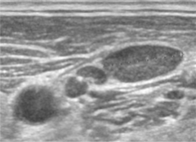
Kikuchi-Fujimoto disease (KFD), also called histiocytic necrotizing lymphadenitis, is more common in young women and typically presents with small, painful, localized cervical lymphadenopathy that resolves spontaneously within a few weeks. Laboratory findings are variable. As many as 40% of KFD cases are reported to be painless, and up to 22% to be generalized lymphadenopathy. Therefore, malignant lymphoma could be a differential diagnosis of KFD. A histopathologic diagnosis is needed when it is difficult to distinguish KFD from lymphoma. KFD typically shows small, highly accumulated cervical lymph nodes on fluorodeoxyglucose positron emission tomography (FDG-PET). This contrasts with malignant lymphoma, which tends to be associated with massive lymphadenopathy. In our case, a 40-year-old Japanese male presented with painless lumps in the right neck, accompanied by fever, night sweats, and loss of appetite. His symptoms and laboratory results worsened over a month. FDG-PET revealed highly accumulated uptake in cervical, mediastinal, and axillary lymph nodes. The PET imaging showed a small, high FDG uptake and contributed to the correct diagnosis of KFD. This case report highlights the importance of FDG-PET, which is a valuable diagnostic tool for KFD as it typically differentiates large clusters of small lymph nodes typical of KFD from normal lymph nodes.
Learning points: Kikuchi-Fujimoto disease (KFD) typically presents with small, painful, localised cervical lymphadenopathy.KFD has atypical patterns showing painless and generalised lymphadenopathy.Fluorodeoxyglucose positron emission tomography (FDG-PET) could be useful for diagnosing not only malignant lymphoma but also KFD.
Keywords: Bentall procedure; Type A aortic dissection; atypical symptoms.
© EFIM 2024.
Conflict of interest statement
Conflicts of Interests: The Authors declare that there are no competing interests.
Figures


References
-
- Perry AM, Choi SM. Kikuchi-Fujimoto Disease: A Review. Arch Pathol Lab Med. 2018;142:1341–1346. - PubMed
-
- Kikuchi M. Lymphadenitis showing focal reticulum cell hyperplasia with nuclear debris and phagocytosis. Nippon Ketsueki Gakkai Zasshi. 1972;35:378–380.
-
- Fujimoto Y, Kozima Y, Hamaguchi K. Cervical necrotizing lymphadenitis: a new clinicopathological agent. Naika. 1972;20:920–927.
-
- Song JY, Lee J, Park DW, Sohn JW, Suh SI, Kim IS, et al. Clinical outcome and predictive factors of recurrence among patients with Kikuchi’s disease. Int J Infect Dis. 2009;13:322–326. - PubMed
LinkOut - more resources
Full Text Sources
Research Materials