

Investigation of dynamical flexibility of D5SIC-DNAM inside DNA duplex in aqueous solution: a systematic classical MD approach
- PMID: 38353005
- PMCID: PMC11080001
- DOI: 10.1039/d3cp05572h
Investigation of dynamical flexibility of D5SIC-DNAM inside DNA duplex in aqueous solution: a systematic classical MD approach
Abstract
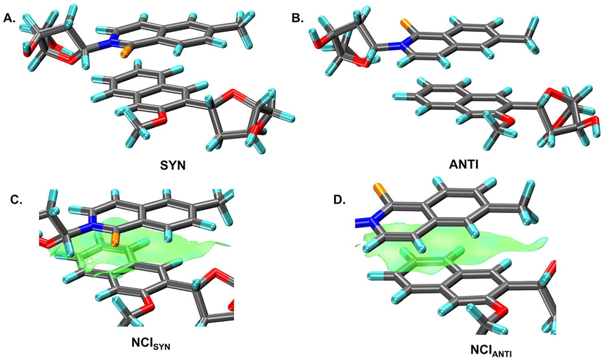
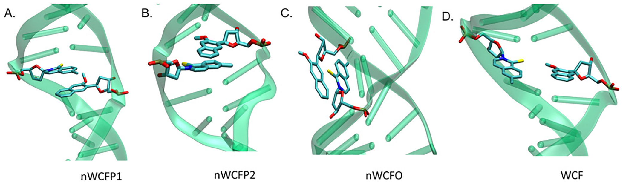
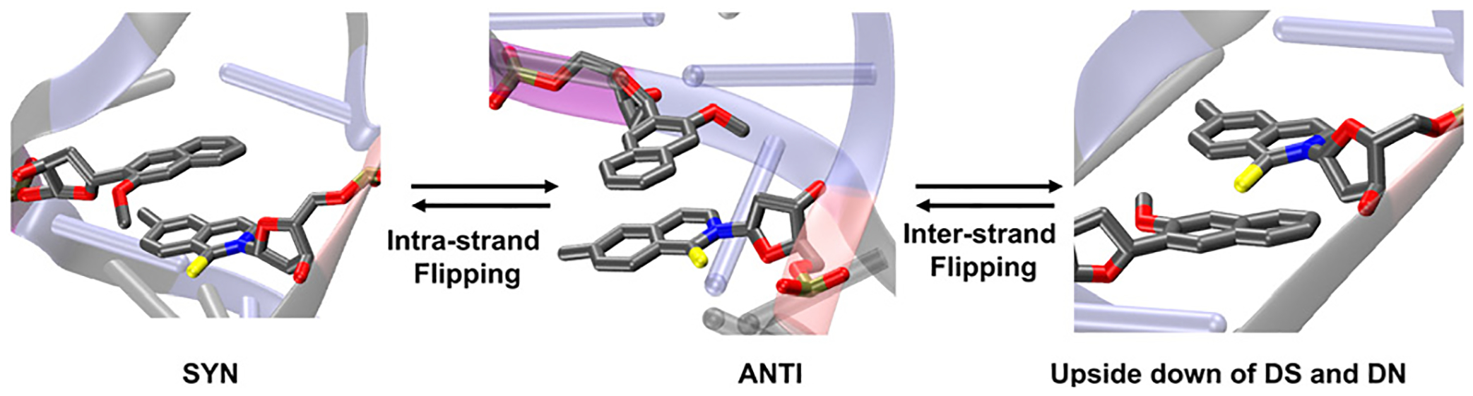
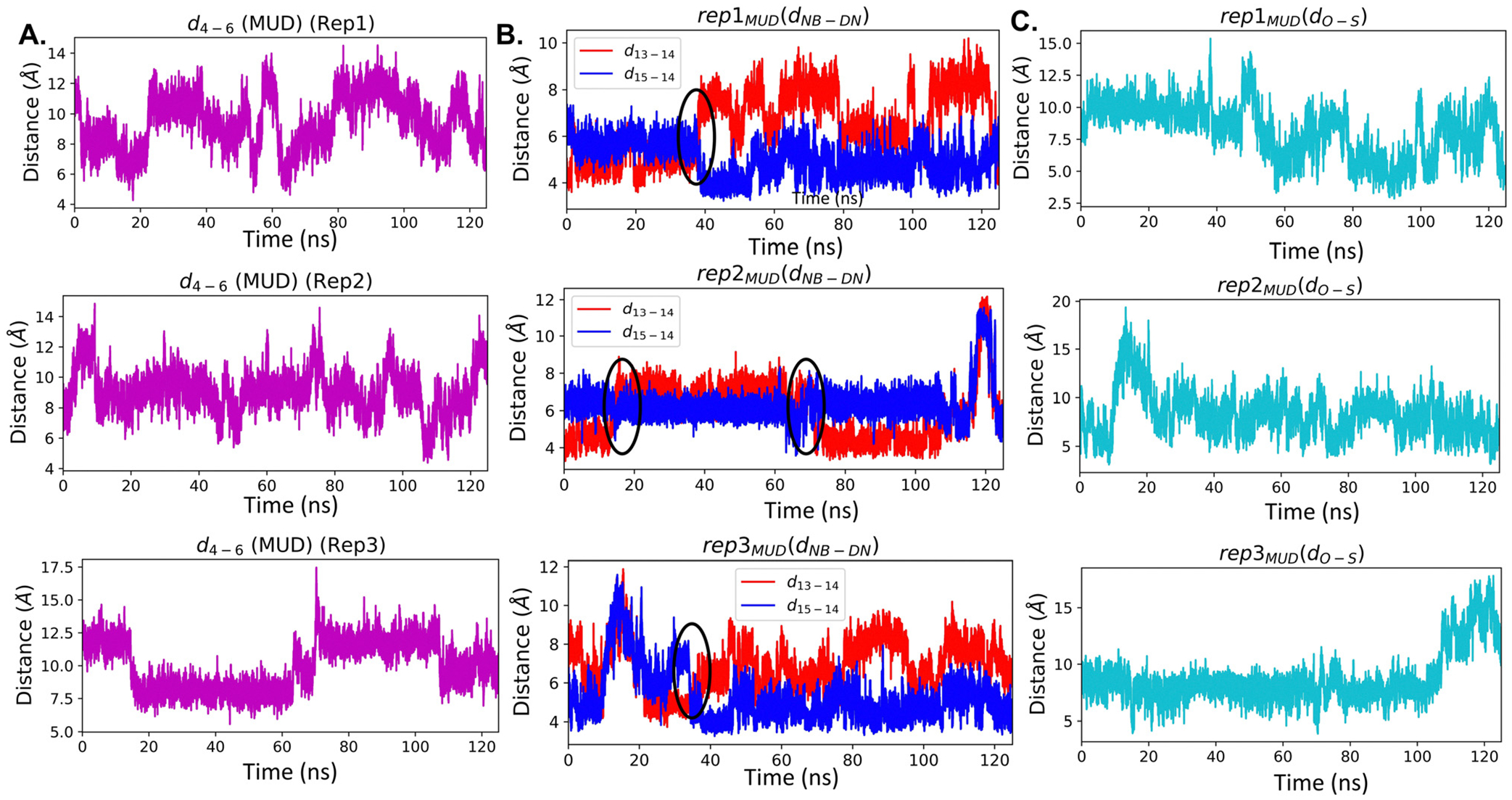
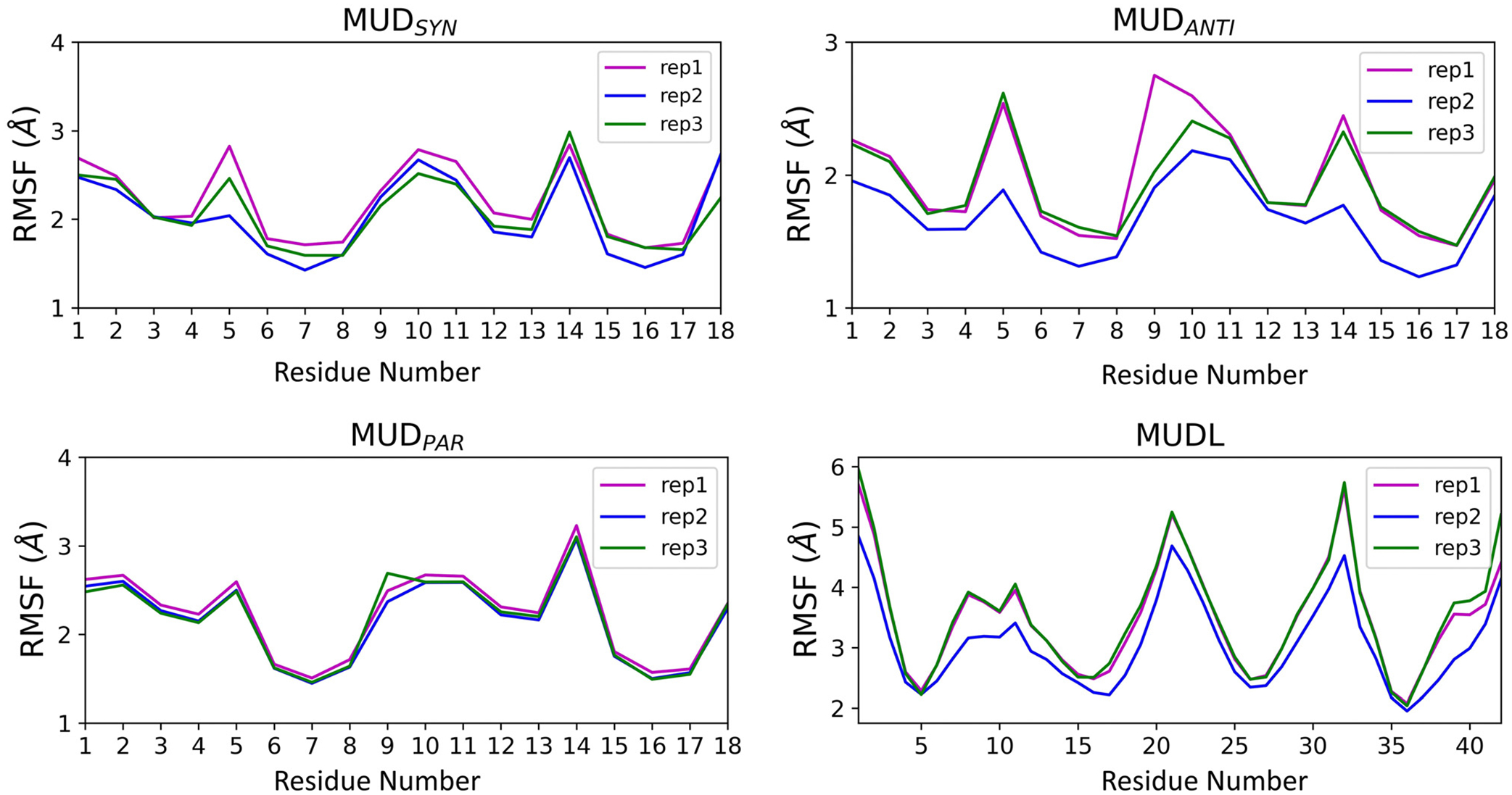
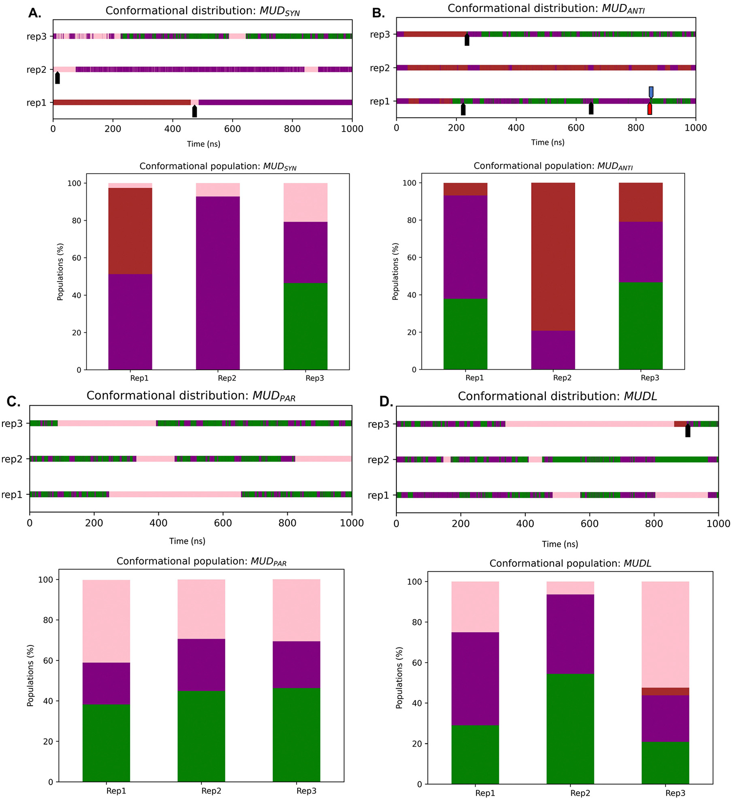
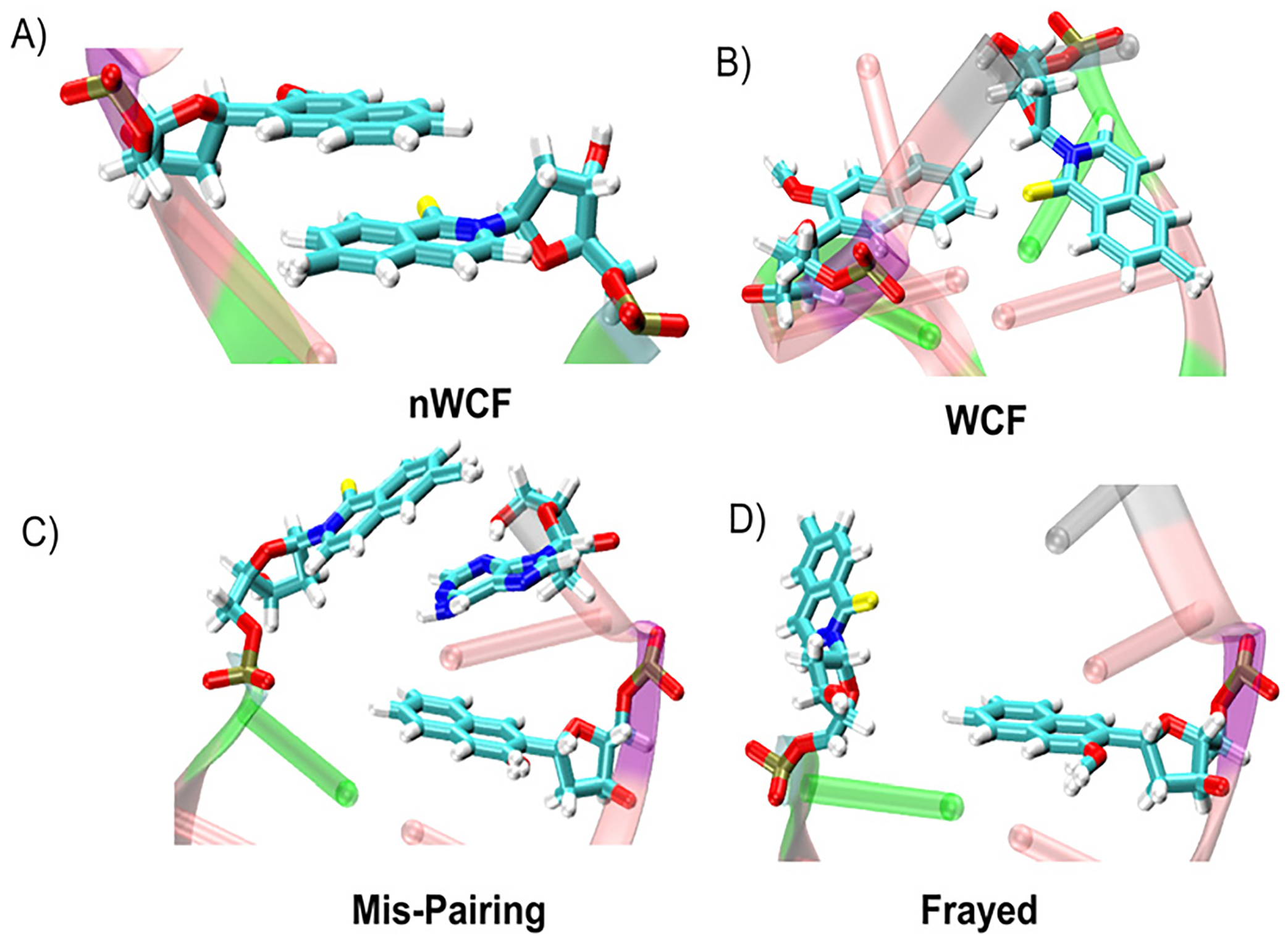
Incorporation of artificial 3rd base pairs (unnatural base pairs, UBPs) has emerged as a fundamental technique in pursuit of expanding the genetic alphabet. 2,6-Dimethyl-2H-isoquiniline-1-thione: D5SIC (DS) and 2-methoxy-3-methylnaphthalene: DNAM (DN), a potential unnatural base pair (UBP) developed by Romesberg and colleagues, has been shown to have remarkable capability for replication within DNA. Crystal structures of a Taq polymerase/double-stranded DNA (ds-DNA) complex containing a DS-DN pair in the 3' terminus showed a parallelly stacked geometry for the pre-insertion, and an intercalated geometry for the post-insertion structure. Unconventional orientations of DS-DN inside a DNA duplex have inspired scientists to investigate the conformational orientations and structural properties of UBP-incorporated DNA. In recent years, computational simulations have been used to investigate the geometry of DS-DN within the DNA duplex; nevertheless, unresolved questions persist owing to inconclusive findings. In this work, we investigate the structural and dynamical properties of DS and DN inside a ds-DNA strand in aqueous solution considering both short and long DNA templates using polarizable, and non-polarizable classical MD simulations. Flexible conformational change of UBP with major populations of Watson-Crick-Franklin (WCF) and three distinct non-Watson-Crick-Franklin (nWCFP1, nWCFP2, nWCFO) conformations through intra and inter-strand flipping have been observed. Our results suggest that a dynamical conformational change leads to the production of diffierent conformational distribution for the systems. Simulations with a short ds-DNA duplex suggest nWCF (P1 and O) as the predominant structures, whereas long ds-DNA duplex simulations indicate almost equal populations of WCF, nWCFP1, nWCFO. DS-DN in the terminal position is found to be more flexible with occasional mispairing and fraying. Overall, these results suggest flexibility and dynamical conformational change of the UBP as well as indicate varied conformational distribution irrespective of starting orientation of the UBP and length og DNA strand.
Conflict of interest statement
Conflicts of interest
There are no conflicts to declare.
Figures

Similar articles
-
Investigation of the stability of D5SIC-DNAM-incorporated DNA duplex in Taq polymerase binary system: a systematic classical MD approach.Phys Chem Chem Phys. 2024 Feb 28;26(9):7287-7295. doi: 10.1039/d3cp05571j. Phys Chem Chem Phys. 2024. PMID: 38353000 Free PMC article.
-
Hydrophobic unnatural base pairs show a Watson-Crick pairing in micro-second molecular dynamics simulations.J Biomol Struct Dyn. 2020 Sep;38(14):4098-4106. doi: 10.1080/07391102.2019.1671898. Epub 2019 Sep 30. J Biomol Struct Dyn. 2020. PMID: 31542995
-
Structural and dynamical instability of DNA caused by high occurrence of d5SICS and dNaM unnatural nucleotides.Phys Chem Chem Phys. 2017 Apr 19;19(16):10571-10580. doi: 10.1039/c7cp01477e. Phys Chem Chem Phys. 2017. PMID: 28394373
-
Toward an Expanded Genome: Structural and Computational Characterization of an Artificially Expanded Genetic Information System.Acc Chem Res. 2017 Jun 20;50(6):1375-1382. doi: 10.1021/acs.accounts.6b00655. Epub 2017 Jun 8. Acc Chem Res. 2017. PMID: 28594167 Free PMC article. Review.
-
Watson-Crick versus Hoogsteen Base Pairs: Chemical Strategy to Encode and Express Genetic Information in Life.Acc Chem Res. 2021 May 4;54(9):2110-2120. doi: 10.1021/acs.accounts.0c00734. Epub 2021 Feb 16. Acc Chem Res. 2021. PMID: 33591181 Review.
Cited by
-
Investigation of the stability of D5SIC-DNAM-incorporated DNA duplex in Taq polymerase binary system: a systematic classical MD approach.Phys Chem Chem Phys. 2024 Feb 28;26(9):7287-7295. doi: 10.1039/d3cp05571j. Phys Chem Chem Phys. 2024. PMID: 38353000 Free PMC article.
-
Comparison of Magnesium and Manganese Ions on the Structural and Catalytic Properties of Human DNA Polymerase Gamma.J Chem Theory Comput. 2025 Jul 3:10.1021/acs.jctc.5c00435. doi: 10.1021/acs.jctc.5c00435. Online ahead of print. J Chem Theory Comput. 2025. PMID: 40607994 Free PMC article.
References
MeSH terms
Substances
Grants and funding
LinkOut - more resources
Full Text Sources
Miscellaneous