. 2024 Jan-Dec:33:9636897241228031.
doi: 10.1177/09636897241228031.

Investigation of Allogeneic Neutrophil Transfusion in Improving Survival Rates of Severe Infection Mice

Affiliations

Investigation of Allogeneic Neutrophil Transfusion in Improving Survival Rates of Severe Infection Mice

Linbin Li et al. Cell Transplant. 2024 Jan-Dec.

Abstract

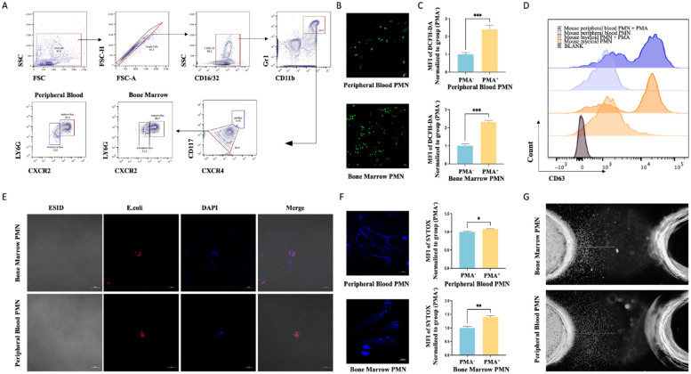
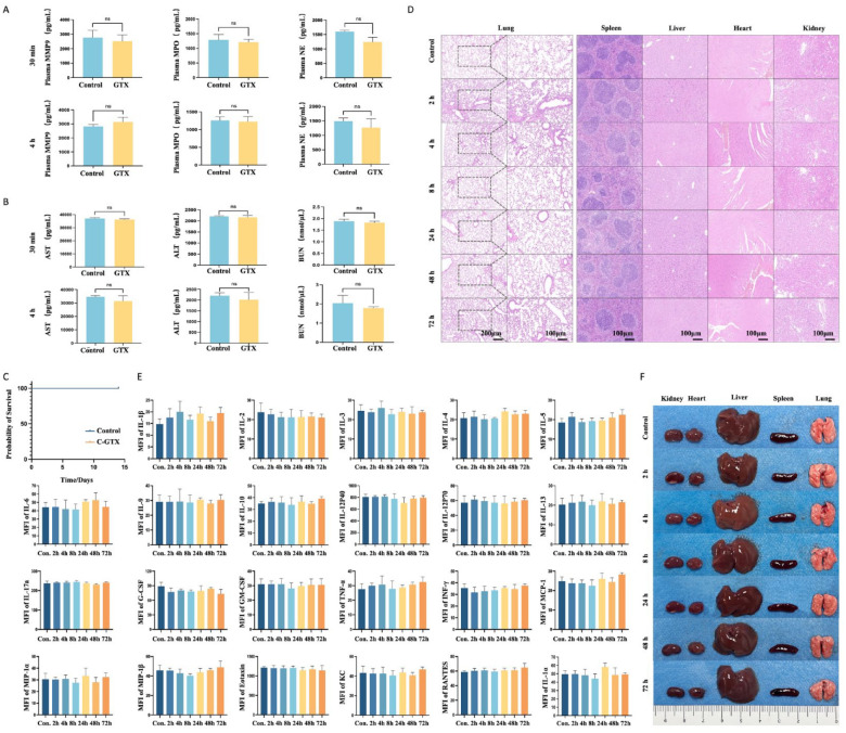
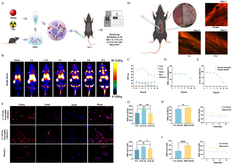
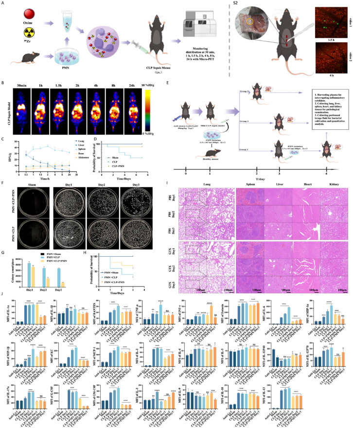
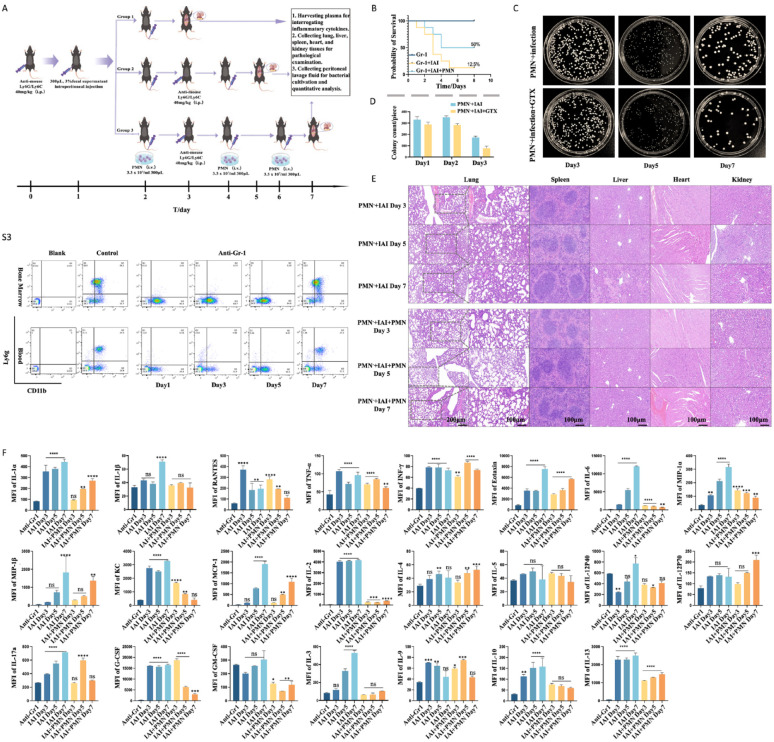
The management of granulocytopenia-associated infections is challenging, and a high mortality rate is associated with traditional supportive therapies. Neutrophils-the primary defenders of the human immune system-have potent bactericidal capabilities. Here, we investigated the dynamic in vivo distribution of neutrophil transfusion and their impact on the treatment outcome of severe granulocytopenic infections. We transfused 89Zr-labeled neutrophils in the C57BL/6 mice and observed the dynamic neutrophil distribution in mice for 24 h using the micro-positron emission tomography (Micro-PET) technique. The labeled neutrophils were predominantly retained in the lungs and spleen up to 4 h after injection and then redistributed to other organs, such as the spleen, liver, and bone marrow. Neutrophil transfusion did not elicit marked inflammatory responses or organ damage in healthy host mice. Notably, allogeneic neutrophils showed rapid chemotaxis to the infected area of the host within 1 h. Tail vein infusion of approximately 107 neutrophils substantially bolstered host immunity, ameliorated the inflammatory state, and increased survival rates in neutrophil-depleted and infected mice. Overall, massive allogeneic neutrophil transfusion had a therapeutic effect in severe infections and can have extensive applications in the future.

Keywords: allogeneic transplantation; infection; investigation; neutrophil; survival rate.


Conflict of interest statement

Declaration of Conflicting InterestsThe author(s) declared no potential conflicts of interest with respect to the research, authorship, and/or publication of this article.

Figures

Figure 1.
Figure 1.
Mouse bone marrow-derived donor neutrophils show complete mature neutrophil functionality. (A) Flow polychrome staining analysis of bone marrow and peripheral blood cells after erythrocyte removal to calculate the proportion of mature neutrophils expressing CXCR2 and LY6G. (B) Neutrophils derive from bone marrow and peripheral blood are purified using the magnetic bead method, and their karyotypes are observed using confocal microscopy after SYTOX staining. (C) Flow cytometry with DCFH-DA staining results show ROS levels after neutrophil stimulation with PMA at 37°C for 30 min. (D) Flow cytometry was used to detect CD63 expression in neutrophils following PMA stimulation (and no stimulation). (E) Confocal microscopy images show phagocytosis after the co-incubation of neutrophils and fluorescent E. coli analogues at 37°C for 2 h. (F) Neutrophils were stimulated with PMA at 37°C for 4 h. The fluorescent expression was detected using flow cytometry after SYTOX staining, and the morphology of neutrophil extracellular traps was visualized using confocal microscopy. (G) Chemotaxis ability of neutrophils after incubating in the agarose chemotaxis model at 37°C for 4 h. FSC: Forward Scattering; SSC: Side Scattering; PMA: phorbol myristate acetate; PMN: polymorphonuclear neutrophil; ROS: reactive oxygen species.
Figure 2.
Figure 2.
Absence of apparent tissue or organ damage in host mice after allogeneic neutrophil transfusion (A) ELISA results show plasma MPO, NE, and MMP9 concentrations in recipient mice. (P > 0.05 for all groups.) (B) ELISA results show plasma AST, ALT, and BUN concentrations in recipient mice. (P > 0.05 for all groups.) (C) Survival rate analysis after neutrophil transfusion in healthy mice. (D) HE staining results show histologic features of major organs in neutrophil allograft recipient mice. (E) Results of Luminex multifactor assay showing the concentrations of plasma inflammatory factors in recipient mice. (P > 0.05 for all groups.) (F) Images of major organs in neutrophil allograft host mice. MFI: mean fluoresence intensity; NE: Neutrophil Elastase; MPO: Myeloper-oxidase; GTX: granulocyte transfusion; ELISA: enzyme-linked immunosorbent assay; AST: aspartate aminotransferase; ALT: alanine aminotransferase; BUN: blood urea nitrogen; HE: hematoxylin and eosin.
Figure 3.
Figure 3.
Dynamic in vivo distribution of transplanted neutrophils and the mechanisms involved. (A) Illustration of the in vivo distribution experiment of 89Zr-oxine labeled neutrophils. (B) Micro-PET monitoring of the dynamic distribution of 89Zr-labeled neutrophils in healthy mice. (C) Percentage injected dose rate per gram of tissue (%ID/g) in the abdominal region and lung, liver, spleen, and bone tissues of the host mice within 24 h. (D) Twenty-four hour of radioactivity retention rate of neutrophils after 89Zr labeling. (E) Twenty-four-hour in vitro survival rate of neutrophils after 89Zr labeling and the 24-h apoptosis rate of normal neutrophils. (F) Confocal microscopy images show the expression of ICAM1 and CD11b after co-culturing neutrophils (3 × 10⁷/ml and 5 × 10⁶/ml) with pulmonary arterial endothelial cells (105/mL) at 37°C for 2 h. (G) Expression of CD11b and ICAM1 on the surface of neutrophils in the lungs of host mice at 2 and 4 h after neutrophil transfusion. (H, I) Flow cytometry-based detection of ICAM1 and CD11b expression after culturing only neutrophils (3 × 10⁷/ml and 5 × 10⁶/ml) for 2 h. WT: wild type; %ID/g: percentage activity of injection dose per gram of tissue; MPAEC: mouse pulmonary arterial endothelial cell; PMN: polymorphnuclear neutrophil.
Figure 4.
Figure 4.
Neutrophil transfusion markedly enhances survival in neutrophil-depleted mice having cecal ligation puncture (CLP)-induced sepsis. (A) Illustration of the experiment indicating in vivo distribution of 89Zr-labeled neutrophils in the CLP-induced sepsis mouse model. (B) Micro-PET image shows the in vivo distribution of 89Zr-labeled neutrophils in the CLP-induced sepsis mouse model. (C) Percentage injected dose rate per gram of tissue (%ID/g) in the abdominal region and lung, liver, spleen, and bone of host mice within 24 h. (D) Seven-day survival rate of mice with severe CLP-induced sepsis (n = 10) after neutrophil transfusion. (E) Schematic of neutrophil transfusion for the treatment of neutrophil-depleted mice having CLP-induced sepsis. (F) Bacterial growth in the peritoneal lavage fluid in neutrophil-depleted mice with CLP-induced sepsis. (G) Bacterial colony enumeration and statistical analysis. (H) Survival rate after neutrophil transfusion in neutrophil-depleted mice with CLP-induced sepsis (treatment group = 10, non-treatment group = 20). (I) Tissue sections of vital organs of mice with CLP-induced sepsis. (J) Luminex multiplex assay results showing inflammatory factors in the plasma of neutrophil-depleted mice having CLP-induced sepsis. MCP: monocyte chemotactic protein; PET: positron emission tomography; NS: none-significant; PMN: polymorphnuclear neutrophil. *P < 0.05; **P < 0.01; ***P < 0.001; ****P < 0.0001, compare with Sham group.
Figure 5.
Figure 5.
Allogeneic neutrophil transfusion improves the survival rate of neutrophil-depleted mice with multi-organ dysfunction caused by abdominal sepsis (A) Neutrophil transfusion strategy for treating granulocytopenic abdominal infection with multiple organ dysfunction in mice. (B) Seven-day survival rate of severe granulocytopenic abdominal infection with multiple organ dysfunction mice treated with neutrophil transfusion (n = 10). (C and D) Bacterial counts and quantitative analysis in abdominal lavage fluid collected from mice with granulocytopenic abdominal infection and multiple organ dysfunction. (E) Histopathologic examination of lungs, spleens, livers, hearts, and kidneys in mice with multiple organ dysfunction resulting from abdominal infection. (F) Results of Luminex multiplex assay for inflammatory cytokines in the plasma of mice with granulocytopenic abdominal infection and multiple organ dysfunction. GTX: granulocyte transfusion; IAI: intra-abdominal infection; NS: none-significant; PMN: polymorphnuclear neutrophil. *P < 0.05; **P < 0.01; ***P < 0.001; ****P < 0.0001, compare with anti-Gr1 group.

Similar articles

Cited by

References

    1. Berlin G, Cherif H, Knutson F, Mattsson J, Axdorph Nygell U. [Granulocyte transfusion—when and how should it be used?]. Lakartidningen. 2018;115:EXUU. - PubMed
    1. White L, Ybarra M. Neutropenic fever. Hematol Oncol Clin North Am. 2017;31(6):981–93. - PubMed
    1. Klastersky J. Empirical treatment of sepsis in neutropenic patients. Hosp Med. 2001;62(2):101–3. - PubMed
    1. Kuderer NM, Dale DC, Crawford J, Cosler LE, Lyman GH. Mortality, morbidity, and cost associated with febrile neutropenia in adult cancer patients. Cancer. 2006;106(10):2258–66. - PubMed
    1. Stroncek DF, Yau YY, Oblitas J, Leitman SF. Administration of G–CSF plus dexamethasone produces greater granulocyte concentrate yields while causing no more donor toxicity than G–CSF alone. Transfusion. 2001;41(8):1037–44. - PubMed

LinkOut - more resources