. 2024 May;11(17):e2308235.
doi: 10.1002/advs.202308235. Epub 2024 Feb 14.

Hybrid Ginseng-derived Extracellular Vesicles-Like Particles with Autologous Tumor Cell Membrane for Personalized Vaccination to Inhibit Tumor Recurrence and Metastasis

Affiliations

Hybrid Ginseng-derived Extracellular Vesicles-Like Particles with Autologous Tumor Cell Membrane for Personalized Vaccination to Inhibit Tumor Recurrence and Metastasis

Haoran Wang et al. Adv Sci (Weinh). 2024 May.

Abstract

Personalized cancer vaccines based on resected tumors from patients is promising to address tumor heterogeneity to inhibit tumor recurrence or metastasis. However, it remains challenge to elicit immune activation due to the weak immunogenicity of autologous tumor antigens. Here, a hybrid membrane cancer vaccine is successfully constructed by membrane fusion to enhance adaptive immune response and amplify personalized immunotherapy, which formed a codelivery system for autologous tumor antigens and immune adjuvants. Briefly, the functional hybrid vesicles (HM-NPs) are formed by hybridizing ginseng-derived extracellular vesicles-like particles (G-EVLPs) with the membrane originated from the resected autologous tumors. The introduction of G-EVLPs can enhance the phagocytosis of autologous tumor antigens by dendritic cells (DCs) and facilitate DCs maturation through TLR4, ultimately activating tumor-specific cytotoxic T lymphocytes (CTLs). HM-NPs can indeed strengthen specific immune responses to suppress tumors recurrence and metastasis including subcutaneous tumors and orthotopic tumors. Furthermore, a long-term immune protection can be obtained after vaccinating with HM-NPs, and prolonging the survival of animals. Overall, this personalized hybrid autologous tumor vaccine based on G-EVLPs provides the possibility of mitigating tumor recurrence and metastasis after surgery while maintaining good biocompatibility.

Keywords: ginseng‐derived nanoparticles; hybrid nanoparticles; long‐term protection; mature DCs; personalized tumor vaccine.


Conflict of interest statement

The authors declare no conflict of interest.

Figures

Figure 1
Figure 1
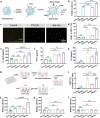
Schematic illustration of the construction and mechanistic process of the cancer vaccine HM‐NPs. a) The preparation procedure of the HM‐NPs. TM‐NPs were obtained from surgically removed tumors on tumor‐bearing mice. Ginseng roots were squeezed to obtain ginseng fluid, and the extraction process was used to prepare G‐EVLPs. Using an extruder, the cancer vaccine HM‐NPs were generated by extruding the mixed G‐EVLPs and TM‐NPs. b) The mechanistic process of HM‐NPs. ① HM‐NPs were subcutaneous injected at the base of tail. ② HM‐NPs enhanced uptake of tumor membrane antigen and dendritic cells (DCs) maturation. ③ DCs migrated to the inguinal lymph nodes after uptake of tumor membrane antigen and maturation. ④ HM‐NPs elicited specific cytotoxic T lymphocytes and B lymphocytes, and formed strengthened anti‐tumor immune memory. ⑤ and ⑥ HM‐NPs activated enhanced personalized adaptive immune response to inhibit the tumor recurrence and tumor metastasis after subcutaneous injection.
Figure 2
Figure 2
Construction and characterization of the cancer vaccine HM‐NPs (B16F10). a) Schematic figure of the manufacture process of the cancer vaccine HM‐NPs. b) Particle size distribution of TM‐NPs, G‐EVLPs and HM‐NPs determined by DLS. c) Particle size and ζ potential of TM‐NPs, G‐EVLPs and HM‐NPs (n = 3). d) Tyndall phenomenon of HM‐NPs with 660 nm laser irradiation on the right. e) Transmission electron microscope (TEM) images of G‐EVLPs (Insert image. enlarged TEM figure of G‐EVLPs. The white arrows indicate the bilayer lipid membrane). f) Transmission electron microscope (TEM) images of HM‐NPs (Insert image I. enlarged TEM figure of HM‐NPs. Insert figure II. enlarged TEM figure of HM‐NPs. The white arrows indicate the bilayer lipid membrane). g) SDS‐PAGE protein analysis of Marker, G‐EVLPs, TM‐NPs and HM‐NPs. h) FRET‐based lipid mixing assay used to monitor the fusion between G‐EVLPs and TMNPs. i) Fluorescence intensity of TM‐NPs (Blank), G‐EVLPs (Blank), G‐EVLPs (with fluorescent dyes) and HM‐NPs (with fluorescent dyes) at Ex/Em = 480 nm/504 nm and Ex/Em = 480 nm/585 nm (n = 3). Data are representative or pooled and are expressed as Mean ± SE. Asterisks indicate statistically significant differences as analyzed by One‐Way ANOVA (∗∗∗p < 0.001, ∗∗p < 0.01, N.S. p > 0.05).
Figure 3
Figure 3
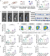
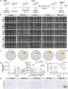
HM‐NPs (B16F10) enhance tumor antigen uptake and activation of immune cells in vitro. a) Schematic illustration of the BMDCs uptake and activation experiments. b) Cellular uptake of DiD‐labeled tumor membrane after a 24‐hour incubation with BMDCs, as assessed by flow cytometry. c) Confocal images of BMDCs for TM‐NPs and HM‐NPs uptake after a 24‐hour incubation (DiD‐labeled tumor membrane). d) Flow cytometry analysis of CD11c+CD86+ BMDCs after incubation with different NPs for 24 hours. e) Secretion of IL‐12p70 in supernatant of the BMDCs medium measured by ELISA kit. f) Secretion of TNF‐α in supernatant of the BMDCs medium measured by ELISA kit. g) Secretion of IL‐6 in supernatant of the BMDCs medium measured by ELISA kit. h) Schematic illustration of the specific immune activation experiments in vitro. i) Percentage of splenic CD8+ T lymphocytes activation. j‐l. LDH concentration in the supernatant after co‐incubation of the splenic T lymphocytes with B16F10 tumor cells j), CT26 tumor cells k) and 4T1 tumor cells l). Data are representative or pooled and are expressed as Mean ± SE. Asterisks indicate statistically significant differences as analyzed by One‐Way ANOVA (∗∗∗p < 0.001, ∗∗p < 0.01, N.S. p > 0.05).
Figure 4
Figure 4
HM‐NPs (B16F10) promote LN accumulation, DCs maturation and splenic T cells activation after vaccination. a) Experimental design to evaluate LN accumulation, DCs maturation and splenic T cells activation. b) Inguinal lymph nodes weight after vaccination (n = 10). c) Fluorescence images after vaccination (DiR‐labeled tumor membrane) (n = 5). d) Fluorescence images of inguinal lymph nodes at after vaccination (DiR‐labeled tumor membrane) (n = 10). e) Fluorescence intensity analysis of inguinal lymph nodes after vaccination (DiR‐labeled tumor membrane). f) Cellular uptake of DiR‐labeled tumor membrane of CD11c+ DCs in inguinal lymph nodes, as assessed by flow cytometry (n = 5). g) Flow cytometry analysis of CD11c+CD86+CD80+ DCs in inguinal lymph nodes. h) Fluorescence intensity analysis of spleen after vaccination (DiR‐labeled tumor membrane). i) Flow cytometry analysis quantification of the CD44hiCD62Llow Tem cells in spleen after vaccination. j–l) IFN‐γ concentration in the supernatant after co‐incubation of the splenic T lymphocytes with B16F10 tumor cells (j), CT26 tumor cells (k) and 4T1 tumor cells (l). Data are representative or pooled and are expressed as Mean ± SE. Asterisks indicate statistically significant differences as analyzed by One‐Way ANOVA (∗∗∗p < 0.001, N.S. p > 0.05).
Figure 5
Figure 5
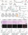
HM‐NPs vaccination induces tumor recurrence suppression in the murine B16F10 tumor model. a) Schematic illustration of the design of the animal experiment. b) Average tumor growth curves of each group in the murine B16F10 tumor model (n = 10). c) Tumor weight of each group in the murine B16F10 tumor model at day 35. d) Body weight of the mice post various treatments (n = 10). e) Tumor complete responses rate of each group (CR: complete responses). f) Survival curves of mice receiving each treatment. g) Photographs are the white filed images of the H&E staining and the immunofluorescence images of CD8+ T of the tumors receiving different treatments. h) Proinflammatory IFN‐γ concentration in the serum of the mice receiving each treatment determined by ELISA assay. i) Proinflammatory TNF‐α concentration in the serum of the mice receiving each treatment determined by ELISA assay. j) Proinflammatory IL‐1β concentration in the serum of the mice receiving each treatment determined by ELISA assay. Data are representative or pooled and are expressed as Mean ± SE. Asterisks indicate statistically significant differences as analyzed by One‐Way ANOVA (∗∗∗p < 0.001, N.S. p > 0.05).
Figure 6
Figure 6
HM‐NPs vaccination inhibits specific tumor recurrence and provides long‐term anti‐tumor protection. a) Schematic illustration of the design of the specific tumor recurrence suppression experiment. b) Tumor volume of CT26 and 4T1 tumors of each group in the murine tumor model. c) Tumor complete responses rate of CT26 and 4T1 tumors of each group (CR: complete responses). d) Schematic illustration of the design of the long‐term anti‐tumor protection experiment. e) Survival curves of mice receiving each treatment. f) Average tumor growth curves of each group in the murine tumor model. g) Body weight of the mice post the two treatments. h) Tumor complete responses rate of 4T1 tumors of each group (CR: complete responses). Data are representative or pooled and are expressed as Mean ± SE. Asterisks indicate statistically significant differences as analyzed by One‐Way ANOVA (∗∗∗p < 0.001, N.S. p > 0.05).
Figure 7
Figure 7
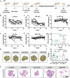
HM‐NPs vaccination inhibits tumor metastasis in murine 4T1 tumor models. a) Schematic illustration of the design of the acute tumor metastasis suppression experiment. b–f) Body weight of each mouse after receiving Saline (b), TM‐NPs (c), G‐EVLPs (d), M‐NPs (e) and HM‐NPs (f). g) Survival curves of mice receiving different treatments. h) Photographs are the white filed images of lung tissues after staining with Bouin's Fluid. i) Quantification of metastatic lesions of the lung tissues. j) Photographs are the panoramic scanning photographs of the H&E staining of lung tissues (Black arrows indicate the metastatic lesions). Data are representative or pooled and are expressed as Mean ± SE. Asterisks indicate statistically significant differences as analyzed by One‐Way ANOVA (N.S. p > 0.05).
Figure 8
Figure 8
HM‐NPs vaccination establishes protection against orthotopic mouse MB49 bladder tumor. a) Schematic illustration of the design of the orthotopic mouse MB49 1‐Luc bladder tumor experiment. b) IVIS bioluminescence imaging of the mice at days 22, 25, 29, 32, and 35 after vaccination with Saline, TM‐NPs, G‐EVLPs, M‐NPs and HM‐NPs respectively. c) Tumor complete responses rate of the orthotopic mouse MB49 tumors of each group (CR: complete responses). d) Bioluminescence intensity analysis of the orthotopic mouse MB49 tumors at days 22, 25, 29, 32, and 35 after vaccination. e) Tumor weight of each group of the orthotopic mouse MB49 tumors at day 36. f) Survival curves of mice receiving each treatment. g) Immunohistochemical images of the CD8+ T cells within tumors post treatments. Data are representative or pooled and are expressed as Mean ± SE. Asterisks indicate statistically significant differences as analyzed by One‐Way ANOVA (N.S. p > 0.05).
Figure 9
Figure 9
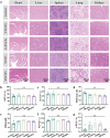
HM‐NPs vaccination exhibits a favorable safety profile. a) Photographs of the H&E staining of different organs of the tumor‐bearing mice receiving each treatment. b) Level of blood aspartate aminotransferase (AST) of the mice receiving each treatment at day 7. c) Level of blood alanine aminotransferase (ALT) of the mice receiving each treatment at day 7. d) Level of blood urea nitrogen (BUN) of the mice receiving each treatment at day 7. e) Level of blood creatinine (CREA) of the mice receiving each treatment at day 7. f) Level of blood alkaline phosphatase (ALP) of the mice receiving each treatment at day 7. g) Level of blood γ‐glutamyl transpeptidase (γ‐GT) of the mice receiving each treatment at day 7. Data are representative or pooled and are expressed as Mean ± SE. Asterisks indicate statistically significant differences as analyzed by One‐Way ANOVA (N.S. p > 0.05).

References

    1. Zhang X., Cui H., Zhang W., Li Z., Gao J., Bioact. Mater. 2023, 22, 491. - PMC - PubMed
    1. a) Cao X., Liang Y., Hu Z., Li H., Yang J., Hsu E. J., Zhu J., Zhou J., Fu Y.‐X., Nat. Commun. 2021, 12, 5866; - PMC - PubMed
    2. b) Wang Z., Meng F., Zhong Z., Adv. Drug Delivery Rev. 2021, 178, 113969. - PubMed
    1. a) Liu C., Liu X., Xiang X., Pang X., Chen S., Zhang Y., Ren E., Zhang L., Liu X., Lv P., Nat. Nanotechnol. 2022, 17, 531; - PubMed
    2. b) Blass E., Ott P. A., Nat. Rev. Clin. Oncol. 2021, 18, 215; - PMC - PubMed
    3. c) Fan Q., Ma Q., Bai J., Xu J., Fei Z., Dong Z., Maruyama A., Leong K. W., Liu Z., Wang C., Sci. Adv. 2020, 6, eabb4639; - PMC - PubMed
    4. d) Lin L., Pei Y., Li Z., Luo D., Interdiscip. Med. 2023, 1, e20220008.
    1. a) Vonderheide R. H., Med. 2023, 4, 395; - PubMed
    2. b) Chiu L.‐C., Lin S.‐M., Lo Y.‐L., Kuo S. C.‐H., Yang C.‐T., Hsu P.‐C., Vaccines 2021, 9, 689; - PMC - PubMed
    3. c) Chen L., Qin H., Zhao R., Zhao X., Lin L., Chen Y., Lin Y., Li Y., Qin Y., Li Y., Sci. Transl. Med. 2021, 13, eabc2816. - PubMed
    1. a) Mota I., Patrucco E., Mastini C., Mahadevan N. R., Thai T. C., Bergaggio E., Cheong T.‐C., Leonardi G., Karaca‐Atabay E., Campisi M., Nat. Cancer 2023, 4, 1016; - PubMed
    2. b) Wang C., Steinmetz N. F., Adv. Funct. Mater. 2020, 30, 2002299; - PMC - PubMed
    3. c) Nejo T., Yamamichi A., Almeida N. D., Goretsky Y. E., Okada H., Semin. Immunol. 2020, 47, 101385. - PubMed

Publication types

Substances

LinkOut - more resources