

Lipid profile of circulating placental extracellular vesicles during pregnancy identifies foetal growth restriction risk
- PMID: 38353485
- PMCID: PMC10865917
- DOI: 10.1002/jev2.12413
Lipid profile of circulating placental extracellular vesicles during pregnancy identifies foetal growth restriction risk
Erratum in
-
Correction to article pagination in the Journal of Extracellular Vesicles.J Extracell Vesicles. 2024 May;13(5):e12443. doi: 10.1002/jev2.12443. J Extracell Vesicles. 2024. PMID: 38695388 Free PMC article. No abstract available.
Abstract
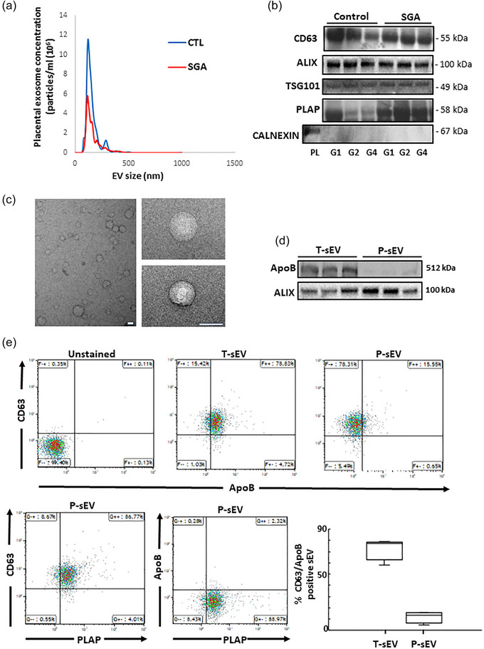
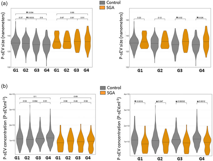
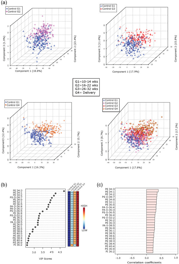
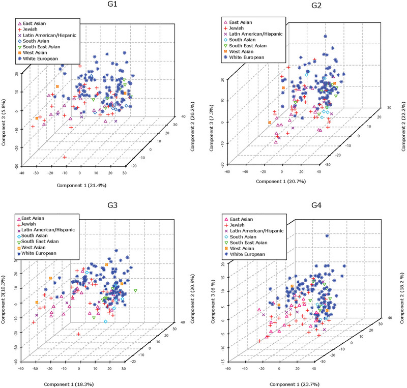
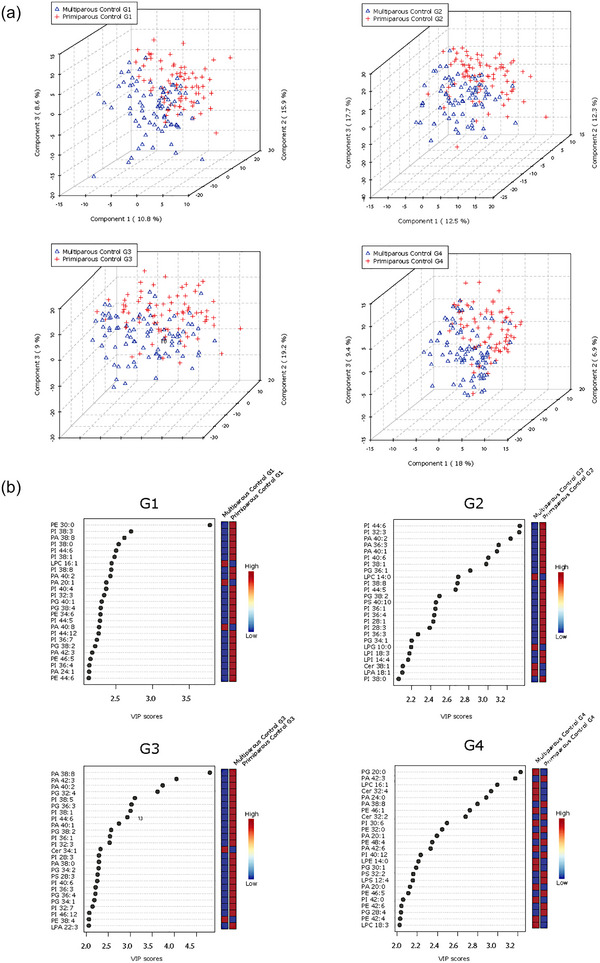
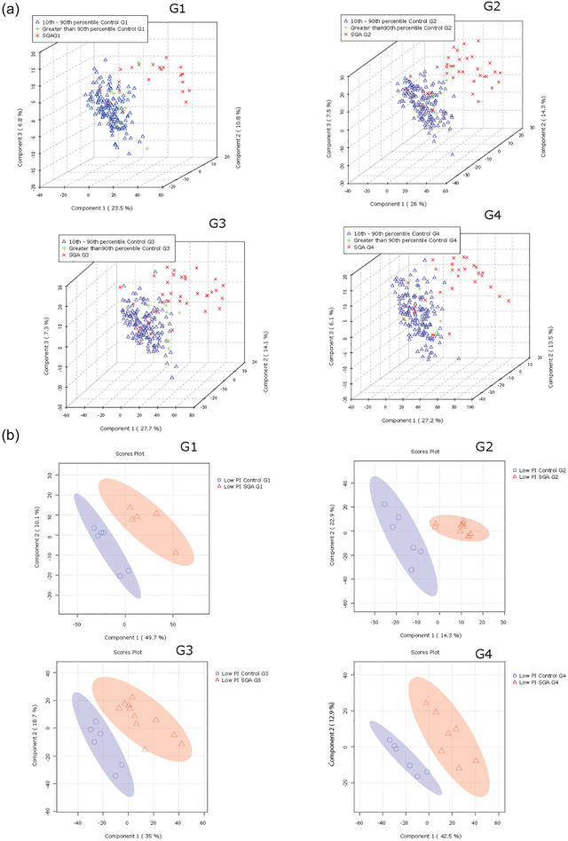
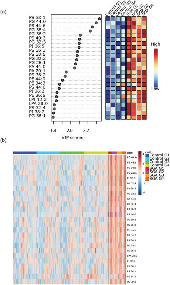
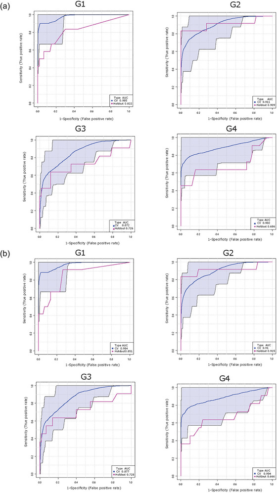
Small-for-gestational age (SGA) neonates exhibit increased perinatal morbidity and mortality, and a greater risk of developing chronic diseases in adulthood. Currently, no effective maternal blood-based screening methods for determining SGA risk are available. We used a high-resolution MS/MSALL shotgun lipidomic approach to explore the lipid profiles of small extracellular vesicles (sEV) released from the placenta into the circulation of pregnant individuals. Samples were acquired from 195 normal and 41 SGA pregnancies. Lipid profiles were determined serially across pregnancy. We identified specific lipid signatures of placental sEVs that define the trajectory of a normal pregnancy and their changes occurring in relation to maternal characteristics (parity and ethnicity) and birthweight centile. We constructed a multivariate model demonstrating that specific lipid features of circulating placental sEVs, particularly during early gestation, are highly predictive of SGA infants. Lipidomic-based biomarker development promises to improve the early detection of pregnancies at risk of developing SGA, an unmet clinical need in obstetrics.
Keywords: SGA pregnancies; lipidomics; placenta; small extracellular vesicles.
© 2024 The Authors. Journal of Extracellular Vesicles published by Wiley Periodicals LLC on behalf of International Society for Extracellular Vesicles.
Conflict of interest statement
All authors declare no competing interests.
Figures
References
-
- Adam, S. , Elfeky, O. , Kinhal, V. , Dutta, S. , Lai, A. , Jayabalan, N. , Nuzhat, Z. , Palma, C. , Rice, G. E. , & Salomon, C. (2017). Review: Fetal‐maternal communication via extracellular vesicles—Implications for complications of pregnancies. Placenta, 54, 83–88. 10.1016/j.placenta.2016.12.001 - DOI - PubMed
-
- Arcangeli, T. , Thilaganathan, B. , Hooper, R. , Khan, K. S. , & Bhide, A. (2012). Neurodevelopmental delay in small babies at term: A systematic review. Ultrasound in Obstetrics & Gynecology : The Official Journal of the International Society of Ultrasound in Obstetrics and Gynecology, 40(3), 267–275. 10.1002/uog.11112 - DOI - PubMed
-
- Bahado‐Singh, R. O. , Yilmaz, A. , Bisgin, H. , Turkoglu, O. , Kumar, P. , Sherman, E. , Mrazik, A. , Odibo, A. , & Graham, S. F. (2019). Artificial intelligence and the analysis of multi‐platform metabolomics data for the detection of intrauterine growth restriction. PLoS One, 14(4), e0214121. 10.1371/journal.pone.0214121 - DOI - PMC - PubMed
-
- Baig, S. , Lim, J. Y. , Fernandis, A. Z. , Wenk, M. R. , Kale, A. , Su, L. L. , Biswas, A. , Vasoo, S. , Shui, G. , & Choolani, M. (2013). Lipidomic analysis of human placental syncytiotrophoblast microvesicles in adverse pregnancy outcomes. Placenta, 34(5), 436–442. 10.1016/j.placenta.2013.02.004 - DOI - PubMed
Publication types
MeSH terms
Substances
Grants and funding
LinkOut - more resources
Full Text Sources
