. 2024 Mar 19;98(3):e0163823.
doi: 10.1128/jvi.01638-23. Epub 2024 Feb 14.

A rapid and versatile reverse genetics approach for generating recombinant positive-strand RNA viruses that use IRES-mediated translation

Affiliations

A rapid and versatile reverse genetics approach for generating recombinant positive-strand RNA viruses that use IRES-mediated translation

Tomokazu Tamura et al. J Virol. .

Abstract

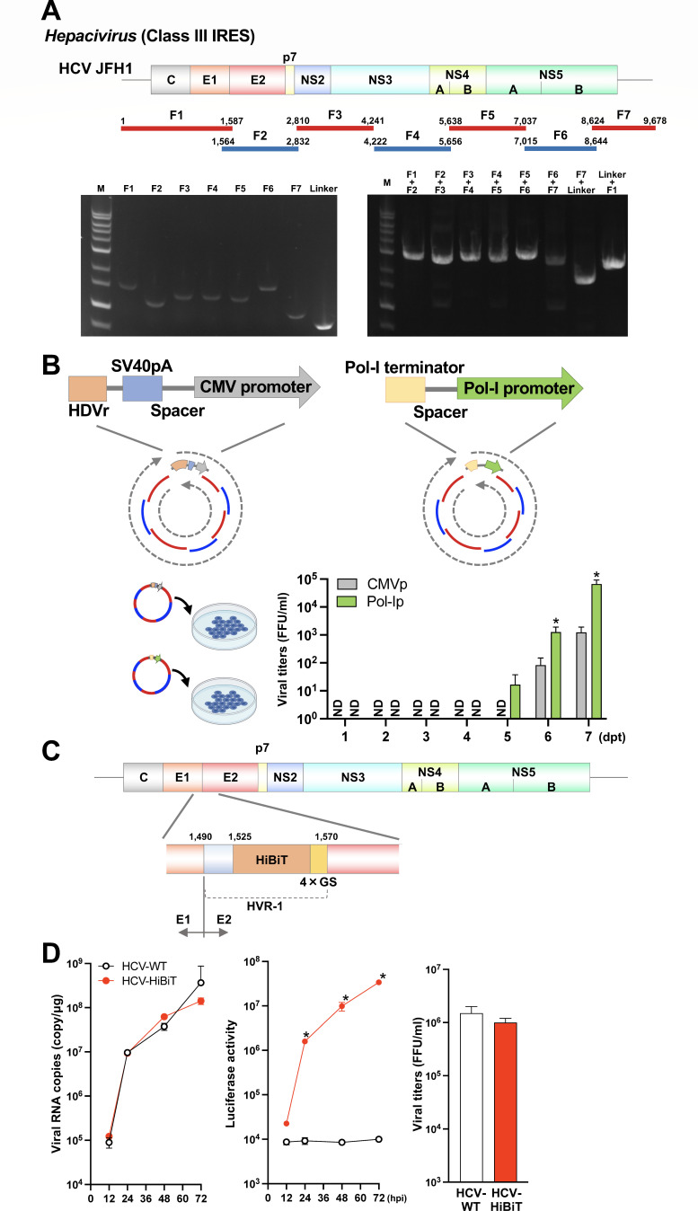
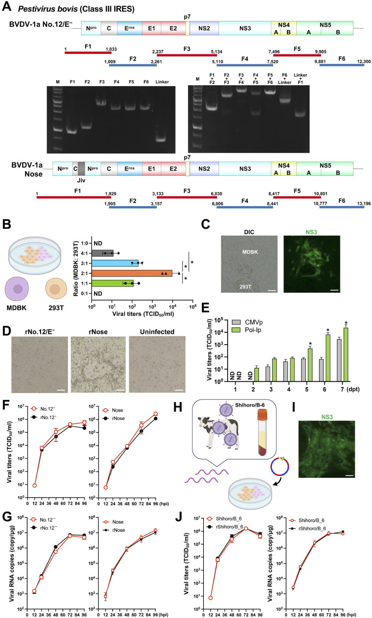
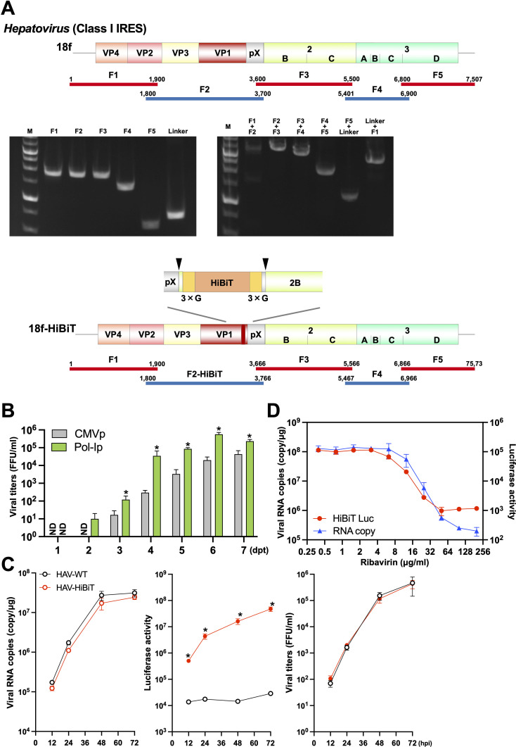
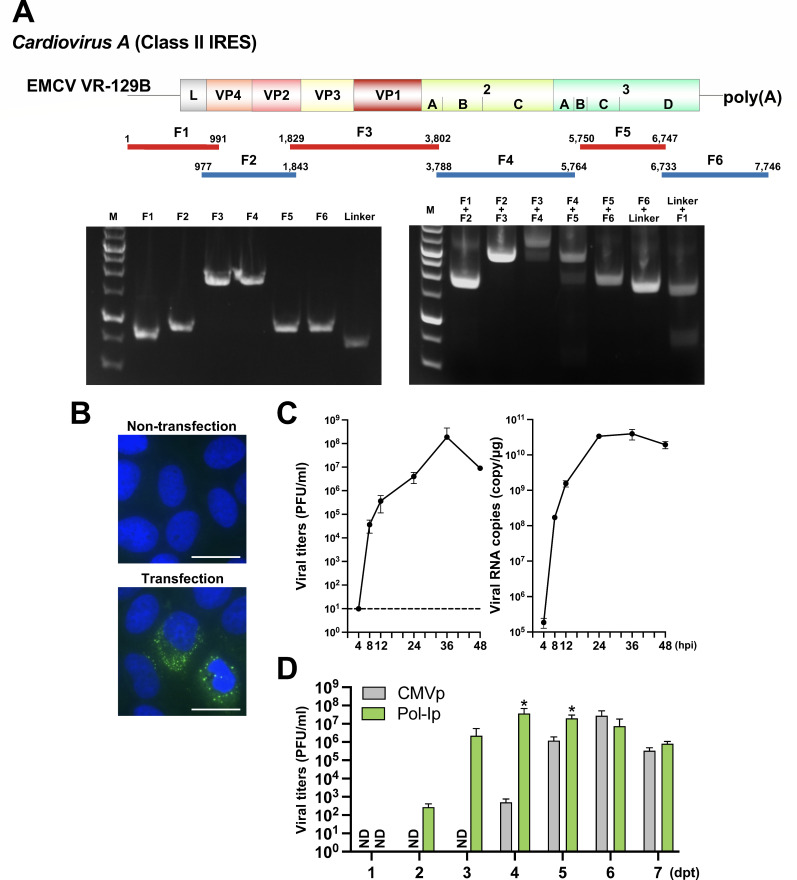
Reverse genetics systems have played a central role in developing recombinant viruses for a wide spectrum of virus research. The circular polymerase extension reaction (CPER) method has been applied to studying positive-strand RNA viruses, allowing researchers to bypass molecular cloning of viral cDNA clones and thus leading to the rapid generation of recombinant viruses. However, thus far, the CPER protocol has only been established using cap-dependent RNA viruses. Here, we demonstrate that a modified version of the CPER method can be successfully applied to positive-strand RNA viruses that use cap-independent, internal ribosomal entry site (IRES)-mediated translation. As a proof-of-concept, we employed mammalian viruses with different types (classes I, II, and III) of IRES to optimize the CPER method. Using the hepatitis C virus (HCV, class III), we found that inclusion in the CPER assembly of an RNA polymerase I promoter and terminator, instead of those from polymerase II, allowed greater viral production. This approach was also successful in generating recombinant bovine viral diarrhea virus (class III) following transfection of MDBK/293T co-cultures to overcome low transfection efficiency. In addition, we successfully generated the recombinant viruses from clinical specimens. Our modified CPER could be used for producing hepatitis A virus (HAV, type I) as well as de novo generation of encephalomyocarditis virus (type II). Finally, we generated recombinant HCV and HAV reporter viruses that exhibited replication comparable to that of the wild-type parental viruses. The recombinant HAV reporter virus helped evaluate antivirals. Taking the findings together, this study offers methodological advances in virology.

Importance: The lack of versatility of reverse genetics systems remains a bottleneck in viral research. Especially when (re-)emerging viruses reach pandemic levels, rapid characterization and establishment of effective countermeasures using recombinant viruses are beneficial in disease control. Indeed, numerous studies have attempted to establish and improve the methods. The circular polymerase extension reaction (CPER) method has overcome major obstacles in generating recombinant viruses. However, this method has not yet been examined for positive-strand RNA viruses that use cap-independent, internal ribosome entry site-mediated translation. Here, we engineered a suitable gene cassette to expand the CPER method for all positive-strand RNA viruses. Furthermore, we overcame the difficulty of generating recombinant viruses because of low transfection efficiency. Using this modified method, we also successfully generated reporter viruses and recombinant viruses from a field sample without virus isolation. Taking these findings together, our adapted methodology is an innovative technology that could help advance virologic research.

Keywords: IRES; RNA virus; cap-independence; reporter virus; reverse genetics.


Conflict of interest statement

The authors declare no conflict of interest.

Figures

Fig 1
Fig 1
Optimization of the current CPER method and generation of the recombinant hepatitis C virus (HCV, class III IRES). (A) A schematic representation of the HCV genome and the gene fragments prepared. A total of seven fragments were amplified and then assembled with a UTR linker fragment by CPER. Gel electrophoresis analysis was conducted to confirm the size of the seven PCR amplicons (left panel). PCR assembly was used to connect neighboring fragments and the size of joined fragments was assessed by gel electrophoresis (right panel). F: fragment; M: 1 kb DNA ladder. (B) A schematic representation of the two UTR linkers was evaluated. The left panel shows the conventional gene cassette of the UTR linker used in CPER that encodes hepatitis D virus ribozyme, SV40 polyA, a 165 nt spacer, and a cytomegalovirus (CMV) promoter. The right panel shows the modified cassette generated in the present study that encodes murine RNA polymerase I terminator, a 165 nt spacer, and a human RNA polymerase I promoter. Each linker was subjected to CPER assembly to evaluate the efficiency of recovery of recombinant HCV as measured by a focus-forming assay (bar graph). Asterisks indicate significant differences (*P < 0.05) versus the results of the CMV promoter. ND: not detected. (C) A schematic representation of the recombinant HCV carrying the HiBiT luciferase gene. The HiBiT luciferase and GS linker sequence were inserted in hypervariable region I (HVR-I) within the HCV E2 envelope protein gene. (D) Huh7.5.1 cells were infected with the parental and recombinant HCV possessing HiBiT at a multiplicity of infection (MOI) of 0.1. Intracellular viral RNA and the luciferase activities in the cells at 12, 24, 48, and 72 hpi (left and middle panels), and the infectious titers in the culture supernatants at 3 dpi (right panel) were determined by qRT-PCR, luciferase assay, and focus-forming assay, respectively. Asterisks indicate significant differences (*P < 0.05) versus the results of the parental virus (HCV-WT). Assays were performed independently in triplicate (B, D). Images were created with BioRender.com.
Fig 2
Fig 2
Generation of recombinant bovine viral diarrhea viruses (BVDV, class III IRES). (A) A schematic representation of the two types of BVDV genomes used (genotype 1a) and the gene fragments prepared. A total of six fragments were amplified and then assembled with the new UTR linker fragment by CPER. Gel electrophoresis analysis was conducted to confirm the size of the six PCR amplicons (left panel). PCR assembly was used to connect neighboring fragments and the size of joined fragments was assessed by gel electrophoresis (right panel). F: fragment; M: 1 kb DNA ladder. BVDV-1a Nose strain possesses a gene duplication in the Npro gene (Npro, C, and cINS). (B) Optimization of co-culturing of two cell lines for the production of recombinant BVDV-1a strain No.12/E. The two cell lines employed in this study were bovine MDBK, which exhibits high utility for BVDV infection, and 293T, which shows high transfection efficiency and protein expression. Ratios of cell numbers and infectious titers are shown as a bar graph. Asterisks indicate significant differences (*P < 0.05) between a pair. (C) Images of transfected cells captured by differential interference contrast (DIC) or immunofluorescence using antiviral NS3 are shown at day 5 post-transfection. Scale bars, 100 µm. (D) Cytopathic effect was observed upon infection with the recombinant BVDVs. Scale bars, 100 µm. (E) Both linkers were individually subjected to CPER assembly to evaluate the efficiency of recovery of recombinant BVDV as measured by a TCID50 assay (bar graph). Asterisks indicate significant differences (*P < 0.05) versus the results of the CMV promoter. ND: not detected. (F, G) MDBK cells were inoculated at an MOI of 0.01 using the parental or recombinant BVDV. Virus titers in supernatants (F) and intracellular viral RNA (G) were determined at the indicated timepoints by TCID50 and qRT-PCR, respectively. (H) Illustration of the CPER method using a crude clinical isolate. Serum was obtained from animals persistently infected with BVDV strain Shihoro/B_6 (genotype 1b). The viral RNA was purified and the resultant viral cDNA was subjected to CPER assembly. The CPER product was transfected into co-cultures at the optimized MDBK:293T ratio of 2:1. (I) Antiviral NS3 immunofluorescence image of transfected cells at day 5 post-transfection. Scale bar, 100 µm. (J) MDBK cells were inoculated with the clinical isolate or recombinant BVDV at an MOI of 0.01. The virus titers in supernatants and intracellular viral RNA were determined at the indicated timepoints. The recombinant viruses are named according to the parental strains from which they were rescued, with “r” added at the start of the name (D, F, G, J). Assays were performed independently in triplicate (B, E, F, G, J). Images were created with BioRender.com.
Fig 3
Fig 3
Generation of the recombinant hepatitis A virus (HAV, class I IRES) and the reporter thereof. (A) A schematic representation of the HAV genome and the gene fragments prepared. A total of five fragments were amplified and then assembled with a UTR linker fragment by CPER. Gel electrophoresis analysis was conducted to confirm the size of the five PCR amplicons (left panel). PCR assembly was used to connect neighboring fragments and the size of joined fragments was assessed by gel electrophoresis (right panel). F: fragment; M: 1 kb DNA ladder. The HiBiT luciferase gene with GS linker was inserted at the pX-VP2B junction. Arrowheads indicate the recognition site of viral 3Cpro protease. (B) Both linkers were individually subjected to CPER assembly to evaluate the efficiency of recovery of recombinant HAV as measured by a focus-forming assay (bar graph). Asterisks indicate significant differences (*P < 0.05) versus the results of the CMV promoter. ND: not detected. (C) Huh7 cells were inoculated with parental (HAV-WT) or recombinant (HAV-HiBiT) virus at an MOI of 0.01. Intracellular viral RNA and luciferase activity as well as the infectious titers of supernatants were determined at the indicated timepoints by qRT-PCR, luciferase assay, or focus-forming assay, respectively. Asterisks indicate significant differences between HAV-HiBiT and HAV-WT (*P < 0.05). (D) Huh7 cells infected with recombinant HAV at an MOI of 0.1 were treated with various concentrations of ribavirin. At 4 dpi, the intracellular viral RNA and luciferase activity were determined by qRT-PCR and luciferase assay, respectively. Assays were performed independently in triplicate (B–D).
Fig 4
Fig 4
Generation of the recombinant EMCV (class II IRES). (A) A schematic representation of the EMCV genome and the gene fragments prepared. A total of six fragments were amplified and then assembled with the new UTR linker fragment by CPER. Gel electrophoresis analysis was conducted to confirm the size of the six PCR amplicons (left panel). PCR assembly was used to connect neighboring fragments and the size of joined fragments was assessed by gel electrophoresis (right panel). F: fragment; M: 1 kb DNA ladder. (B) Huh7 cells were inoculated with 100 µL of culture supernatants collected from CPER-transfected cells. At 8 hpi, the cells were subjected to immunofluorescent staining of double-stranded RNA (dsRNA; green). Nuclei were counterstained with DAPI (blue). Scale bars, 25 µm. (C) Huh7 cells were inoculated with recombinant EMCV at an MOI of 0.01. The virus titers in supernatants and intracellular viral RNA were determined at the indicated timepoints by plaque-forming assay and qRT-PCR, respectively. (D) Both linkers were individually subjected to CPER assembly to evaluate the efficiency of recovery of recombinant EMCV as measured by a plaque-forming assay (bar graph). Asterisks indicate significant differences (*P < 0.05) versus the results of the CMV promoter. ND: not detected. Assays were performed independently in triplicate (C, D).

Similar articles

Cited by

References

    1. Thomas SJ, Yoon IK. 2019. A review of Dengvaxia: development to deployment. Hum Vaccin Immunother 15:2295–2314. doi:10.1080/21645515.2019.1658503 - DOI - PMC - PubMed
    1. Yeh MT, Bujaki E, Dolan PT, Smith M, Wahid R, Konz J, Weiner AJ, Bandyopadhyay AS, Van Damme P, De Coster I, Revets H, Macadam A, Andino R. 2020. Engineering the live-attenuated polio vaccine to prevent reversion to virulence. Cell Host Microbe 27:736–751. doi:10.1016/j.chom.2020.04.003 - DOI - PMC - PubMed
    1. Mueller S, Wimmer E. 1998. Expression of foreign proteins by poliovirus polyprotein fusion: analysis of genetic stability reveals rapid deletions and formation of cardioviruslike open reading frames. J Virol 72:20–31. doi:10.1128/JVI.72.1.20-31.1998 - DOI - PMC - PubMed
    1. Elliott G, O’Hare P. 1999. Live-cell analysis of a green fluorescent protein-tagged herpes simplex virus infection. J Virol 73:4110–4119. doi:10.1128/JVI.73.5.4110-4119.1999 - DOI - PMC - PubMed
    1. Manicassamy B, Manicassamy S, Belicha-Villanueva A, Pisanelli G, Pulendran B, García-Sastre A. 2010. Analysis of in vivo dynamics of influenza virus infection in mice using a GFP reporter virus. Proc Natl Acad Sci U S A 107:11531–11536. doi:10.1073/pnas.0914994107 - DOI - PMC - PubMed

MeSH terms