. 2024 Mar 5;12(3):e0249323.
doi: 10.1128/spectrum.02493-23. Epub 2024 Feb 14.

Engineering recombinant replication-competent bluetongue viruses expressing reporter genes for in vitro and non-invasive in vivo studies

Affiliations

Engineering recombinant replication-competent bluetongue viruses expressing reporter genes for in vitro and non-invasive in vivo studies

Sergio Utrilla-Trigo et al. Microbiol Spectr. .

Abstract

Bluetongue virus (BTV) is the causative agent of the important livestock disease bluetongue (BT), which is transmitted via Culicoides bites. BT causes severe economic losses associated with its considerable impact on health and trade of animals. By reverse genetics, we have designed and rescued reporter-expressing recombinant (r)BTV expressing NanoLuc luciferase (NLuc) or Venus fluorescent protein. To generate these viruses, we custom synthesized a modified viral segment 5 encoding NS1 protein with the reporter genes located downstream and linked by the Porcine teschovirus-1 (PTV-1) 2A autoproteolytic cleavage site. Therefore, fluorescent signal or luciferase activity is only detected after virus replication and expression of non-structural proteins. Fluorescence or luminescence signals were detected in cells infected with rBTV/Venus or rBTV/NLuc, respectively. Moreover, the marking of NS2 protein confirmed that reporter genes were only expressed in BTV-infected cells. Growth kinetics of rBTV/NLuc and rBTV/Venus in Vero cells showed replication rates similar to those of wild-type and rBTV. Infectivity studies of these recombinant viruses in IFNAR(-/-) mice showed a higher lethal dose for rBTV/NLuc and rBTV/Venus than for rBTV indicating that viruses expressing the reporter genes are attenuated in vivo. Interestingly, luciferase activity was detected in the plasma of viraemic mice infected with rBTV/NLuc. Furthermore, luciferase activity quantitatively correlated with RNAemia levels of infected mice throughout the infection. In addition, we have investigated the in vivo replication and dissemination of BTV in IFNAR (-/-) mice using BTV/NLuc and non-invasive in vivo imaging systems.IMPORTANCEThe use of replication-competent viruses that encode a traceable fluorescent or luciferase reporter protein has significantly contributed to the in vitro and in vivo study of viral infections and the development of novel therapeutic approaches. In this work, we have generated rBTV that express fluorescent or luminescence proteins to track BTV infection both in vitro and in vivo. Despite the availability of vaccines, BTV and other related orbivirus are still associated with a significant impact on animal health and have important economic consequences worldwide. Our studies may contribute to the advance in orbivirus research and pave the way for the rapid development of new treatments, including vaccines.

Keywords: IVIS; bluetongue virus (BTV); fluorescent protein; luciferase; non-structural protein 1 (NS1); reporter gene; reverse genetics.


Conflict of interest statement

The authors declare no conflict of interest.

Figures

Fig 1
Fig 1
In vitro characterization of reporter-expressing rBTV. (a) Diagrammatic representation of the rescued recombinant viruses. Venus and NLuc reporter genes were included as fusion proteins by means of a 2A picornavirus peptide fused to the 3′ end of the segment 5 of BTV-1. (b) Viral replication kinetics of synthetic viruses in Vero cells. Confluent monolayers were infected with each BTV at an MOI of 0.01, and the virus supernatants were titrated as described in Materials and Methods assays were performed in triplicate (n = 3). Statistical differences between replication curves of rBTV-4 and rBTV-8 expressing Venus or NLuc and their corresponding wild-type viruses at different post-infection times were observed (Holm-Šídák method). *P < 0.05; **P < 0.0021; ***P < 0.0002; ****P < 0.0001. The error bars correspond to the standard deviation. (c) NLuc activity measured in tissue culture supernatants from cells infected with rBTV-1, rBTV-1/NLuc, rBTV-4, rBTV-4/NLuc, rBTV-8, or rBTV-8/NLuc. RLU: relative light units. Assays were performed in triplicate (n = 3). The error bars correspond to the standard deviation. (d) Plaque phenotype. Representative pictures of viral plaques in Vero cells (6-well-plate format) infected with the indicated viruses are shown. Virus lysis plaques stained with crystal violet. The area of 10 plaques was measured and statistics were performed comparing each of the rescued viruses with the WT by unpaired t-test (*P < 0.05). (e) Analysis of protein expression. Vero cells were mock infected or infected (MOI of 0.01) with rBTV-1, rBTV-1/NLuc, rBTV-1/Venus, rBTV-4, rBTV-4/NLuc, rBTV-4/Venus, rBTV-8, rBTV-8/NLuc, or rBTV-8/Venus. Protein expression was examined by Western blotting at 48 h.p.i using specific antibodies for GFP (Venus), NLuc, and NS2. Actin was used as a loading control. The numbers on the left indicate the molecular size of the protein markers in kilodaltons (kDa). The NS1-2A-reporter and the reporter (Nluc or Venus) proteins are indicated with black and gray arrows.
Fig 2
Fig 2
Formation of microtubules in cells infected with reporter-expressing rBTV. (a) Confocal microscopy. Indirect immunofluorescence of Vero cells infected (MOI of 0.1) with (a) rBTV-1, rBTV-4, rBTV-8, or non-infected and (b) rBTV-1/Venus, rBTV-4/Venus, rBTV-8/Venus, or non-infected. NS1 was detected using a mouse polyclonal hyperimmune serum against NS1 (red). NS2-Nt was detected using a mouse MAb (red). Venus expression was directly visualized in green. Nuclei were stained with DAPI (blue). Bars, 20 µm. (c–e) Electron microscopy. Semiconfluent Vero cell monolayers were infected with rBTV-1 (b) or rBTV-1/Venus (MOI of 1) (c and d). After 24 h, cells were fixed and at 24 h and processed for transmission electron microscopy. Arrow heads indicate longitudinal (black) and transversal cross (red) sections of NS1 microtubules, which are shown at higher magnification in the upper insets. Black and blue arrows in d indicate BTV particles. The lower inset in panel d shows BTV particles budding at the plasma membrane virions. Bars, 500 nm (c–e), 100 nm (insets in c–e).
Fig 3
Fig 3
Genetic stability of reporter-expressing BTV in vitro. (a) After generation of virus working stocks (P2), recombinant viruses were passaged up to five times in Vero cells (P3–P7). Stability is expressed as the percentage of plaques (n = 30) that expressed NLuc (b) or Venus (c) reporter genes in each passage.
Fig 4
Fig 4
Pathogenesis and replication of NLuc-expressing rBTVs in Imice. Groups of IFNAR(−/−) mice (n = 5) were inoculated with a dose of 10, 100, or 1,000 PFU of rBTV-1, rBTV-1/NLuc, rBTV-4, rBTV-8, or rBTV-8/NLuc. For rBTV-4/NLuc, doses of 100, 1,000, or 10,000 PFU were used in the experiment. (a, e, i) Survival rates after infection. Statistical differences between curves of NLuc-expressing rBTV and their corresponding wild-type recombinant viruses were calculated by Log-rank test (P < 0.05). (b, f, j) RNAemia analyzed by RT-qPCR after viral inoculation. Expression of mRNA of segment 5 was quantified at 3, 5, 7, 10, and 14 d.p.i. Results were expressed as Ct (left y axis). The real-time RT-qPCR specific for BTV segment 5 was performed as described by Toussaint et al. (83). Cut-off Ct ≥ 38 (dotted gray line). Points represent individual Ct value for each mouse, and lines of the corresponding color represent the mean Ct value of each group. No statistical differences were found between groups as calculated by multiple t test analysis using the Holm-Šídák method. (c, g, k) Luminescence activity in sera from rBTV inoculated mice. Points represent individual signal for each mouse and lines of the corresponding color represent the mean signal value of each group. No statistical differences were found between groups as calculated by multiple t test analysis using the Holm-Šídák method. (d, h, l) Correlation between RNAemia levels and luminescence signals after viral inoculation as calculated by Spearman’s rank order correlation.
Fig 5
Fig 5
Evaluation of vaccine efficacy by measuring NLuc expression in plasma samples. Groups of IFNAR(−/−) mice (n = 10) were inoculated with two doses of a commercial inactivated vaccine (iBTV-8) against BTV-8 and challenged with 1,000 PFU of rBTV-8/NLuc. A group of mice was non-immunized (control). (a) Survival rates after infection. Statistical differences between curves were calculated by Log-rank test (***P < 0.0002). (b) RNAemia analyzed by RT-qPCR after viral inoculation. Expression of mRNA of segment 5 was quantified at 3, 5, 7, 10, and 14 d.p.i. Results were expressed as Ct (left y axis). The real-time RT-qPCR specific for BTV segment 5 was performed as described by Toussaint et al. (83). Cut-off Ct ≥ 38 (dotted gray line). Points represent individual Ct value for each mouse, and lines of the corresponding color represent the mean Ct value of each group. Differences between groups were calculated by multiple t test analysis using the Holm-Šídák method. (c) Luminescence activity in sera from inoculated mice. Points represent individual signal for each mouse, and lines of the corresponding color represent the mean signal value of each group. Differences between groups were calculated by multiple t test analysis using the Holm-Šídák method. *P < 0.05; ***P < 0.0002; ****P < 0.0001. (d) Correlation between RNAemia levels and luminescence signals after viral inoculation as calculated by Spearman’s rank order correlation.
Fig 6
Fig 6
In vivo kinetics of rBTV-1/NLuc infection by real-time monitoring of NLuc expression. IFNAR(−/−) mice were mock-infected (one male and one female) or inoculated with 103 PFU of rBTV-1/NLuc (two males and two females). NLuc activity in the whole mouse was evaluated on the indicated days p.i. (1, 3, 5, and 6). (a) Images of the mice for each time point show the radiance (number of photons per second per square centimeter per steradian [p/sec/cm2/sr]). (b) Bioluminescence values were quantified, and the total flux [in log10 (number of photons per second) (p/s)] from ROIs (Region of interest) is presented.
Fig 7
Fig 7
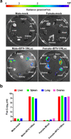
rBTV-1/NLuc propagation in animal organs. Mice from day 6 post-infection (Fig. 6) mock infected (one male and one female) or infected (one male and one female) were sacrificed, and the brain, spleen, lungs, heart, kidney, liver, and testis/ovaries were removed in one piece, washed, and examined by IVIS to assess for NLuc activity. Images of whole organs (a) and NLuc quantification (b) are indicated. Radiance (number of photons per second per square centimeter per steradian [p/sec/cm2/sr]). The average total flux [in log10 (number of photons per second) (p/s)] is shown.

Similar articles

Cited by

References

    1. Maclachlan NJ, Drew CP, Darpel KE, Worwa G. 2009. The pathology and pathogenesis of bluetongue. J Comp Pathol 141:1–16. doi:10.1016/j.jcpa.2009.04.003 - DOI - PubMed
    1. Rushton J, Lyons N. 2015. Economic impact of bluetongue: a review of the effects on production. Vet Ital 51:401–406. doi:10.12834/VetIt.646.3183.1 - DOI - PubMed
    1. Gethmann J, Probst C, Conraths FJ. 2020. Economic impact of a bluetongue serotype 8 epidemic in Germany. Front Vet Sci 7:65. doi:10.3389/fvets.2020.00065 - DOI - PMC - PubMed
    1. Du Toit RM. 1944. The transmission of blue-tongue and horse-sickness by Culicoides. J Vet Sc Anim Ind 19:7–16. http://hdl.handle.net/2263/59110.
    1. Ratinier M, Caporale M, Golder M, Franzoni G, Allan K, Nunes SF, Armezzani A, Bayoumy A, Rixon F, Shaw A, Palmarini M. 2011. Identification and characterization of a novel non-structural protein of bluetongue virus. PLoS Pathog 7:e1002477. doi:10.1371/journal.ppat.1002477 - DOI - PMC - PubMed