. 2024 May;36(19):e2308377.
doi: 10.1002/adma.202308377. Epub 2024 Feb 22.

Enhancing CAR Macrophage Efferocytosis Via Surface Engineered Lipid Nanoparticles Targeting LXR Signaling

Affiliations

Enhancing CAR Macrophage Efferocytosis Via Surface Engineered Lipid Nanoparticles Targeting LXR Signaling

Skylar T Chuang et al. Adv Mater. 2024 May.

Abstract

The removal of dying cells, or efferocytosis, is an indispensable part of resolving inflammation. However, the inflammatory microenvironment of the atherosclerotic plaque frequently affects the biology of both apoptotic cells and resident phagocytes, rendering efferocytosis dysfunctional. To overcome this problem, a chimeric antigen receptor (CAR) macrophage that can target and engulf phagocytosis-resistant apoptotic cells expressing CD47 is developed. In both normal and inflammatory circumstances, CAR macrophages exhibit activity equivalent to antibody blockage. The surface of CAR macrophages is modified with reactive oxygen species (ROS)-responsive therapeutic nanoparticles targeting the liver X receptor pathway to improve their cell effector activities. The combination of CAR and nanoparticle engineering activated lipid efflux pumps enhances cell debris clearance and reduces inflammation. It is further suggested that the undifferentiated CAR-Ms can transmigrate within a mico-fabricated vessel system. It is also shown that our CAR macrophage can act as a chimeric switch receptor (CSR) to withstand the immunosuppressive inflammatory environment. The developed platform has the potential to contribute to the advancement of next-generation cardiovascular disease therapies and further studies include in vivo experiments.

Keywords: CAR macrophage; atherosclerosis; efferocytosis; lipid nanoparticle; β‐cyclodextrin.


Figures

Figure 1.
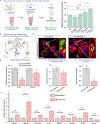
Figure 1.. Nano-immunoengineered CAR-M for potential atherosclerosis therapy.
(a) Schematic illustration of CAR-transduced THP-1 monocytes anchored with β-CD LNPs. The inset shows a confocal micrograph of CAR-monocyte carrying β-CD LNP backpacks. Scale bar is 5 μm; (a2) schematic depiction and transmission electron micrograph of a β-CD LNP. Scale bar is 100 nm; (a3) design of the anti-CD47 CAR construct; (a4) the β-CD LNP loaded CAR-Ms (CAR-M/β-CD LNP) could locate the inflamed lesion, traverse, and mediate targeted clearance of lesion apoptotic cells with elevated CD47 expression (CD47Hi ACs). (b) The ingested cholesterol-rich materials (ACs and CC) could be scavenged by β-CD released from cell surface LNPs due to ROS-mediated degradation. Oxysterols converted from cholesterol facilitated by β-CD could activate the LXR signaling pathway and increase key downstream genes such as Abca1, Abcg1, and Mertk to promote cholesterol efflux and suppression of inflammation. Scale bars are 5 μm.
Figure 2.
Figure 2.. In-vitro surface anchoring of β-CD LNP onto monocytes.
(a) Schematic illustration of β-CD LNP loading onto THP-1 cells via CD45 targeting. (b) Size and zeta potential change before and after CD45 antibody conjugation and antibody quantification. (c) Loading efficiency of the β-CD LNPs based on a density of 1.0 × 10⁶ cells/mL. (d) Confocal microscopy images of THP-1 cells (red) with β-CD LNP backpacks (green). Scale bar equals 20 μm. (e) THP-1 monocytes could be loaded with the indicated doses of β-CD LNPs and analyzed by flow cytometry. (f-g) The effect of CD45 antibody targeting on cell-associated LNPs was evaluated on day 0 and day 2 by (f) flow cytometry and (g) confocal microscopy. Data represent mean ± SEM; n = 3; ***p < 0.001 by unpaired two-tailed Student’s t test. Scale bars equal 5 μm. (h) Surface β-CD LNPs containing DSPE-PEG-biotin could be detected using a secondary reporter (AlexaFluor 647-streptavidin) by flow cytometry. (i) Representative dot-plot of surface β-CD LNP on THP-1 cells over 2 days analyzed by flow cytometry.
Figure 3.
Figure 3.. β-CD LNP loading mediates protection and potentiates efferocytosis in macrophages.
(a) Schematic depicting in vitro CC dissolution of THP-1 cells containing β-CD LNP backpacks. Data are mean ± SEM; n = 3; *p < 0.05, ***p < 0.001 by one-way ANOVA with Tukey post-hoc analysis; n.s. means no significance. (b) THP-1 cells with or without β-CD LNP backpacks were challenged with various inflammatory mediators (LPS and IFN-γ; CC; oxLDL) to evaluate the protective effects of β-CD. (c) Confocal microscopy images of THP-1 macrophages (red) examined 16 h after incubation with CC (green). Scale bar equals 20 μm. (d) Canonical inflammasome genes in THP-1 macrophages after 3 h incubation with CC determined by RT-qPCR. (e) DCFDA fluorescence of THP-1 macrophages as a measure of intracellular ROS levels after 24 h of stimulation with LPS (100 ng/mL) and IFN-γ (50 ng/mL). (f) Nile red fluorescence of THP-1 to assess lipid content in THP-1 macrophages. (g) Gene expression changes in THP-1 macrophages with or without β-CD LNP backpacks incubated with apoptotic Jurkat cells (ACs) determined by RT-qPCR. Data represent mean ± SEM; n = 3; *p < 0.05, **p < 0.01, ***p < 0.001 by unpaired two-tailed Student’s t test.
Figure 4.
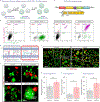
Figure 4.. CAR-M engineering and phagocytosis activity.
(a) Schematic diagram illustrating the process generating CAR-Ms from THP-1 cells. (b) Lentiviral construct used for CAR transduction. (c) Verification of surface expression of anti-CD47 CAR in CAR-Ms. (d) The anti-CD47 CAR represents a chimeric switch receptor (CSR) that reverses existing inhibitory signal mediated by the SIRPα-CD47 axis to enhance phagocytosis of CD47Hi ACs. (e) Confocal micrographs depicting phagocytosis of CD47Hi ACs in control macrophages, macrophages with CD47-blocked ACs, and CAR-Ms after 1 h incubation at 37 °C. Scale bar equals 20 μm. (f) Representative fluorescence microscopy images of control macrophages and CAR-Ms incubated with ACs for 2 h at 37 °C. The cells were washed three times prior to imaging. Scale bar equals 100 μm. (g-i) Quantitative analysis of partial (g), full (h), and total (i) engulfment of CD47Hi AC using CellTagging after 2 h co-culture at 37 °C. Data are mean ± SEM; n = 5; *p < 0.05, **p < 0.01 by unpaired two-tailed Student’s t test.
Figure 5.
Figure 5.. CAR-M phagocytosis under inflammatory condition.
(a) In addition to increasing CD47 expression in lesion cells, chronic inflammation also reduces the capacity of macrophages to clear ACs via ectodomain shedding. Efferocytic receptors (ex. MerTK) can be cleaved by metalloproteinase ADAM17, leading to reduced efferocytosis overall. (b) Representative fluorescence micrographs depicting phagocytosis of standard ACs (without inducing CD47 elevation) by M1-control macrophages or CAR-Ms after 2 h incubation. Arrowheads point to fully internalized ACs. Scale bar equals 100 μm. (c-e) Quantification of (c) partial, (d) full, and (e) total internalization of standard ACs by either M1 control macrophages or CAR-Ms by CellTagging. (f) Quantitative analysis of phagocytosis of standard ACs by control macrophages or CAR-Ms pretreated with either TNF-α alone or TNF-α and LPS. Data are mean ± SEM; n = 3; *p < 0.05, **p < 0.01 by unpaired two-tailed Student’s t test. (g) Combined M1 macrophages with CD47Hi ACs co-culture to simulate the phagocytosis in the atherosclerotic lesion environment. Scale bar equals 100 μm. (h) Suppression of TNF-α expression in macrophages upon efferocytosis with CD47Hi ACs characterized by RT-qPCR. Data are mean ± SEM; n = 3; *p < 0.05, **p < 0.01 by one-way ANOVA with Tukey post-hoc analysis.
Figure 6.
Figure 6.. Assessment of CAR-M phagocytosis combined with β-CD LNP.
(a) The various conditions selected to evaluate macrophage phagocytosis of CD47Hi AC. (b-d) Quantitative analysis via CellTagging of (b) partial, (c) full, and (d) total engulfment after 1 h incubation of macrophage with CD47Hi AC at 37°C. (e) The activation of LXR signaling targets Abca1 and Abcg1 as well as efferocytosis targets Il-10 and Mertk in control macrophages with β-CD LNPs or CAR-Ms with β-CD LNPs after co-culture with CD47Hi AC for 2 h 37 °C. (f) Time lapse fluorescence imaging of CAR-Ms with CD47Hi AC for 1 h. Scale bar equals 45 μm. (g) (top) 3D rendering of the microfabricated vessel-on-a-chip device constructed using photolithography; (bottom) HUVECs were seeded in the top channel and visualized by microscopy. Scale bars equal 100 μm. (h) Experimental timeline for HUVEC activation and CAR-M co-culture. (i) Fluorescence microscopy images examining the adherence of control THP-1 cells and CAR-Ms to TNF-α-activated HUVECs in the device. Scale bars equal 20 μm. (j) Confocal microscopy image and quantitation of CAR-Ms in the GelMA hydrogel layer indicating transendothelial migration. The CAR-Ms were fluorescently labeled with β-CD LNPs (red). Scale bar equals 100 μm. Data are mean ± SEM; n = 3, **p < 0.01, ***p < 0.001 by one-way ANOVA with Tukey post-hoc analysis; n.s. means not significant.

References

    1. a)Chao MP, Majeti R, Weissman IL, Nature Reviews Cancer 2012, 12, 58; - PubMed
    2. b)Feng M, Marjon KD, Zhu F, Weissman-Tsukamoto R, Levett A, Sullivan K, Kao KS, Markovic M, Bump PA, Jackson HM, Choi TS, Chen J, Banuelos AM, Liu J, Gip P, Cheng L, Wang D, Weissman IL, Nature Communications 2018, 9, 3194. - PMC - PubMed
    1. Voll RE, Herrmann M, Roth EA, Stach C, Kalden JR, Girkontaite I, Nature 1997, 390, 350. - PubMed
    1. Kojima Y, Volkmer JP, McKenna K, Civelek M, Lusis AJ, Miller CL, Direnzo D, Nanda V, Ye J, Connolly AJ, Schadt EE, Quertermous T, Betancur P, Maegdefessel L, Matic LP, Hedin U, Weissman IL, Leeper NJ, Nature 2016, 536, 86. - PMC - PubMed
    1. Stocker R, Keaney JF Jr., Physiol Rev 2004, 84, 1381. - PubMed
    1. a)Duewell P, Kono H, Rayner KJ, Sirois CM, Vladimer G, Bauernfeind FG, Abela GS, Franchi L, Nuñez G, Schnurr M, Espevik T, Lien E, Fitzgerald KA, Rock KL, Moore KJ, Wright SD, Hornung V, Latz E, Nature 2010, 464, 1357; - PMC - PubMed
    2. b)Rajamäki K, Lappalainen J, Oörni K, Välimäki E, Matikainen S, Kovanen PT, Eklund KK, PLoS One 2010, 5, e11765. - PMC - PubMed

LinkOut - more resources