

Optimizing the management of thyroid specimens to efficiently generate whole slide images for diagnosis
- PMID: 38353775
- PMCID: PMC11271424
- DOI: 10.1007/s00428-024-03762-3
Optimizing the management of thyroid specimens to efficiently generate whole slide images for diagnosis
Abstract
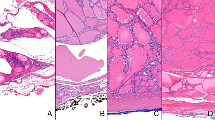
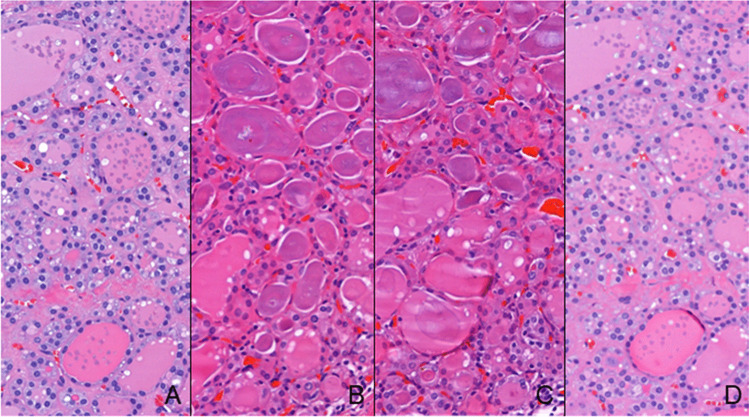
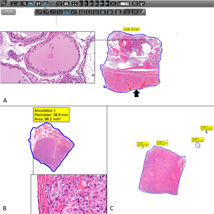
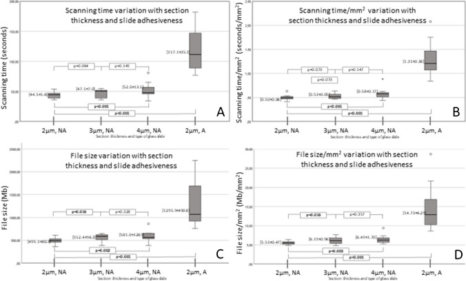
Transition from optical to digital observation requires an additional procedure in the pathology laboratory, the scanning of glass slides, leading to increased time and digital archive consumption. Thyroid surgical samples often carry the need to collect several tissue fragments that generate many slides to be scanned. This study evaluated the impact of using different inking colours for the surgical margin, section thickness, and glass slide type, in the consumption of time and archive. The series comprehended 40 nodules from 30 patients, including 34 benign nodules in follicular nodular disease, 1 NIFTP, and 5 papillary carcinomas. In 12 nodules, the dominant pattern was microfollicular/solid and in 28 it was macrofollicular. Scanning times/mm2 were longer in red-inked fragments in comparison to green (p = 0.04) and black ones (p = 0.024), and in blue-inked in comparison to green ones (p = 0.043). File sizes/mm2 were larger in red-inked fragments in comparison to green (p = 0.008) and black ones (p = 0.002). The dominant pattern microfollicular/solid was associated with bigger file size/mm2 in comparison with the macrofollicular one (p < 0.001). All scanner outputs increase significantly with the thickness of the section. All scanning outputs increase with the usage of adhesive glass slides in comparison to non-adhesive ones. Small interventions in thyroid sample management that can help optimizing the digital workflow include to prefer black and green inking colours for the surgical margins and 2 µm section in non-adhesive glass slides for increased efficiency.
Keywords: Digital pathology; Digital workflow; Efficiency; Specimen inking; Whole slide image.
© 2024. The Author(s).
Conflict of interest statement
C.E. consulted for Mindpeak, MSD, and Leica. A.P. consults for Indica Labs. Other authors declare no conflict of interest.
Figures
Similar articles
-
Digital image-assisted quantitative nuclear analysis improves diagnostic accuracy of thyroid fine-needle aspiration cytology.Cancer Cytopathol. 2019 Aug;127(8):501-513. doi: 10.1002/cncy.22120. Epub 2019 May 31. Cancer Cytopathol. 2019. PMID: 31150162
-
Preoperative Cytologic Diagnosis of Noninvasive Follicular Thyroid Neoplasm with Papillary-Like Nuclear Features: A Prospective Analysis.Thyroid. 2016 Oct;26(10):1466-1471. doi: 10.1089/thy.2016.0280. Epub 2016 Sep 8. Thyroid. 2016. PMID: 27457786
-
Study on the Transformation Process of Thyroid Fine-Needle Aspiration Liquid-Based Cytology to Whole-Slide Image.Cytopathology. 2025 Mar;36(2):106-114. doi: 10.1111/cyt.13468. Epub 2025 Jan 8. Cytopathology. 2025. PMID: 39780471 Free PMC article.
-
Pathology quiz case 2. Diagnosis: Papillary thyroid carcinoma arising in the setting of black thyroid.Arch Otolaryngol Head Neck Surg. 2012 Nov;138(11):1093-5. doi: 10.1001/2013.jamaoto.363b. Arch Otolaryngol Head Neck Surg. 2012. PMID: 23165392 Review. No abstract available.
-
Surgical management of thyroid disease.Otolaryngol Clin North Am. 2010 Apr;43(2):273-83, viii. doi: 10.1016/j.otc.2010.01.004. Otolaryngol Clin North Am. 2010. PMID: 20510714 Review.
Cited by
-
Digital transformation of pathology - the European Society of Pathology expert opinion paper.Virchows Arch. 2025 Mar 31. doi: 10.1007/s00428-025-04090-w. Online ahead of print. Virchows Arch. 2025. PMID: 40164935
-
Impact of Tissue Thickness on Computational Quantification of Features in Whole Slide Images for Diagnostic Pathology.Endocr Pathol. 2025 Apr 8;36(1):10. doi: 10.1007/s12022-025-09855-2. Endocr Pathol. 2025. PMID: 40198470 Free PMC article.
References
-
- Pinto DG, Bychkov A, Tsuyama N, Fukuoka J, Eloy C (2023) Real-world implementation of digital pathology: results from an intercontinental survey. Lab Invest 103(12):100261. 10.1016/j.labinv.2023.100261 - PubMed
-
- Caputo A, Macri L, Gibilisco F, Vatrano S, Taranto C, Occhipinti E, Santamaria F, Arcoria A, Scillieri R, Fraggetta F (2023) Validation of full-remote reporting for cervicovaginal cytology. The Caltagirone-Acireale distributed lab. J Am Soc Cytopathol. 10.1016/j.jasc.2023.06.001 10.1016/j.jasc.2023.06.001 - DOI - PubMed
MeSH terms
LinkOut - more resources
Full Text Sources
Medical
