. 2024 Feb 14;35(1):13.
doi: 10.1007/s10856-024-06781-3.

Bioactive potential of Bio-C Temp demonstrated by systemic mineralization markers and immunoexpression of bone proteins in the rat connective tissue

Affiliations

Bioactive potential of Bio-C Temp demonstrated by systemic mineralization markers and immunoexpression of bone proteins in the rat connective tissue

Camila Soares Lopes et al. J Mater Sci Mater Med. .

Abstract

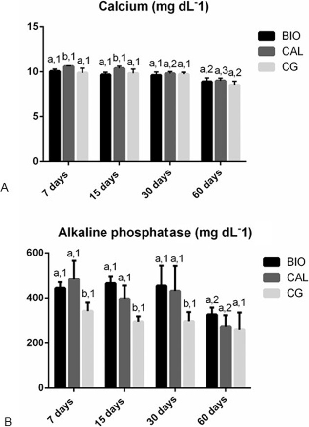
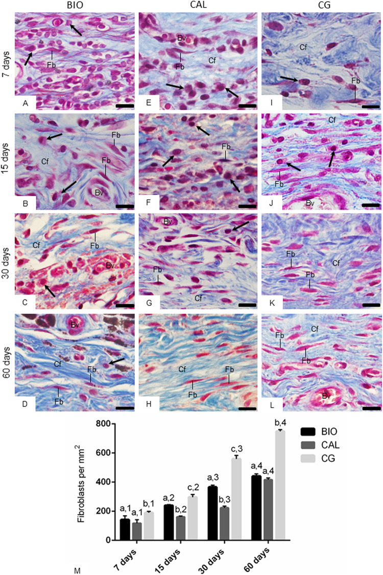
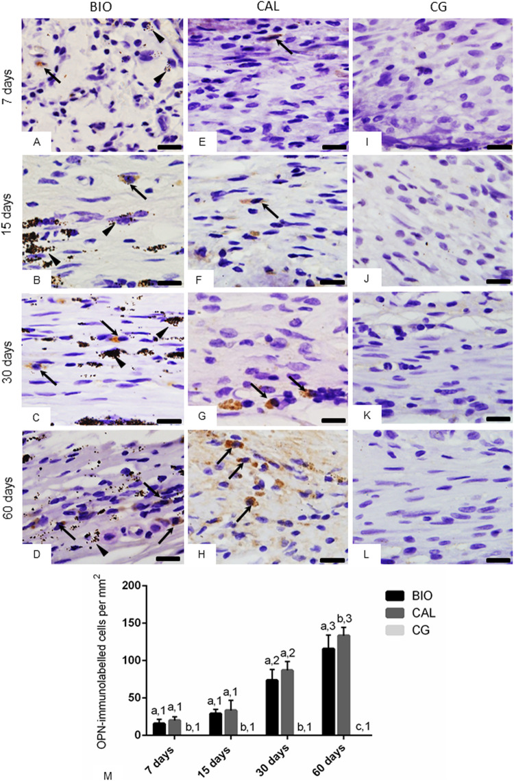
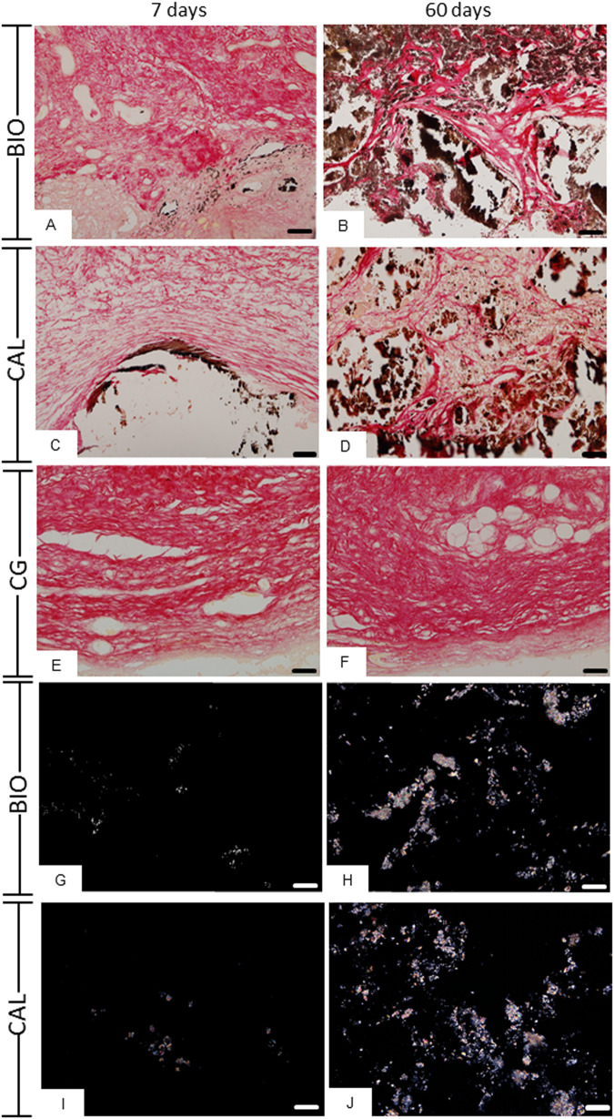
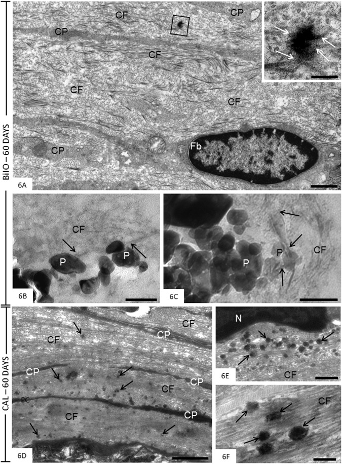
Intracanal medications are used in endodontic treatment due to their antibacterial activity and ability to induce the periapical repair. Among the intracanal medications, the Calen (CAL; SS. White, Brazil) is a calcium hydroxide-based medication that provides an alkaline pH and releases calcium, exerting an antimicrobial activity. Bio-C Temp (BIO; Angelus, Brazil), a ready-to-use bioceramic intracanal medication, was designed to stimulate the mineralized tissues formation. Here, we investigated the bioactive potential of BIO in comparison to the CAL in the rat subcutaneous. Polyethylene tubes filled with medications, and empty tubes (control group, CG) were implanted in the subcutaneous tissue of rats. After 7, 15, 30 and 60 days, the blood was collected for calcium (Ca+2) and alkaline phosphatase (ALP) measurement, and the capsules around the implants were processed for morphological analyses. The data were submitted to two-way ANOVA and Tukey test (p < 0.05). At 7, 15 and 30 days, the ALP level was grater in BIO and CAL than in CG (p < 0.0001). At 7 and 15 days, greater Ca+2 level was seen in the serum of CAL samples. From 7 to 60 days, an increase in the number of fibroblasts, osteocalcin- and osteopontin-immunolabelled cells was observed in BIO and CAL groups (p < 0.0001). In all periods, BIO and CAL specimens showed von Kossa-positive structures. Moreover, ultrastructural analysis revealed globules of mineralization in the capsules around the BIO and CAL specimens. Thus Bio-C Temp caused an increase in the ALP, osteocalcin and osteopontin, which may have allowed the formation of calcite, suggesting bioactive potential.


Conflict of interest statement

The authors declare no competing interests.

Figures

Fig. 1
Fig. 1
Graphics showing the values (expressed as mean ± standard deviation) of the concentration of calcium (A), and alkaline phosphatase (B) in the serum of the Bio-C Temp (BIO), Calen (CAL) and CG specimens at 7, 15, 30 and 60 days. In each period, the comparison among the groups is indicated by superscript letters; different letters = significant difference. The superscript numbers indicate the analysis of each group over time; different numbers = significant difference. Tukey’s test (p ≤ 0.05)
Fig. 2
Fig. 2
Photomicrographs showing portions of the capsules adjacent to the opening of the implanted tubes of the BIO (AD), CAL (EH) and CG (IL) groups show an increase in fibroblasts (Fb) and collagen fibers (in blue) over time. Arrows, inflammatory cells; Bv, blood vessels; Cf, collagen fibers. Masson’s trichrome. Bars: 18 μm. Figure 2M Graphic showing the number of fibroblasts per mm2 (expressed as mean ± standard deviation) from BIO, CAL and CG groups at 7, 15, 30 and 60 days. In each period, the comparison among the groups is indicated by superscript letters; different letters = significant difference. The superscript numbers indicate the analysis of each group over time; different numbers = significant difference. Tukey’s test (p ≤ 0.05)
Fig. 3
Fig. 3
A–L Photomicrographs showing portions of sections submitted to immunohistochemistry for detection of OCN (in brown colour) and counterstained with haematoxylin. In BIO (AD) and CAL (EH) some elliptical and/or fusiform cells exhibit immunolabelling in their cytoplasm (arrows). None immunolabelled cell is observed in the CG specimens (IL). Arrowhead: material particles; BV, blood vessels. Bars: 18 μm. Figure 3M—Graphic showing the number of OCN-immunolabelled cells per mm2 (expressed as mean ± standard deviation) from BIO, CAL and CG groups at 7, 15, 30 and 60 days. In each period, the comparison among the groups is indicated by superscript letters; different letters = significant difference. The superscript numbers indicate the analysis of each group over time; different numbers = significant difference. Tukey’s test (p ≤ 0.05)
Fig. 4
Fig. 4
A–L Photomicrographs showing portions of the sections submitted to immunohistochemistry for detection of OPN (in brown colour) and counterstained with haematoxylin. At 7 and 15 days, few immunolabelled cells (arrows), mainly fibroblasts, are observed in the capsules of BIO (A, B) and CAL (E, F). At 30 and 60 days, several strongly immunolabelled cells (arrows) are present in the capsules of BIO (C, D) and CAL (G, H) groups. Note absence of immunolabelled cells in the capsules of CG specimens (IL). Arrowhead: material particles. Bars: 18 μm. Figure 4M Graphic showing the number of OPN-immunolabelled cells per mm2 (expressed as mean ± standard deviation) from BIO, CAL and CG groups at 7, 15, 30 and 60 days. In each period, the comparison among the groups is indicated by superscript letters; different letters = significant difference. The superscript numbers indicate the analysis of each group over time; different numbers = significant difference. Tukey’s test (p ≤ 0.05)
Fig. 5
Fig. 5
Photomicrographs showing portions of capsules adjacent to the opening of the tubes implanted in the subcutaneous for 7 (A, C, E, G, I) and 60 days (B, D, F, H, J). A–F Sections submitted to the von Kossa reaction and counterstained with picrosirius-red. von Kossa-positive structures (black colour) are observed dispersed in the capsules of BIO (A, B) and CAL (C, D) specimens. In (E, F), no positive structures are observed in CG. Figure 5G–J Photomicrographs showing unstained sections analyzed under polarized light. Note the presence of birefringent structures in the BIO and CAL groups at 7 and 60 days. Bars: 52 μm
Fig. 6
Fig. 6
A–F Electron micrographs of portions of capsules after 60 days of implantation in the connective tissue of the subcutaneous. A–C (BIO specimens) The Fig. 6A shows an electron-opaque structure (outlined area) in the collagen-rich extracellular matrix. The inset, high magnification of outlined area, reveals that some collagen fibrils are in continuity with this electron-opaque structure (arrows). Fb, fibroblast; CP, cytoplasmic processes; CF, collagen fibrils. In (B, C), the BIO particles (P) are surrounded by thin collagen fibrils (CF); some fibrils (arrows) appear to be in close juxtaposition to the material particles (P). Bars: 2.5 μm (Fig. 6A), 0.5 μm (Fig. 6A, inset) and 200 nm (Fig. 6B, C). D–F (CAL specimens). In the Fig. 6D, a portion of capsule exhibits bundles of densely packed collagen (CF) and thin cytoplasmic processes (CP) adjacent to the opening of the tube (I) implanted in the subcutaneous connective tissue. Several electron-opaque round/ovoid shaped structures (arrows) are dispersed by rich-fibrous matrix. Figure 6E and F—high magnification of portions of capsules exhibiting electron-opaque structures (arrows). Round, ovoid or irregularly shaped structures (arrows) with variable electron-opacity are observed amongst collagen fibrils (CF). N, nucleus. Bars: 2 μm (Fig. 6D), 1 μm (Fig. 6E) and 500 nm (Fig. 6F)

References

    1. Holland R, Gomes-Filho JE, Cintra LTA, Queiroz ÍOA, Estrela C. Factors affecting the periapical healing process of endodontically treated teeth. J Appl Oral Sci. 2017;25:465–76. 10.1590/1678-7757-2016-0464 - PMC - PubMed
    1. Mohammadi Z, Dummer PM. Properties and applications of calcium hydroxide in endodontics and dental traumatology. Int Endod J. 2011;44:697–730. 10.1111/j.1365-2591.2011.01886 - PubMed
    1. Estrela C, Sydney GB, Bammann LL, Felippe-Júnior O. Mechanism of action of calcium and hydroxyl ions of calcium hydroxide on tissue and bacteria. Braz Dent J. 1995;6:85–90. - PubMed
    1. Mizuno M, Banzai Y. Calcium ion release from calcium hydroxide stimulated fibronectin gene expression in dental pulp cells and the differentiation of dental pulp cells to mineralized tissue forming cells by fibronectin. Int Endod J. 2008;41:933–8. 10.1111/j.1365-2591.2008.01420.x - PubMed
    1. Alsubait S, Alsaad N, Alahmari S, Alfaraj F, Alfawaz H, Alqedairi A. The effect of intracanal medicaments used in Endodontics on the dislocation resistance of two calcium silicate-based filling materials. BMC Oral Health. 2020;20:57. 10.1186/s12903-020-1044-6 - PMC - PubMed