. 2024 Feb 20;121(8):e2309465121.
doi: 10.1073/pnas.2309465121. Epub 2024 Feb 14.

Two-pore channels regulate endomembrane tension to enable remodeling and resolution of phagolysosomes

Affiliations

Two-pore channels regulate endomembrane tension to enable remodeling and resolution of phagolysosomes

Sarah R Chadwick et al. Proc Natl Acad Sci U S A. .

Abstract

Phagocytes promptly resolve ingested targets to replenish lysosomes and maintain their responsiveness. The resolution process requires that degradative hydrolases, solute transporters, and proteins involved in lipid traffic are delivered and made active in phagolysosomes. It also involves extensive membrane remodeling. We report that cation channels that localize to phagolysosomes were essential for resolution. Specifically, the conductance of Na+ by two-pore channels (TPCs) and the presence of a Na+ gradient between the phagolysosome lumen and the cytosol were critical for the controlled release of membrane tension that permits deformation of the limiting phagolysosome membrane. In turn, membrane deformation was a necessary step to efficiently transport the cholesterol extracted from cellular targets, permeabilizing them to hydrolases. These results place TPCs as regulators of endomembrane remodeling events that precede target degradation in cases when the target is bound by a cholesterol-containing membrane. The findings may help to explain lipid metabolism dysfunction and autophagic flux impairment reported in TPC KO mice and establish stepwise regulation to the resolution process that begins with lysis of the target.

Keywords: glycocalyx; phagocytosis; solute transport.


Conflict of interest statement

Competing interests statement:The authors declare no competing interest.

Figures

Fig. 1.
Fig. 1.
Two-pore channels are not required for phagocytosis. (AI) BMDM from wt, TPC1−/−, TPC2−/−, and TPC1/2−/− mice challenged with indicated targets for 15 min. In (A), (D), and (G), representative images of BMDM (F-actin, yellow) and targets (inside/outside labeling, blue/magenta as indicated). (Scale bar, 20 µm.) Dots represent individual fields containing >5 cells. n = 3. Here and elsewhere, box and whisker plots represent minimum to maximum values with median center lines. Here and elsewhere, significance was assessed by one-way ANOVA followed by Tukey’s test for multiple comparisons.
Fig. 2.
Fig. 2.
TPC activity is required for phagosome resolution. (A and B) RAW 264.7 cells expressing TPC1-tdTomato or TPC2-mCherry challenged with Alexa Fluor 488 anti-IgG labeled sRBC for indicated times. xy plane, Left. Extended focus, Left. (Scale bar, 5 µm.) Quantification in (B) for individual phagosomes. n = 3. (C and D) wt and TPC1/2−/− BMDM challenged with sRBC. Resolution assessed up to 4 h. (Scale bar, 10 µm.) Phagosome volume quantified in (D). Dots represent individual fields containing >3 cells, >9 phagosomes. n = 3. (E) As in (C), using 25× magnification. (Scale bar, 25 µm.) (F) wt and TPC1/2−/− BMDM and TPC1/2−/− BMDM expressing wtTPC2-mCherry or TPC2(N257A)-mCherry, quantified as in (D). Dots represent individual phagosomes. >30 cells, >100 phagosomes. n = 2.
Fig. 3.
Fig. 3.
TPCs are not required for phagosome maturation. (A) LAMP-1 acquisition. Dots represent individual fields containing >5 cells. n = 3, >150 phagosomes, error bars = SD. (B and C) Phagosomal pH, 1 h post-phagocytosis measured by ratiometric fluorescence microscopy of FITC-labeled RBC or zymosan. Dots represent individual phagosomes, n = 3, >100 phagosomes. (D) BMDM challenged with IgG-opsonized silica beads coated with DQ-BSA. (Scale bar, 5 µm.) (E and F) Western blot. n = 3, error bars = SD.)
Fig. 4.
Fig. 4.
TPC-mediated Na+ transport relieves endomembrane tension to generate the PTN. (A) TEM of RBC-containing phagosomes, 2 h post-internalization. (B) Quantification of intraluminal vesicles (ILVs) per section. n = 3, dots represent sections, error bars = SD. (C) Phagosomes containing IgG-opsonized silica beads coated with DQ-BSA. (Scale bar, 5 µm.) (D and E) LysoFlipper-TR fluorescence lifetime, 1 h post-phagocytosis. In (E), dots represent measurements of individual phagosomes, >45 phagosomes per condition. Violins indicate median and quartiles, and width represents data distribution. (F) BMDM challenged with sRBC for 4 h with or without ouabain. n = 3. (G and H) BMDM depleted of Ca2+ (300 nM thapsigargin and Ca2+-free media) or Na+ (substituted for NMG+) 30 min after phagocytosis for an additional 2 h. n = 3. (Scale bar, 10 µm.)
Fig. 5.
Fig. 5.
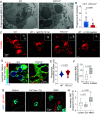
TPC activity and the lysosome-to-cytosol Na+ gradient drive lysosome tubulation. (A) wt and TPC1/2−/− BMDM incubated with 30 mM sucrose and 3 kDa TMR-dextran overnight, chased for >1 h, followed by 1 h of invertase + MCSF. (B) Predicted effect of inhibitors. (C) HeLa cells expressing TPC2-mCherry or TPC2(N257A). Cells were treated with 10 µM tetrandrine, 2 mM ouabain, or 10 µM BAPTA-AM for 30 min. (Scale bar, 2 µm.)
Fig. 6.
Fig. 6.
A cholesterol barrier to target lysis and phagolysosome resolution in TPC1/2−/− macrophages. (A and B) Target permeability to 3 kDa dextran after 1 h. Delivery of the dextran from lysosomes is followed by lysis of the RBC, allowing its access, as quantified in (B). n = 3, >40 phagosomes per time point, points indicate mean values, area fill indicates ± SD. (Scale bar, 2 µm.) (C and D) As in (A and B), treatment with YM201636 or ouabain after 30 min. (E and F) Staining for cathepsin B and quantified as in (B). (G and H) Targets in which cholesterol was pre-extracted using 5 mM MβCD. (Scale bar, 10 µm.) Quantified as in (B). (I and J) Resolution of cholesterol-extracted sRBC. Representative images in (I), quantified in (J). n = 3, dots represent sum RBC surface area from individual fields with >80 phagosomes normalized to vehicle at 1 h. (Scale bar, 10 µm.)
Fig. 7.
Fig. 7.
NPC1/2 are required for target lysis. (A and B) Target permeability to 3 kDa dextran after 2 h in the presence of 1 µg/mL U18666A or vehicle added for the last 1.5 h. (Scale bar, 2 µm.) n = 3, >80 phagosomes. (C) Target size for BMDM with or without 1 µg/mL U18666A given after 30 min. n = 3, >80 phagosomes. (D) RAW 264.7 cells expressing mCherry-NPC2 challenged with Alexa Fluor 647-labeled IgG-opsonized sRBC with or without the addition of tetrandrine after 1 h. When overexpressed, NPC2 permeates the phagocytic target even in the presence of tetrandrine (below). (Scale bar, 10 µm.) (E) BMDM treated with 1 µg/mL U18666A or vehicle and stained with filipin. (F and G) Filipin staining of BMDM given 40 µg/mL LDL for 24 h. In (G), the sum fluorescence per field is normalized to cell number. Significance was assessed by one-way ANOVA followed by Tukey’s test. (Scale bar, 10 µm.)
Fig. 8.
Fig. 8.
The lysosome glycocalyx presents a barrier to phagolysosome resolution in TPC1/2−/− macrophages. (A) Western blot for LAMP-2 after endoglycosidase H (EndoH) treatment for indicated times. (B and C) Target permeability to 3 kDa dextran after 1 h (B) and over time with 25 mU EndoH. (Scale bar, 2 µm.) (D and E) wt and TPC1/2−/− BMDM given 25 mU EndoH for 2 h prior to phagocytosis of sRBC. Phagosome volume measured over time. (Scale bar, 25 µm.) (F) wt and TPC1/2−/− BMDM 1 h postphagocytosis of sRBC before and after hyperosmotic shock. Phagosome volume measured over time. Individual fields (5 to 10 cells) normalized to control at 30 min, n = 3, significance assessed by an unpaired t test. (G) Model.

References

    1. Franken L., Schiwon M., Kurts C., Macrophages: Sentinels and regulators of the immune system. Cell Microbiol. 18, 475–487 (2016). - PubMed
    1. Khanna S., et al. , Macrophage dysfunction impairs resolution of inflammation in the wounds of diabetic mice. PLoS One 5, e9539 (2010). - PMC - PubMed
    1. Uderhardt S., Martins A. J., Tsang J. S., Lammermann T., Germain R. N., Resident macrophages cloak tissue microlesions to prevent neutrophil-driven inflammatory damage. Cell 177, 541–555.e517 (2019). - PMC - PubMed
    1. Ren Y., Savill J., Apoptosis: The importance of being eaten. Cell Death Differ. 5, 563–568 (1998). - PubMed
    1. Lancaster C. E., et al. , Phagosome resolution regenerates lysosomes and maintains the degradative capacity in phagocytes. J. Cell Biol. 220, e202005072 (2021). - PMC - PubMed

LinkOut - more resources