

Lesion mapping and functional characterization of hemiplegic children with different patterns of hand manipulation
- PMID: 38354671
- PMCID: PMC10944177
- DOI: 10.1016/j.nicl.2024.103575
Lesion mapping and functional characterization of hemiplegic children with different patterns of hand manipulation
Abstract
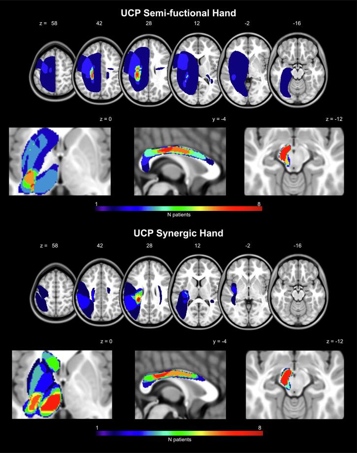
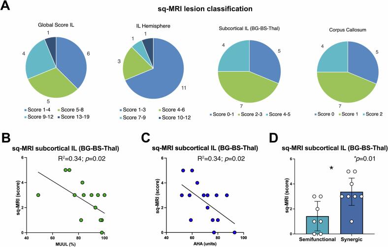
Brain damage in children with unilateral cerebral palsy (UCP) affects motor function, with varying severity, making it difficult the performance of daily actions. Recently, qualitative and semi-quantitative methods have been developed for lesion classification, but studies on mild to moderate hand impairment are lacking. The present study aimed to characterize lesion topography and preserved brain areas in UCP children with specific patterns of hand manipulation. A homogeneous sample of 16 UCP children, aged 9 to 14 years, was enrolled in the study. Motor assessment included the characterization of the specific pattern of hand manipulation, by means of unimanual and bimanual measures (Kinematic Hand Classification, KHC; Manual Ability Classification System, MACS; House Functional Classification System, HFCS; Melbourne Unilateral Upper Limb Assessment, MUUL; Assisting Hand Assessment, AHA). The MRI morphological study included multiple methods: (a) qualitative lesion classification, (b) semi-quantitative classification (sq-MRI), (c) voxel-based morphometry comparing UCP and typically developed children (VBM-DARTEL), and (d) quantitative brain tissue segmentation (q-BTS). In addition, functional MRI was used to assess spared functional activations and cluster lateralization in the ipsilesional and contralesional hemispheres of UCP children during the execution of simple movements and grasping actions with the more affected hand. Lesions most frequently involved the periventricular white matter, corpus callosum, posterior limb of the internal capsule, thalamus, basal ganglia and brainstem. VMB-DARTEL analysis allowed to detect mainly white matter lesions. Both sq-MRI classification and q-BTS identified lesions of thalamus, brainstem, and basal ganglia. In particular, UCP patients with synergic hand pattern showed larger involvement of subcortical structures, as compared to those with semi-functional hand. Furthermore, sparing of gray matter in basal ganglia and thalamus was positively correlated with MUUL and AHA scores. Concerning white matter, q-BTS revealed a larger damage of fronto-striatal connections in patients with synergic hand, as compared to those with semi-functional hand. The volume of these connections was correlated to unimanual function (MUUL score). The fMRI results showed that all patients, but one, including those with cortical lesions, had activation in ipsilesional areas, regardless of lesion timing. Children with synergic hand showed more lateralized activation in the ipsilesional hemisphere both during grasping and simple movements, while children with semi-functional hand exhibited more bilateral activation during grasping. The study demonstrates that lesion localization, rather than lesion type based on the timing of their occurrence, is more associated with the functional level of hand manipulation. Overall, the preservation of subcortical structures and white matter can predict a better functional outcome. Future studies integrating different techniques (structural and functional imaging, TMS) could provide further evidence on the relation between brain reorganization and specific pattern of manipulation in UCP children.
Keywords: Action execution; Cerebral palsy; Functional reorganization; Lesion classification; Upper limb; Voxel-based morphometry.
Copyright © 2024 The Author(s). Published by Elsevier Inc. All rights reserved.
Conflict of interest statement
Declaration of competing interest The authors declare that they have no known competing financial interests or personal relationships that could have appeared to influence the work reported in this paper.
Figures


Similar articles
-
Short-Term Memory Impairment.2024 Jun 8. In: StatPearls [Internet]. Treasure Island (FL): StatPearls Publishing; 2025 Jan–. 2024 Jun 8. In: StatPearls [Internet]. Treasure Island (FL): StatPearls Publishing; 2025 Jan–. PMID: 31424720 Free Books & Documents.
-
Prescription of Controlled Substances: Benefits and Risks.2025 Jul 6. In: StatPearls [Internet]. Treasure Island (FL): StatPearls Publishing; 2025 Jan–. 2025 Jul 6. In: StatPearls [Internet]. Treasure Island (FL): StatPearls Publishing; 2025 Jan–. PMID: 30726003 Free Books & Documents.
-
Hand function after neonatal stroke: A graph model based on basal ganglia and thalami structure.Neuroimage Clin. 2024;41:103568. doi: 10.1016/j.nicl.2024.103568. Epub 2024 Jan 22. Neuroimage Clin. 2024. PMID: 38277807 Free PMC article.
-
The Black Book of Psychotropic Dosing and Monitoring.Psychopharmacol Bull. 2024 Jul 8;54(3):8-59. Psychopharmacol Bull. 2024. PMID: 38993656 Free PMC article. Review.
-
Magnetic resonance perfusion for differentiating low-grade from high-grade gliomas at first presentation.Cochrane Database Syst Rev. 2018 Jan 22;1(1):CD011551. doi: 10.1002/14651858.CD011551.pub2. Cochrane Database Syst Rev. 2018. PMID: 29357120 Free PMC article.
Cited by
-
Impact of Brain Lesion Characteristics on Motor Function and Cortical Reorganization in Hemiplegic Cerebral Palsy.Medicina (Kaunas). 2025 Jan 24;61(2):205. doi: 10.3390/medicina61020205. Medicina (Kaunas). 2025. PMID: 40005322 Free PMC article.
References
Publication types
MeSH terms
LinkOut - more resources
Full Text Sources
Medical
Miscellaneous
