Review
. 2024 Feb 13;57(2):206-222.
doi: 10.1016/j.immuni.2024.01.010.

LAG-3, TIM-3, and TIGIT: Distinct functions in immune regulation

Affiliations
Review

LAG-3, TIM-3, and TIGIT: Distinct functions in immune regulation

Nicole Joller et al. Immunity. .

Abstract

LAG-3, TIM-3, and TIGIT comprise the next generation of immune checkpoint receptors being harnessed in the clinic. Although initially studied for their roles in restraining T cell responses, intense investigation over the last several years has started to pinpoint the unique functions of these molecules in other immune cell types. Understanding the distinct processes that these receptors regulate across immune cells and tissues will inform the clinical development and application of therapies that either antagonize or agonize these receptors, as well as the profile of potential tissue toxicity associated with their targeting. Here, we discuss the distinct functions of LAG-3, TIM-3, and TIGIT, including their contributions to the regulation of immune cells beyond T cells, their roles in disease, and the implications for their targeting in the clinic.

Keywords: autoimmunity; cancer; checkpoint receptor; immunotherapy.


Conflict of interest statement

Declaration of interests N.J. is a paid consultant for Almirall, Anaptysbio, and Merck. A.C.A. is a member of the scientific advisory board for Tizona Therapeutics, Trishula Therapeutics, Compass Therapeutics, Zumutor Biologics, ImmuneOncia, and Excepgen, which have interests in cancer immunotherapy. A.C.A. is a paid consultant for iTeos Therapeutics and Larkspur Biosciences. V.K.K. has an ownership interest in and is a member of the scientific advisory board for Tizona Therapeutics, Bicara Therapeutics, Compass Therapeutics, Larkspur Biosciences, and Trishula Therapeutics. V.K.K. is a co-founder of and has an ownership interest in Celsius Therapeutics. N.J., A.C.A., and V.K.K. are inventors on patents related to TIM-3 and TIGIT. A.C.A.’s and V.K.K.’s interests were reviewed and managed by Mass General Brigham in accordance with their conflict of interest policies.

Figures
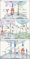
Figure 1.
Figure 1.. The LAG-3, TIM-3, and TIGIT pathways
A)LAG-3: LAG-3 can bind to MHC II, LSECtin, GALECTIN-3 (GAL-3), FGL1, and in cis to the CD3-TCR complex within the immunological synapse. EP repeats in the cytoplasmic tail of LAG-3 result in local acidification of the cytosol triggering the dissociation of phospho-Lck from either the CD4 or CD8 co-receptors, thereby limiting proximal TCR signaling. In addition, LAG-3 contains a membrane proximal FXXL motif followed by a KIEELE motif in its cytoplasmic tail, both of which have been implicated in inhibitory function. The extracellular domain of LAG-3 can be shed as soluble LAG-3 (sLAG-3) upon cleavage by the metalloproteases ADAM10 and ADAM17 as a means to rapidly control LAG-3 activity. (B)TIM-3: GALECTIN-9 (GAL-9), HMGB1, CEACAM1, and Phosphatidyl Serine (PtdSer) bind to TIM-3. T cell (Left): In the absence of TIM-3 ligand, the adaptor BAT3 is bound to the region containing Y256 and Y263 in the TIM-3 cytoplasmic tail. BAT3 recruits phospho-LCK to promote TCR signaling. Upon ligand-binding, Y256 and Y263 become phosphorylated. This triggers dissociation of BAT3, thereby allowing FYN to bind and TIM-3-mediated inhibition to ensue. Additionally, TIM-3 may recruit the phosphatases CD45 and CD148 into the synapse to inhibit TCR signaling. DC (Right): TIM-3 binding to HMGB1 prevents HMGB1-DNA complex binding to RAGE and HMGB1 binding to TLR2,4, and IL1R, thereby limiting DC activation via these pathways. TIM-3 further inhibits activation of the inflammasome and cGAS-STING by as yet unknown mechanisms. BAT3 is expressed in DCs and restrains tolerogenic programs but little else is known regarding BAT3 and its effect on TIM-3 signaling in DCs. (C)TIGIT: TIGIT and CD226 share the ligands CD112 and CD155 but bind them with different affinities (indicated by arrow thickness). TIGIT additionally binds the FAP2 protein expressed by F. nucleatum. CD112 and CD155 also serve as ligands for CD112R and CD96, respectively, completing a complex receptor-ligand network. TIGIT inhibits positive signaling via CD226 through competition for its ligands and by disrupting CD226 homodimerization, thereby disabling ligand binding and phosphorylation of the ITT motif in the CD226 tail. TIGIT further contains an ITT-like and an ITIM motif, which become phosphorylated upon ligand binding, recruiting the adapters GRB2 and β-arrestin and, subsequently, SHIP1 into the immunological synapse to inhibit TCR signaling. Lastly, TIGIT binding to CD155 results in phosphorylation of the ITIM motif in the cytoplasmic tail of CD155, delivering an inhibitory signal into the CD155-expressing cell.
Figure 2.
Figure 2.. Cellular and Stage specification
(A)Cellular specification: Expression profile of LAG-3, TIM-3, and TIGIT on different immune cell subsets. The relevant references are included in the respective sections in the text. (B) Stage specification: Checkpoint receptor expression at different stages of immune activation can tune T cell activation and differentiation. During acute T cell activation, LAG-3 and TIGIT are co-expressed with PD-1 whereas TIM-3 expression comes later when cells are fully differentiated and commited to the CD4+ Th1 lineage or to fully cytotoxic CD8+ T cells. During chronic activation, TIGIT is expressed together with PD-1 in stem-like T cells, highlighting unique roles for these checkpoint receptors in regulating this important cell population. LAG-3 expression follows on effector-like cells and finally TIM-3 is expressed in terminally differentiated dysfunctional T cells.
Figure 3.
Figure 3.. Functional and Anatomic specification
A) Functional specification: Cellular and stage specification of checkpoint receptors determines specialized functions in immune regulation. LAG-3 limits T cell priming due to its expression early during T cell activation . TIM-3 has a specialized role in determining DC function through regulation of DNA-sensing and inflammasome activation and in regulating Type 1 Immunity due to its expression in terminally differentiated IFN-γ-secreting CD4+ and CD8+ T cells. TIGIT has a specialized role in promoting Treg and regulatory B cell responses as well as dampening inflammation by selectively inhibiting Type 1/ Type 17 immunity and thereby shifting the balance towards Type 2 immune responses ,,. B)Anatomic specification: LAG-3-, TIM-3-, and TIGIT-driven regulation may dominate in different tissues reflecting ligand expressions or the activity of receptor-expressing cell types. LAG-3 may be particularly active in the heart and pancreas; loss of LAG-3 has been associated with myocarditis and LAG-3 expression with poor prognosis in pancreatic cancer . TIM-3 may be critically important for controlling inflammation in the CNS and in the gut; TIM-3 expression has been shown to be defective in patients with Multiple sclerosiss , and the TIM-3 ligands GALECTIN-9 and CEACAM-1 are highly expressed in the gut ,. TIGIT may be active in the lung and skin, where the balance of Type 1/Type 17 vs Type 2 immunity is important ,. Further, TIGIT function in regulatory T cells and B cells seem critical for controlling inflammation in the gut and CNS ,.

References

    1. Anderson AC, Joller N, and Kuchroo VK (2016). Lag-3, Tim-3, and TIGIT: Co-inhibitory Receptors with Specialized Functions in Immune Regulation. Immunity 44. 10.1016/j.immuni.2016.05.001. - DOI - PMC - PubMed
    1. Woo SR, Turnis ME, Goldberg MV, Bankoti J, Selby M, Nirschl CJ, Bettini ML, Gravano DM, Vogel P, Liu CL, et al. (2012). Immune inhibitory molecules LAG-3 and PD-1 synergistically regulate T-cell function to promote tumoral immune escape. Cancer Res 72, 917–927. 10.1158/0008-5472.CAN-11-1620. - DOI - PMC - PubMed
    1. Triebel F, Jitsukawa S, Baixeras E, Roman-Roman S, Genevee C, Viegas-Pequignot E, and Hercend T (1990). LAG-3, a novel lymphocyte activation gene closely related to CD4. J Exp Med 171, 1393–1405. 10.1084/jem.171.5.1393. - DOI - PMC - PubMed
    1. Grosso JF, Goldberg MV, Getnet D, Bruno TC, Yen H-R, Pyle KJ, Hipkiss E, Vignali DAA, Pardoll DM, and Drake CG (2009). Functionally Distinct LAG-3 and PD-1 Subsets on Activated and Chronically Stimulated CD8 T Cells. The Journal of Immunology 182, 6659–6669. 10.4049/jimmunol.0804211. - DOI - PMC - PubMed
    1. Lino AC, Dang VD, Lampropoulou V, Welle A, Joedicke J, Pohar J, Simon Q, Thalmensi J, Baures A, Flühler V, et al. (2018). LAG-3 Inhibitory Receptor Expression Identifies Immunosuppressive Natural Regulatory Plasma Cells. Immunity 49, 120–133.e9. 10.1016/j.immuni.2018.06.007. - DOI - PMC - PubMed

Publication types

MeSH terms

Substances

LinkOut - more resources