. 2024 Apr 4;70(2):82-95.
doi: 10.1262/jrd.2023-087. Epub 2024 Feb 15.

Efficient derivation of embryonic stem cells and primordial germ cell-like cells in cattle

Affiliations

Efficient derivation of embryonic stem cells and primordial germ cell-like cells in cattle

Atsushi Shirasawa et al. J Reprod Dev. .

Abstract

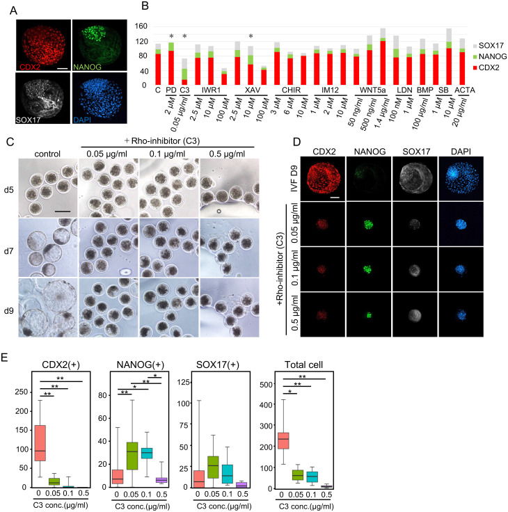
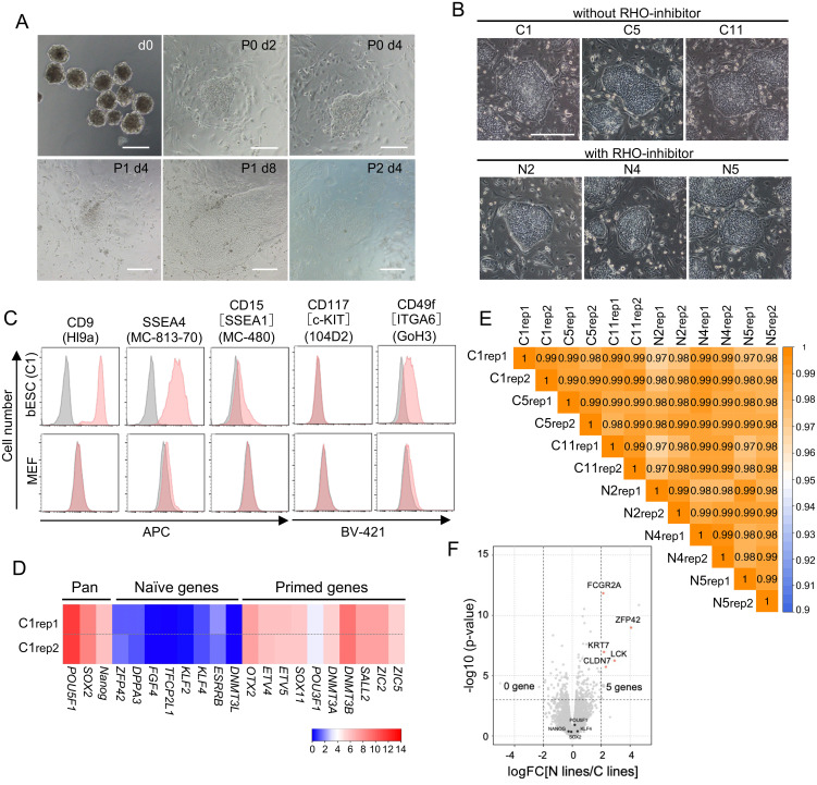
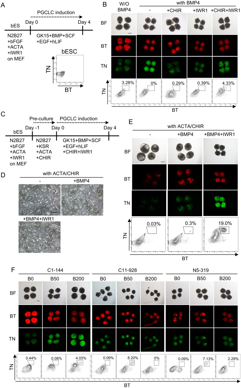
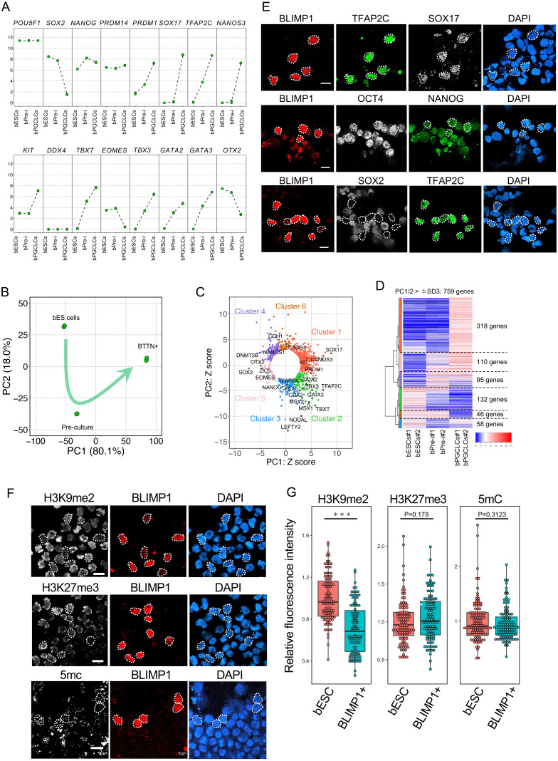
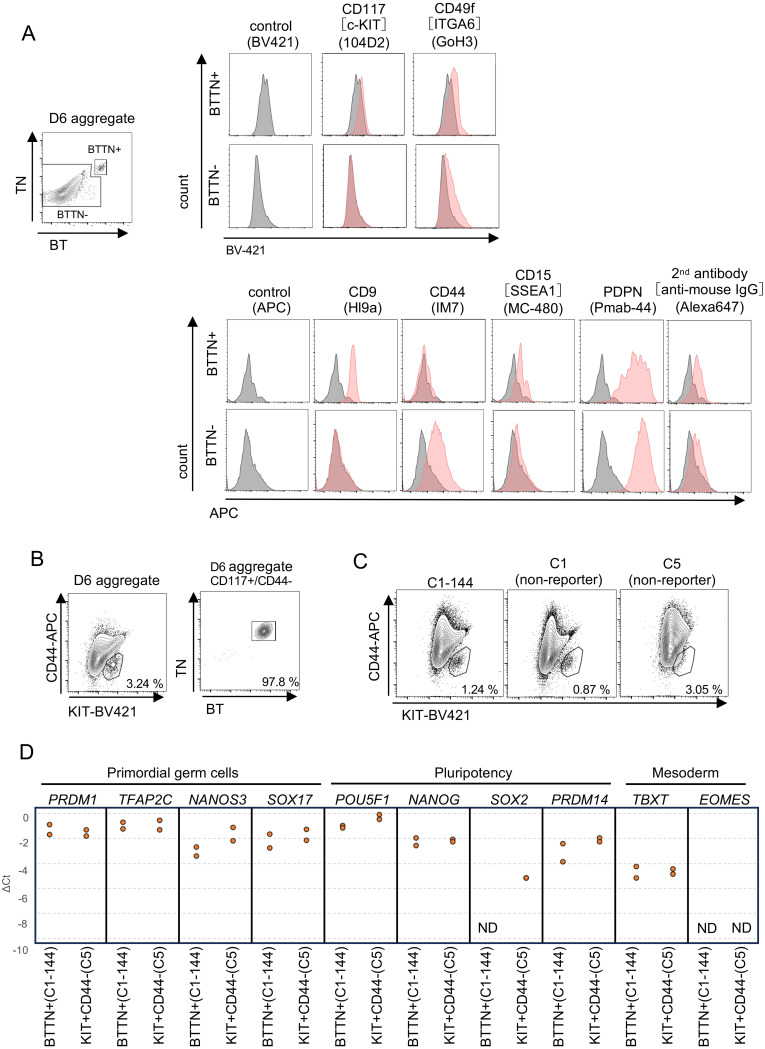
The induction of the germ cell lineage from pluripotent stem cells (in vitro gametogenesis) will help understand the mechanisms underlying germ cell differentiation and provide an alternative source of gametes for reproduction. This technology is especially important for cattle, which are among the most important livestock species for milk and meat production. Here, we developed a new method for robust induction of primordial germ cell-like cells (PGCLCs) from newly established bovine embryonic stem (bES) cells. First, we refined the pluripotent culture conditions for pre-implantation embryos and ES cells. Inhibition of RHO increased the number of epiblast cells in the pre-implantation embryos and dramatically improved the efficiency of ES cell establishment. We then determined suitable culture conditions for PGCLC differentiation using bES cells harboring BLIMP1-tdTomato and TFAP2C-mNeonGreen (BTTN) reporter constructs. After a 24-h culture with bone morphogenetic protein 4 (BMP4), followed by three-dimensional culture with BMP4 and a chemical agonist and WNT signaling chemical antagonist, bES cells became positive for the reporters. A set of primordial germ cells (PGC) marker genes, including PRDM1/BLIMP1, TFAP2C, SOX17, and NANOS3, were expressed in BTTN-positive cells. These bovine PGCLCs (bPGCLCs) were isolated as KIT/CD117-positive and CD44-negative cell populations. We anticipate that this method for the efficient establishment of bES cells and induction of PGCLCs will be useful for stem cell-based reproductive technologies in cattle.

Keywords: Blastocyst; Pluripotent stem cells; Primordial germ cells.


Conflict of interest statement

We declare that there are no conflicts of interest that could be perceived as prejudicing the impartiality of the reported research.

Figures

Fig. 1.
Fig. 1.
In vitro culture of bovine embryos with RHO inhibitor. (A) Immunofluorescent images of D9 bovine blastocysts stained with CDX2, NANOG, and SOX17 antibodies. Scale bar: 200 µm. (B) Number of CDX2-, NANOG-, and SOX17-positive cells after culturing bovine embryos with the respective reagents. The abbreviations are as follows: C, Control (n = 118); PD, PD039521 2 μM (n = 40); C3, C3 (Rhoi) 0.05 ng/ml (n = 18); IWR1 2.5 μM (n = 30), 10 μM (n = 31), and 100 μM (n = 6); XAV, XAV939 2.5 μM (n = 31), 10 μM (n = 44), and 100 μM (n = 8); CHIR, CHIR99021 3 μM (n = 54), 6 μM (n = 31), and 15 μM (n = 32); IM12, IM12 (GSK3-i) 1 μM (n = 73), 2 μM (n = 27), and 10 μM (n = 30); WNT5a, WNT5a 50 ng/ml (n = 21), 500 ng/ml (n = 41), and 1.4 μg/ml (n = 8); LDN, LDN193189 100 nM (n = 5) and 1 μM (n = 8); BMP, BMP4 100 ng/ml (n = 7); SB43, SB431542 1 μM (n = 15) and 10 μM (n = 9); and ACTA, Activin A 20 ng/ml (n = 7). Asterisks indicate the tests with significantly more NANOG-positive cells than control (P < 0.05). (C) Immunofluorescent images of D5-9 bovine embryos cultured with several concentrations of RHO-inhibitors. Scale bar: 200 µm. (D) Immunofluorescent image of bovine embryos cultured with C3. Scale bar: 200 µm. (E) Number of cells positive for CDX2, NANOG or SOX17 in bovine D9 embryos cultured with C3 at the indicated concentration of 0 μg/ml (n = 17), 0.05 μg/ml (n = 18), 0.1 μg/ml (n = 17), and 0.5 μg/ml (n = 18). * P < 0.05, ** P < 0.01 by Turkey-Kramer test.
Fig. 2.
Fig. 2.
bES cell establishment. (A) Culture of bovine inner cell masses and the procedure for establishment of ES cells. Scale bar: 200 µm. (B) Colony morphology of bES cells established without (C1, C5, C11) or with C3 (N2, N4, N5). Scale bar: 200 µm. (C) FACS analysis of surface marker protein expression in bES cells and MEF. Red and grey histograms show the cells with and without antibody, respectively. (D) Expression of pluripotency-associated gene expression in bES cells. Two biologically independent samples were used for this analysis. (E) Pluripotency status of the established ES cell lines. Two biologically independent samples of each bESs cell were used for this analysis. The heatmap shows correlation coefficients of gene expression for pluripotency status. (F) Volcano plot comparing the gene expression of bES cells established without (all C lines) or with C3 (all N lines).
Fig. 3.
Fig. 3.
bPGCLC induction. (A) Scheme for the direct induction of bPGCLCs from bES cells. (B) PGCLC induction with a WNT agonist plus WNT antagonist. Shown are bright-field (BF), fluorescence images and FACS analysis of D4 aggregates of bES cells. BT, BLIMP1-tdTomato. TN, TFAP2C-mNeonGreen. Scale bar: 200 µm. (C) Scheme for the induction of bPGCLCs under the culture conditions indicated. (D) bES cells after 24 h of incubation under the conditions indicated. (E) PGCLC induction with BMP4 and a WNT antagonist. Shown are bright-field (BF) and fluorescence images and FACS patterns of D4 aggregates of bES cells after 24 h of pre-incubation. BT, BLIMP1-tdTomato. TN, TFAP2C-mNeonGreen. Scale bar: 200 µm. (F) PGCLC induction from various cell lines. C1-144 yielded the most BTTN-positive cells when the BMP4 concentration was 200 ng/ml, while C11-928 and N5-319 were best when the BMP4 concentration was 50 ng/ml. The concentration of CHIR was 6 µM in the all experiments. FACS analyses were done on day 4 of PGCLC induction. BT, BLIMP1-tdTomato. TN, TFAP2C-mNeonGreen. Scale bar: 200 µm.
Fig. 4.
Fig. 4.
Characteristic analysis of bovine BTTN-positive cells. (A) Gene expression dynamics during PGCLC differentiation. Shown are each value and the averaged values of gene expression based on transcriptome analyses. (B) PCA of transcriptomes of bES cell derivatives. (C) Scatterplot of the z scores of the genes for the PC1 and PC2. Genes with a radius of > 3 standard deviations (SDs) (759 genes) are shown. (D) Heatmap representation of the expression dynamics of the 759 genes (SD > 3, I) highly contributing to PC1 or PC2. Unsupervised hierarchical clustering analysis showed that the expression dynamics can be subdivided into six clusters. (E) Fluorescence immunostaining image of sorted bovine BTTN-positive cells. BTTN-negative cells are bES cells. BTTN-positive cells co-express SOX17, OCT4, and NANOG, but not SOX2. Scale bar: 20 µm. (F) Fluorescence immunostaining images of epigenomic markers in sorted bovine BTTN-positive cells. BTTN- negative cells are bES cells. Scale bar: 20 µm. (G) Quantification of the epigenetic modifications in BLIMP1-positive cells. The plots show the values relative to the averaged value in BLIMP1-negative bES cells. *** P < 0.001 by Student’s t-test.
Fig. 5.
Fig. 5.
Separation of bPGCLCs by surface markers. (A) FACS analysis of surface marker protein expression in BTTN-positive and -negative cells in D6 aggregate. Red and grey histograms show the cells with antibody and without antibody, respectively. BT, BLIMP1-tdTomato. TN, TFAP2C-mNeonGreen. (B) Separation of induced PGCLCs (C1-144 ES cell) by surface markers KIT/CD117 and CD44. BT, BLIMP1-tdTomato. TN, TFAP2C-mNeonGreen. (C) Separation of PGCLCs derived from non-reporter bES cells by surface markers. (D) Comparison of gene expression in BTTN+ cells and KIT+/CD44– cells. Shown are ΔCt values determined by qPCR analysis.

Similar articles

Cited by

References

    1. Saitou M, Hayashi K. Mammalian in vitro gametogenesis. Science 2021; 374: eaaz6830. - PubMed
    1. Shono M, Kishimoto K, Hikabe O, Hayashi M, Semi K, Takashima Y, Sasaki E, Kato K, Hayashi K. Induction of primordial germ cell-like cells from common marmoset embryonic stem cells by inhibition of WNT and retinoic acid signaling. Sci Rep 2023; 13: 3186. - PMC - PubMed
    1. Oikawa M, Kobayashi H, Sanbo M, Mizuno N, Iwatsuki K, Takashima T, Yamauchi K, Yoshida F, Yamamoto T, Shinohara T, Nakauchi H, Kurimoto K, Hirabayashi M, Kobayashi T. Functional primordial germ cell-like cells from pluripotent stem cells in rats. Science 2022; 376: 176–179. - PubMed
    1. Hayashi M, Zywitza V, Naitou Y, Hamazaki N, Goeritz F, Hermes R, Holtze S, Lazzari G, Galli C, Stejskal J, Diecke S, Hildebrandt TB, Hayashi K. Robust induction of primordial germ cells of white rhinoceros on the brink of extinction. Sci Adv 2022; 8: eabp9683. - PMC - PubMed
    1. Kobayashi T, Castillo-Venzor A, Penfold CA, Morgan M, Mizuno N, Tang WWC, Osada Y, Hirao M, Yoshida F, Sato H, Nakauchi H, Hirabayashi M, Surani MA. Tracing the emergence of primordial germ cells from bilaminar disc rabbit embryos and pluripotent stem cells. Cell Rep 2021; 37: 109812. - PubMed