. 2024 Feb 14;11(2):ENEURO.0444-22.2023.
doi: 10.1523/ENEURO.0444-22.2023. Print 2024 Feb.

Chronic Treatment with Serotonin Selective Reuptake Inhibitors Does Not Affect Regrowth of Serotonin Axons Following Amphetamine Injury in the Mouse Forebrain

Affiliations

Chronic Treatment with Serotonin Selective Reuptake Inhibitors Does Not Affect Regrowth of Serotonin Axons Following Amphetamine Injury in the Mouse Forebrain

Haley N Janowitz et al. eNeuro. .

Abstract

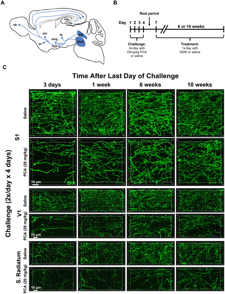
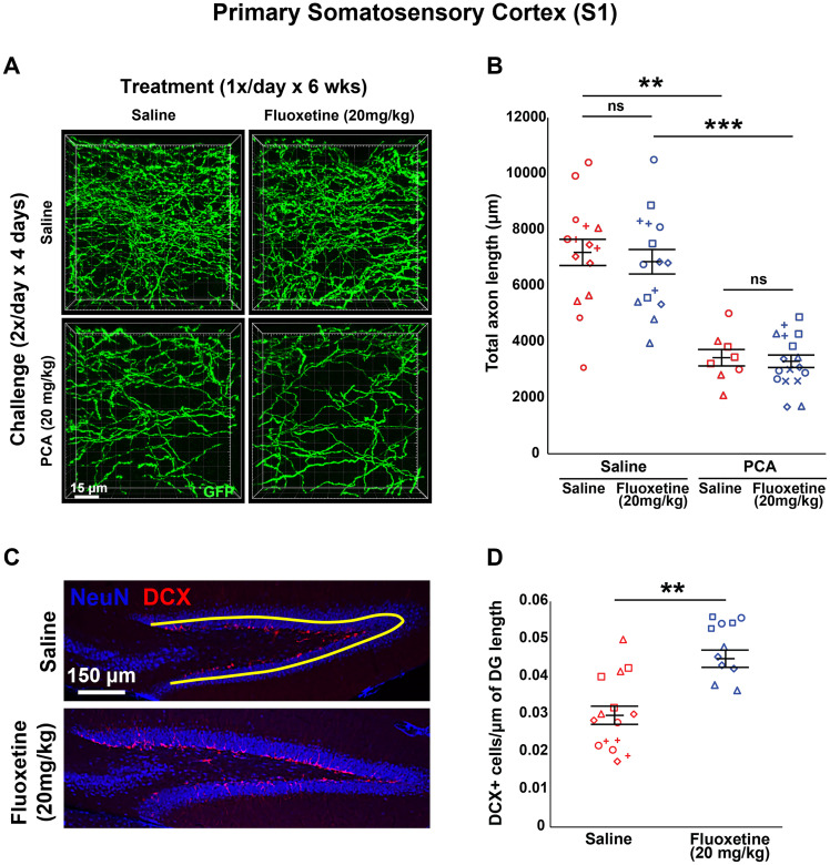
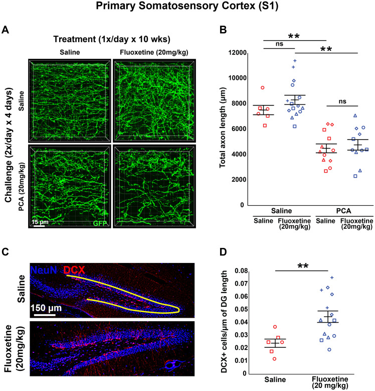
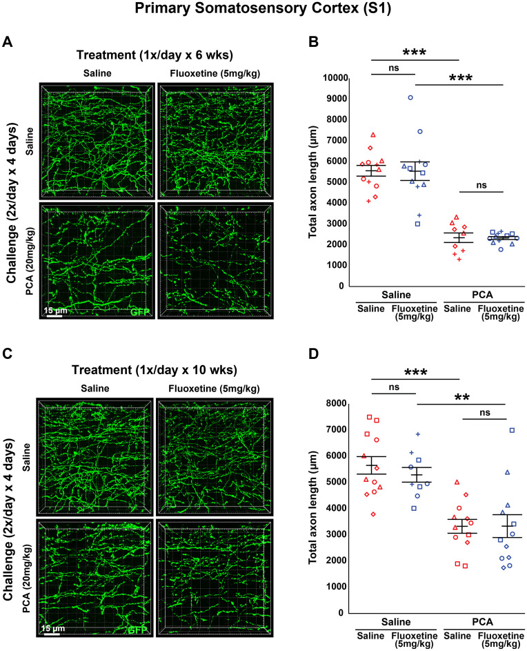
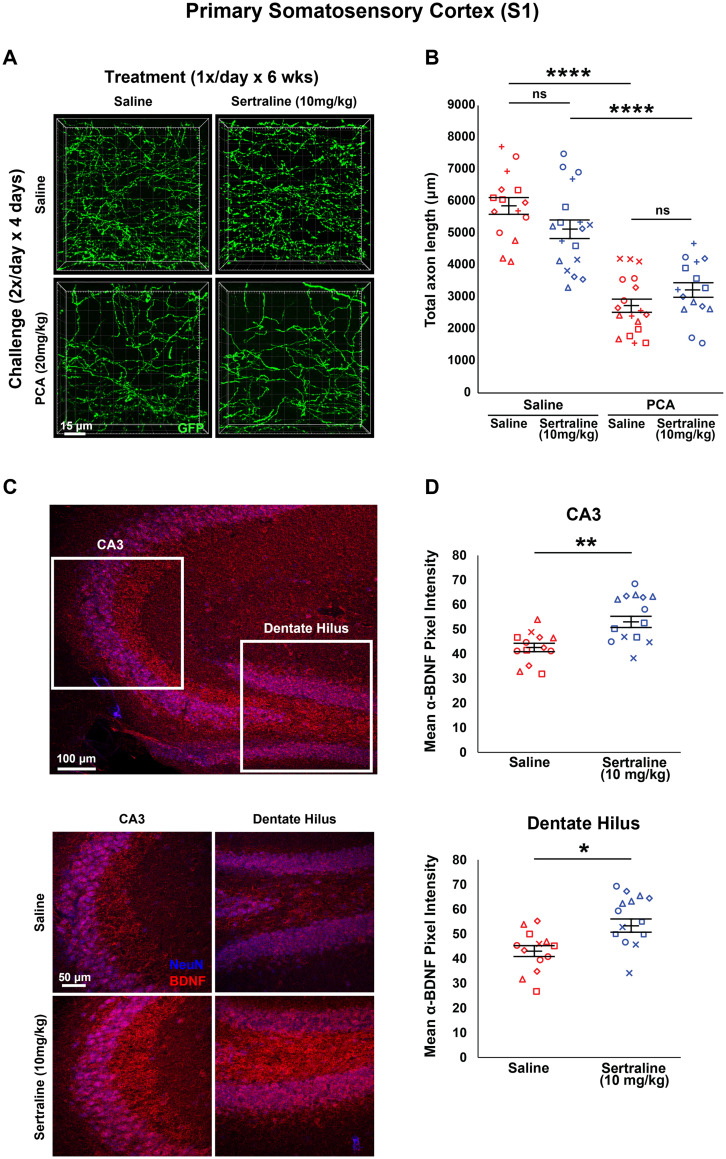
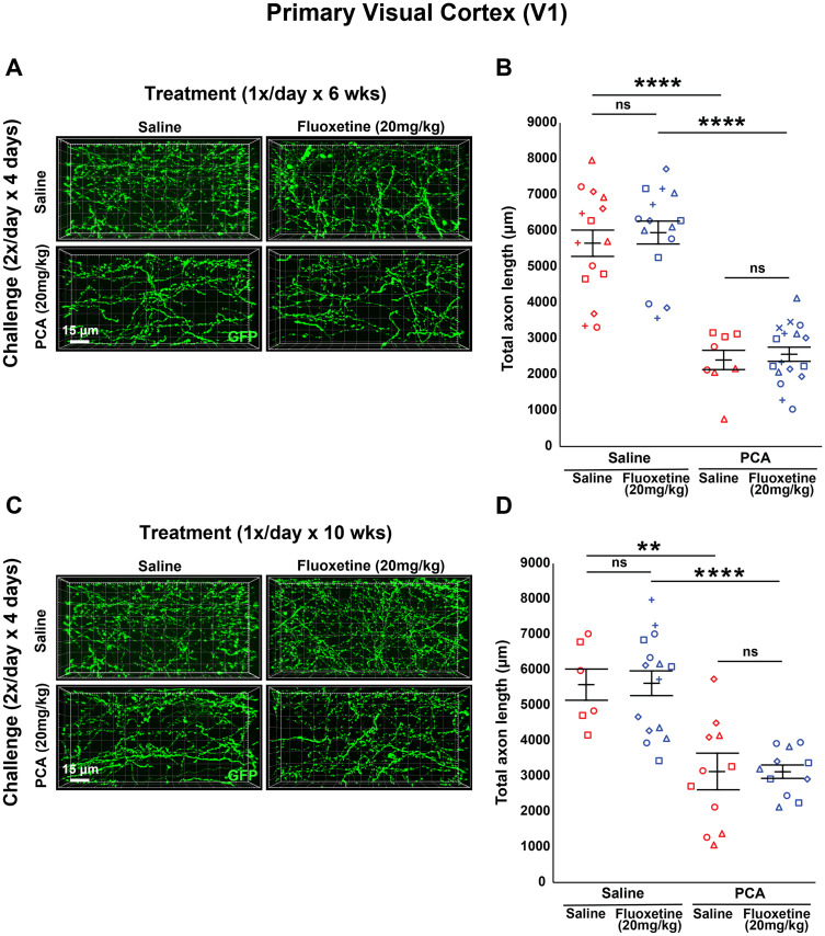
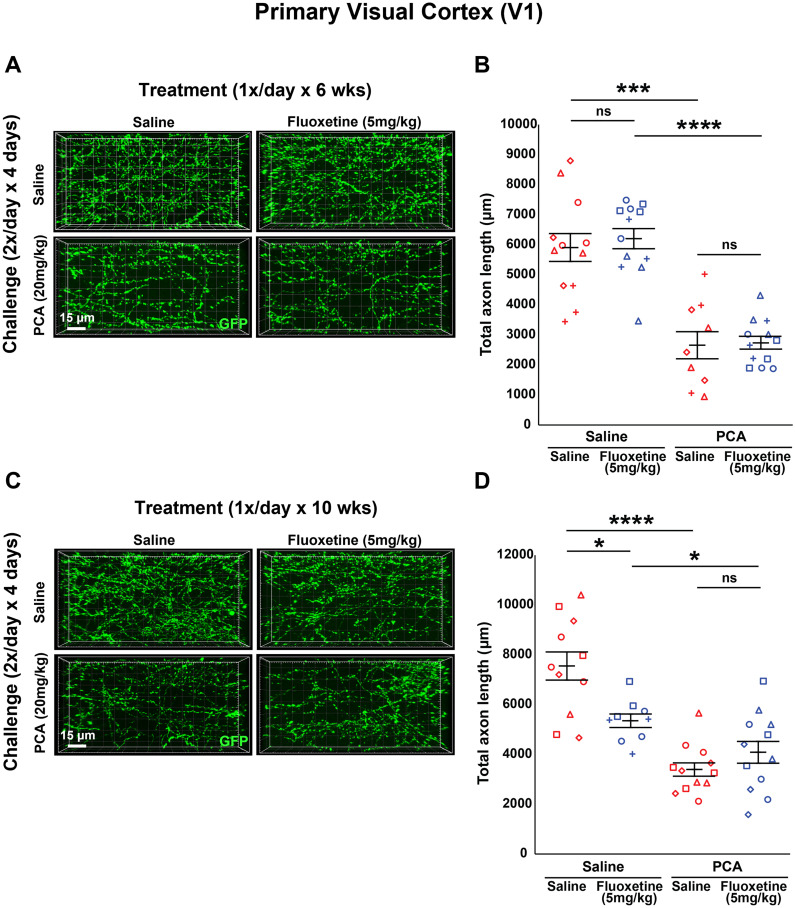
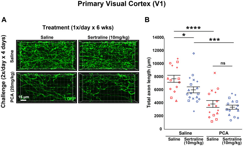
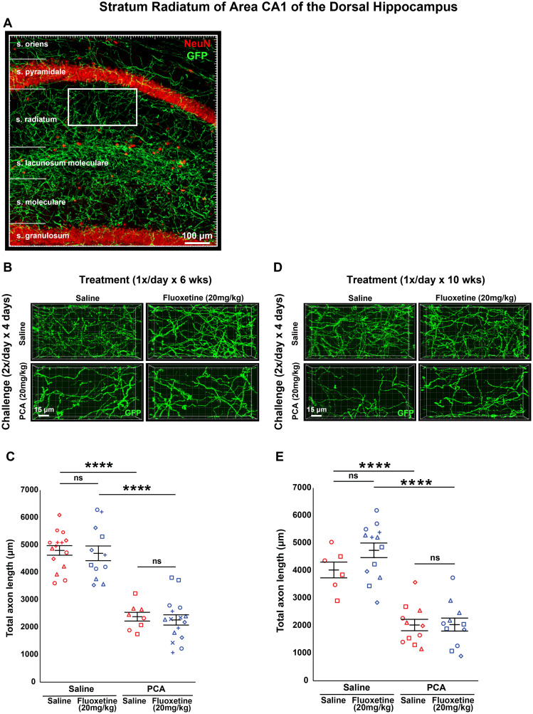
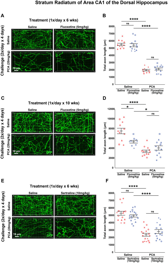
A current hypothesis to explain the limited recovery following brain and spinal cord trauma stems from the dogma that neurons in the mammalian central nervous system lack the ability to regenerate their axons after injury. Serotonin (5-HT) neurons in the adult brain are a notable exception in that they can slowly regrow their axons following chemical or mechanical lesions. This process of regrowth occurs without intervention over several months and results in anatomical recovery that approximates the preinjured state. During development, serotonin is a trophic factor, playing a role in both cell survival and axon growth. Additionally, some studies have shown that stroke patients treated after injury with serotonin selective reuptake inhibitors (SSRIs) appeared to have improved recovery. To test the hypothesis that serotonin can influence the regrowth of 5-HT axons, mice received a high dose of para-chloroamphetamine (PCA) to induce widespread retrograde degeneration of 5-HT axons. Then, after a short rest period to avoid any interaction with the acute injury phase, SSRIs were administered daily for 6 or 10 weeks. Using immunohistochemistry in 5-HT transporter-GFP BAC transgenic mice, we determined that while PCA led to a rapid initial decrease in total 5-HT axon length in the somatosensory cortex, visual cortex, or area CA1 of the hippocampus, treatment with either fluoxetine or sertraline (two different SSRIs) did not affect the recovery of axon length. These results suggest that chronic SSRI treatment does not affect the regrowth of 5-HT axons and argue against SSRIs as a potential therapy following brain injury.

Keywords: brain injury; fluoxetine; regeneration; serotonin; serotonin selective reuptake inhibitors; sertraline.


Conflict of interest statement

The authors declare no competing financial interests.

Figures

Figure 1.
Figure 1.
5-HT axons innervating the adult mouse forebrain regrow following a chemical lesion with PCA. A, Schematic diagram of a sagittal section of an adult mouse brain shows some of the diverse projection patterns of the anterior group of 5-HT neurons. Abbreviations: DR, dorsal raphe; MnR, median raphe; th, thalamus; amg,amygdala; hip, hippocampus; hyp, hypothalamus; stri, striatum; ac, nucleus accumbens; and ob, olfactory bulb. B, The experimental timeline shows a PCA/saline challenge period followed, after a 2-day-long rest period, by an SSRI/saline treatment period. C, Representative confocal stack images of anti-GFP fluorescence in layer 1 of primary somatosensory cortex (S1), layer 1 of primary visual cortex (V1), and stratum radiatum of area CA1 of the dorsal hippocampus in sagittal slices from Slc6a4-GFP mice treated with either PCA or saline for 4 d and then allowed to recover for 3 d, 1 week, 6 weeks, or 10 weeks.
Figure 2.
Figure 2.
Daily injections of 20 mg/kg fluoxetine for 6 weeks following injury with PCA does not affect 5-HT axon regrowth in layer 1 of somatosensory cortex (S1). A, B, Slc6a4-GFP mice were initially treated with either PCA or saline for 4 d, allowed 2 d of recovery, and then followed by daily injections of either fluoxetine or saline for 6 weeks. A, Representative images of each combination of conditions. B, Total axon length was determined via histological analysis. Images were acquired using confocal microscopy and layer 1 of area S1 was analyzed in 3D with IMARIS software. Each animal had three technical replicates to account for within-animal variability. Each animal is represented by a different plot symbol. Mean and SE are plotted. Group data were analyzed using a mixed effect repeated-measures ANOVA. N = 9 males and 10 females. F(1,30) = 52.8, p < 0.0001 for the effect of PCA in fluoxetine treated animals. F(1,21) = 28.4, p < 0.0001 for the effect of PCA in saline-treated animals. F(1,23) = 0.106, p = 0.748 for the effect of fluoxetine in PCA treated animals. F(1,28) = 0.257, p = 0.616 for the effect of fluoxetine in saline-treated animals. C, D, Total number of DCX immuno-positive cells in the DG were measured using histological analysis. C, Representative images of each condition with an example tracing of DG length in yellow. D, Images were acquired using widefield microscopy and analyzed with IMARIS. The total number of DCX immune-positive cells was normalized to the length of the DG to account for small variations in the medial–lateral location of each sagittal slice. Each animal had three technical replicates to account for within-animal variability. Each animal is represented by a different plot symbol. Mean and SE are plotted. Group data were analyzed using a mixed effect repeated-measures ANOVA. F(1,27) = 18.648, p < 0.001.
Figure 3.
Figure 3.
Daily injections of 20 mg/kg fluoxetine for 10 weeks following injury with PCA does not affect 5-HT axon regrowth in layer 1 of primary somatosensory cortex. A, B, Slc6a4-GFP mice were initially treated with either PCA or saline for 4 d, allowed 2 d of recovery, and then followed by daily injections of either fluoxetine or saline for 10 weeks. A, Representative images of each combination of conditions. B, Each animal is represented by a different plot symbol. Mean and SE are plotted. Group data were analyzed using a mixed effect repeated-measures ANOVA. N = 3 males and 12 females. F(1,24) = 37.5, p < 0.0001 for the effect of PCA in fluoxetine treated animals. F(1,16) = 25.7, p < 0.001 for the effect of PCA in saline-treated animals. F(1,21) = 0.236, p = 0.632 for the effect of fluoxetine in PCA-treated animals. F(1,19) = 1.419, p = 0.237 for the effect of fluoxetine in saline-treated animals. C, D, Total number of DCX immuno-positive cells in the DG were measured using histological analysis. C, Representative images of each condition with an example tracing of DG length in yellow. D, Each animal is represented by a different plot symbol. Mean and SE are plotted. Group data were analyzed using a mixed effect repeated-measures ANOVA. F(1,18) = 7.447, p < 0.05.
Figure 4.
Figure 4.
Daily injections of 5 mg/kg fluoxetine following injury with PCA do not affect 5-HT axon regrowth in layer 1 of primary somatosensory cortex. A, B, Slc6a4-GFP mice were initially treated with either PCA or saline for 4 d, allowed 2 d of recovery, and then followed by daily injections of either fluoxetine or saline for 6 weeks. A, Representative images of each combination of conditions. B, Each animal is represented by a different plot symbol. Mean and SE are plotted. Group data were analyzed using a mixed effect repeated-measures ANOVA. N = 5 males and 10 females. F(1,22) = 37.539, p < 0.0001 for the effect of PCA in fluoxetine-treated animals. F(1,19) = 73.835, p < 0.0001 for the effect of PCA in saline-treated animals. F(1,19) = 0.004, p = 0.953 for the effect of fluoxetine in PCA-treated animals. F(1,22) = 0.002, p = 0.969 for the effect of fluoxetine in saline-treated animals. C, D, Slc6a4-GFP mice were initially treated with either PCA or saline for 4 d, allowed 2 d of recovery, and then followed by daily injections of either fluoxetine or saline for 10 weeks. C, Representative images of each combination of conditions. D, Each animal is represented by a different plot symbol. Mean and SE are plotted. Group data were analyzed using a mixed effect repeated-measures ANOVA. N = 6 males and 9 females. F(1,19) = 11.171, p < 0.01 for the effect of PCA in fluoxetine treated animals. F(1,22) = 27.48, p < 0.0001 for the effect of PCA in saline-treated animals. F(1,22) = 0.000135, p = 0.991 for the effect of fluoxetine in PCA-treated animals. F(1,19) = 0.574, p = 0.458 for the effect of fluoxetine in saline-treated animals.
Figure 5.
Figure 5.
Daily injections of 10 mg/kg sertraline following injury with PCA do not affect 5-HT axon regrowth in layer 1 of primary somatosensory cortex. A, B, Slc6a4-GFP mice were initially treated with either PCA or saline for 4 d, allowed 2 d of recovery, and then followed by daily injections of either sertraline or saline for 6 weeks. A, Representative images of each combination of conditions. B, Each animal is represented by a different plot symbol. Mean and SE are plotted. Group data were analyzed using a mixed effect repeated-measures ANOVA. N = 8 males and 14 females. F(1,31) = 23.361, p < 0.0001 for the effect of PCA in fluoxetine-treated animals. F(1,31) = 84.883, p < 0.0001 for the effect of PCA in saline-treated animals. F(1,31) = 2.401, p = 0.131 for the effect of fluoxetine in PCA-treated animals. F(1,31) = 3.151, p = 0.086 for the effect of fluoxetine in saline-treated animals. C, D, Intensity of BDNF immunoreactivity in the CA3 region of the hippocampus and the hilus of the dentate gyrus was measured using histological analysis. C, Representative images of each condition in each brain region. Location of each region outlined in a lower magnification image. D, Images were acquired using confocal microscopy and the mean pixel intensity of the anti-BDNF channel was measured using ImageJ software. Each animal had three technical replicates to account for within-animal variability. Each animal is represented by a different plot symbol. Mean and SE are plotted. Group data were analyzed using a mixed effect repeated-measures ANOVA. F(1,18) = 7.447, p < 0.05. F(1,28) = 11.302, p < 0.01 for the effect on BDNF intensity in CA3. F(1,28) = 7.555, p < 0.05 for the effect on BDNF intensity in the hilus and arms of the dentate gyrus.
Figure 6.
Figure 6.
Daily injections of 20 mg/kg fluoxetine following injury with PCA do not affect 5-HT axon regrowth in layer 1 of primary visual cortex (V1). A, B, Slc6a4-GFP mice were initially treated with either PCA or saline for 4 d, allowed 2 d of recovery, and then followed by daily injections of either fluoxetine or saline for 6 weeks. A, Representative images of each combination of conditions. B, Total axon length was determined via histological analysis. Images were acquired using confocal microscopy and layer 1 of area V1 was analyzed in 3D with IMARIS software. Each animal had three technical replicates to account for within-animal variability. Each animal is represented by a different plot symbol. Mean and SE are plotted. Group data were analyzed using a mixed effect repeated-measures ANOVA. N = 9 males and 10 females. F(1,29) = 74.3, p < 0.0001 for the effect of PCA in fluoxetine-treated animals. F(1,22) = 36, p < 0.0001 for the effect of PCA in saline-treated animals. F(1,23) = 0.202, p = 0.657 for the effect of fluoxetine in PCA-treated animals. F(1,28) = 0.218, p = 0.644 for the effect of fluoxetine in saline-treated animals. C, D, Slc6a4-GFP mice were initially treated with either PCA or saline for 4 d, allowed 2 d of recovery, and then followed by daily injections of either fluoxetine or saline for 10 weeks. C, Representative images of each combination of conditions. D, Each animal is represented by a different plot symbol. Mean and SE are plotted. Group data were analyzed using a mixed effect repeated-measures ANOVA. N = 3 males and 12 females. F(1,24) = 30.1, p < 0.0001 for the effect of PCA in fluoxetine-treated animals. F(1,15) = 12.7, p < 0.01 for the effect of PCA in saline-treated animals. F(1,20) = 0.028, p = 0.869 for the effect of fluoxetine in PCA-treated animals. F(1,19) = 0.003, p = 0.954 for the effect of fluoxetine in saline-treated animals.
Figure 7.
Figure 7.
Daily injections of 5 mg/kg fluoxetine following injury with PCA do not affect 5-HT axon regrowth in layer 1 of primary visual cortex. A, B, Slc6a4-GFP mice were initially treated with either PCA or saline for 4 d, allowed 2 d of recovery, and then followed by daily injections of either fluoxetine or saline for 6 weeks. A, Representative images of each combination of conditions. B, Each animal is represented by a different plot symbol. Mean and SE are plotted. Group data were analyzed using a mixed effect repeated-measures ANOVA. N = 5 males and 10 females. F(1,22) = 70.1, p < 0.0001 for the effect of PCA in fluoxetine-treated animals. F(1,19) = 21.8, p < 0.001 for the effect of PCA in saline-treated animals. F(1,19) = 0.029, p = 0.866 for the effect of fluoxetine in PCA-treated animals. F(1,22) = 0.247, p = 0.624 for the effect of fluoxetine in saline-treated animals. C, D, Slc6a4-GFP mice were initially treated with either PCA or saline for 4 d, allowed 2 d of recovery, and then followed by daily injections of either fluoxetine or saline for 10 weeks. C, Representative images of each combination of conditions. D, Each animal is represented by a different plot symbol. Mean and SE are plotted. Group data were analyzed using a mixed effect repeated-measures ANOVA. N = 6 males and 9 females. F(1,19) = 4.66, p < 0.05 for the effect of PCA in fluoxetine-treated animals. F(1,21) = 42.4, p < 0.0001 for the effect of PCA in saline-treated animals. F(1,22) = 1.66, p = 0.211 for the effect of fluoxetine in PCA-treated animals. F(1,18) = 9.64, p = 0.006 for the effect of fluoxetine in saline-treated animals.
Figure 8.
Figure 8.
Daily injections of 10 mg/kg sertraline following injury with PCA do not affect 5-HT axon regrowth in layer 1 of primary visual cortex. A, B, Slc6a4-GFP mice were initially treated with either PCA or saline for 4 d, allowed 2 d of recovery, and then followed by daily injections of either sertraline or saline for 6 weeks. A, Representative images of each combination of conditions. B, Each animal is represented by a different plot symbol. Mean and SE are plotted. Group data were analyzed using a mixed effect repeated-measures ANOVA. N = 8 males and 14 females. F(1,31) = 19.6, p < 0.001 for the effect of PCA in sertraline-treated animals. F(1,27) = 24.4, p < 0.0001 for the effect of PCA in saline-treated animals. F(1,28) = 0.592, p = 0.448 for the effect of sertraline in PCA-treated animals. F(1,30) = 5.42, p = 0.027 for the effect of sertraline in saline-treated animals.
Figure 9.
Figure 9.
Daily injections of 20 mg/kg fluoxetine following injury with PCA do not affect 5-HT axon regrowth in area CA1 of the dorsal hippocampus. A, Representative tiled confocal stack image of anti-GFP and anti-NeuN (neuronal nuclei marker) fluorescence of area CA1, with each layer indicated. An exemplary region of interest within the more dorsal portion of stratum radiatum is denoted by the white box. B, C, Slc6a4-GFP mice were initially treated with either PCA or saline for 4 d, allowed 2 d of recovery, and then followed by daily injections of either fluoxetine or saline for 6 weeks. B, Representative images of each combination of conditions. C, Total axon length was determined via histological analysis. Images were acquired using confocal microscopy and the stratum radiatum of the CA1 region of the hippocampus was analyzed in 3D with IMARIS software. Each animal had three technical replicates to account for within-animal variability. Each animal is represented by a different plot symbol. Mean and SE are plotted. Group data were analyzed using a mixed effect repeated-measures ANOVA. N = 9 males and 10 females. F(1,26) = 54.5, p < 0.0001 for the effect of PCA in fluoxetine-treated animals. F(1,21) = 75.4, p < 0.0001 for the effect of PCA in saline-treated animals. F(1,22) = 0.152, p = 0.700 for the effect of fluoxetine in PCA-treated animals. F(1,25) = 0.111, p = 0.742 for the effect of fluoxetine in saline-treated animals. D, E, Slc6a4-GFP mice were initially treated with either PCA or saline for 4 d, allowed 2 d of recovery, and then followed by daily injections of either fluoxetine or saline for 10 weeks. D, Representative images of each combination of conditions. E, Each animal is represented by a different plot symbol. Mean and SE are plotted. Group data were analyzed using a mixed effect repeated-measures ANOVA. N = 3 males and 12 females. F(1,22) = 51.2, p < 0.0001 for the effect of PCA in fluoxetine-treated animals. F(1,15) = 28.5, p < 0.0001 for the effect of PCA in saline-treated animals. F(1,20) = 0.002, p = 0.962 for the effect of fluoxetine in PCA-treated animals. F(1,17) = 2.39, p = 0.141 for the effect of fluoxetine in saline-treated animals.
Figure 10.
Figure 10.
Daily injections of 5 mg/kg fluoxetine or 10 mg/kg sertraline following injury with PCA do not affect 5-HT axon regrowth in area CA1 of the dorsal hippocampus. A, B, Slc6a4-GFP mice were initially treated with either PCA or saline for 4 d, allowed 2 d of recovery, and then followed by daily injections of either fluoxetine or saline for 6 weeks. A, Representative images of each combination of conditions. B, Each animal is represented by a different plot symbol. Mean and SE are plotted. Group data were analyzed using a mixed effect repeated-measures ANOVA. N = 5 males and 10 females. F(1,22) = 138, p < 0.0001 for the effect of PCA in fluoxetine-treated animals. F(1,18) = 257, p < 0.0001 for the effect of PCA in saline-treated animals. F(1,19) = 0.56, p = 0.463 for the effect of fluoxetine in PCA-treated animals. F(1,21) = 0.13, p = 0.722 for the effect of fluoxetine in saline-treated animals. C, E, Slc6a4-GFP mice were initially treated with either PCA or saline for 4 d, allowed 2 d of recovery, and then followed by daily injections of either fluoxetine or saline for 10 weeks. C, Representative images of each combination of conditions. D, Each animal is represented by a different plot symbol. Mean and SE are plotted. Group data were analyzed using a mixed effect repeated-measures ANOVA. N = 6 males and 9 females. F(1,19) = 41.8, p < 0.0001 for the effect of PCA in fluoxetine-treated animals. F(1,22) = 65.9, p < 0.0001 for the effect of PCA in saline-treated animals. F(1,22) = 0.088, p = 0.769 for the effect of fluoxetine in PCA-treated animals. F(1,19) = 0.083, p = 0.776 for the effect of fluoxetine in saline-treated animals. E, F, Slc6a4-GFP mice were initially treated with either PCA or saline for 4 d, allowed 2 d of recovery, and then followed by daily injections of either sertraline or saline for 6 weeks. E, Representative images of each combination of conditions. F, Each animal is represented by a different plot symbol. Mean and SE are plotted. Group data were analyzed using a mixed effect repeated-measures ANOVA. N = 8 males and 14 females. F(1,31) = 47.2, p < 0.0001 for the effect of PCA in sertraline-treated animals. F(1,30) = 73.5, p < 0.0001 for the effect of PCA in saline-treated animals. F(1,30) = 1.74, p = 0.196 for the effect of sertraline in PCA-treated animals. F(1,31) = 1.28, p = 0.266 for the effect of sertraline in saline-treated animals.

Similar articles

Cited by

References

    1. Agrawal L, Vimal SK, Shiga T (2019) Role of serotonin 4 receptor in the growth of hippocampal neurons during the embryonic development in mice. Neuropharmacology 158:107712. 10.1016/j.neuropharm.2019.107712 - DOI - PubMed
    1. Alamri F, al Shoyaib A, Syeara N, Paul A, Jayaraman S, Karamyan S, Arumugam T, Karamyan V (2021) Delayed atomoxetine or fluoxetine treatment coupled with limited voluntary running promotes motor recovery in mice after ischemic stroke. Neural Regen Res 16:1244–1251. 10.4103/1673-5374.301031 - DOI - PMC - PubMed
    1. Almeida OP, et al. (2021) Depression outcomes among patients treated with fluoxetine for stroke recovery: the AFFINITY randomized clinical trial. JAMA Neurol 78:1072–1079. 10.1001/jamaneurol.2021.2418 - DOI - PMC - PubMed
    1. Andreska T, et al. (2020) Induction of BDNF expression in layer II/III and layer V neurons of the motor cortex is essential for motor learning. J Neurosci 40:6289–6308. 10.1523/JNEUROSCI.0288-20.2020 - DOI - PMC - PubMed
    1. Bombardier CH, Fann JR, Temkin NR, Esselman PC, Barber J, Dikmen SS (2010) Rates of major depressive disorder and clinical outcomes following traumatic brain injury. JAMA 303:1938–1945. 10.1001/jama.2010.599 - DOI - PMC - PubMed

LinkOut - more resources