

Microvesicles from stored red blood cells induce P-selectin and von Willebrand factor release from endothelial cells via a protein kinase C-dependent mechanism
- PMID: 38355315
- PMCID: PMC10997436
- DOI: 10.1016/j.transci.2024.103890
Microvesicles from stored red blood cells induce P-selectin and von Willebrand factor release from endothelial cells via a protein kinase C-dependent mechanism
Abstract
Introduction: The use of packed red blood cells (pRBCs) for resuscitation is limited by the red blood cell storage lesion, a series of biochemical and physiological changes that occur during the storage and aging of blood. Microvesicles (MVs) shed from pRBCs during this process are one component of the red blood cell storage lesion and lead to acute lung injury and pulmonary vascular microthrombi. We hypothesized that MVs from stored pRBCs lead to the release of P-selectin and von Willebrand factor (vWF) from endothelial cells and that this mechanism is mediated via activation of protein kinase C (PKC) or protein kinase A (PKA).
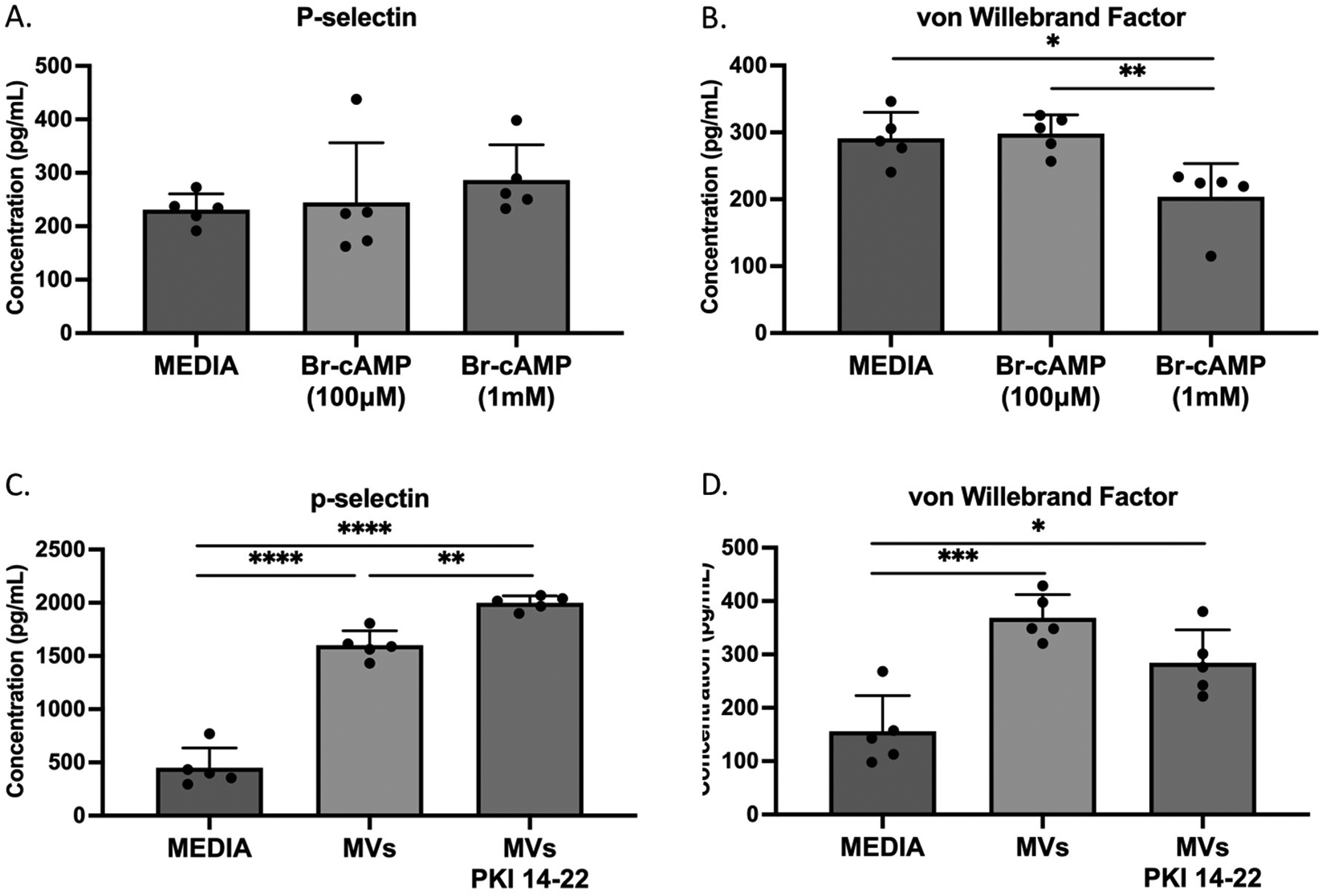
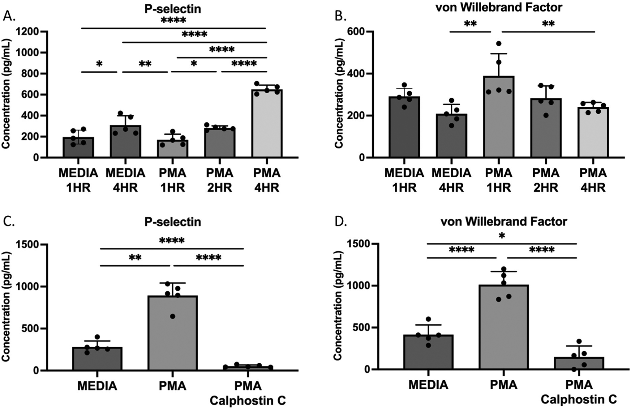
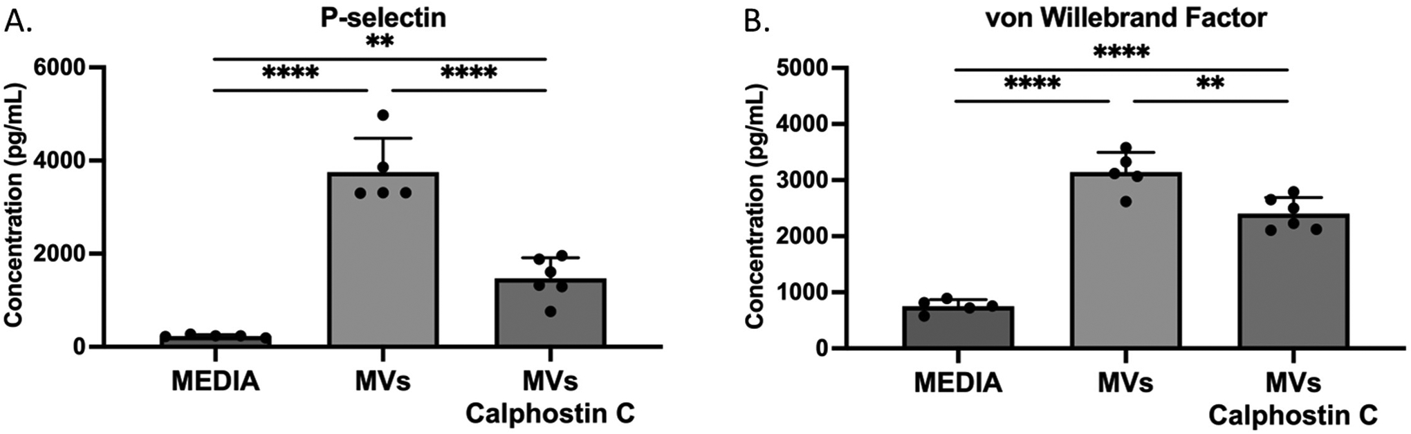
Methods: Leukoreduced, platelet-poor murine pRBCs were isolated from C57BL/6 8-12 week-old male mice via cardiac puncture, prepared via centrifugation using a Ficoll gradient, and stored for up to 14 days, the equivalent of 42 days of storage in humans. MVs were isolated from the stored pRBC units via sequential high-speed centrifugation. Murine lung endothelial cells (MLECs) were cultured and grown to confluence, then treated with MVs and either calphostin C, a PKC inhibitor (10 μg/mL), or PKI 14-22 amide, a PKA inhibitor (10 μM). The supernatant was collected after 1 h. P-selectin and vWF A2 concentrations were quantified via ELISA. Immunofluorescent staining for vWF was performed on MLECs. Statistical analysis was performed via unpaired t-test or ANOVA as indicated and reported as mean ± SD. Concentration is reported as pg/mL.
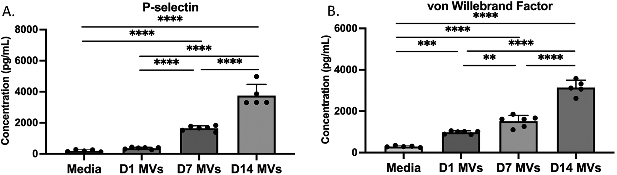
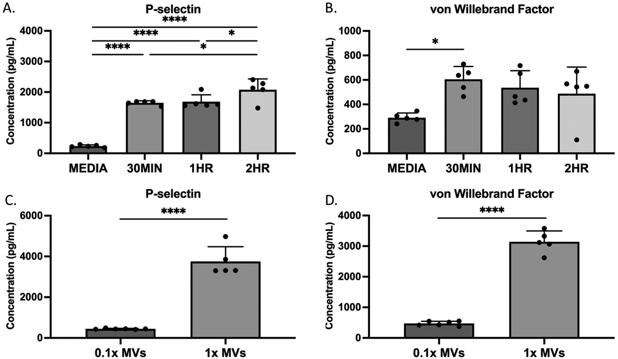
Results: MLECs treated with MVs isolated from stored pRBCs demonstrated increased release of P-selectin and vWF A2 in a dose-dependent fashion. MLECs treated with MVs prepared from stored as compared to fresh pRBCs demonstrated increased release of P-selectin (3751 ± 726 vs 359 ± 64 pg/mL, p < 0.0001) and vWF A2 (3141 ± 355 vs 977 ± 75 pg/mL, p < 0.0001) with increasing duration of storage. The treatment of MVs with calphostin C decreased the amount of P-selectin (1471 ± 444 vs 3751 ± 726 pg/mL, p < 0.0001) and VWF A2 (2401 ± 289 vs 3141 ± 355 pg/mL, p = 0.0017) released into the supernatant by MLECs compared to MVs alone. The treatment of MVs with PKI 14-22 increased the amount of P-selectin released compared to MVs alone (1999 ± 67 vs 1601 ± 135 pg/mL, p = 0.0018).
Conclusions: MVs from stored pRBCs stimulate the release of P-selectin and VWF A2 from endothelial cells. The effect of MVs increases with both dose of MVs and age of stored pRBCs from which they are formed. This mechanism is dependent on activation of PKC and inhibition of this enzyme represents a potentially significant strategy to modulate the inflammatory response to resuscitation with stored pRBCs.
Keywords: Endothelial cells; Microvesicles; P-selectin; Red blood cells; von Willebrand factor.
Copyright © 2024 Elsevier Ltd. All rights reserved.
Conflict of interest statement
Declaration of Competing Interest The authors declare that they have no conflicts of interest related to this study.
Figures
Similar articles
-
Microparticles from aged packed red blood cell units stimulate pulmonary microthrombus formation via P-selectin.Thromb Res. 2020 Jan;185:160-166. doi: 10.1016/j.thromres.2019.11.028. Epub 2019 Nov 26. Thromb Res. 2020. PMID: 31821908 Free PMC article.
-
Leukoreduction of packed red blood cells attenuates proinflammatory properties of storage-derived microvesicles.J Surg Res. 2018 Mar;223:128-135. doi: 10.1016/j.jss.2017.09.052. Epub 2017 Dec 22. J Surg Res. 2018. PMID: 29433864 Free PMC article.
-
Amitriptyline Decreases Mouse Lung Endothelial Cell Inflammatory Responses to Packed Red Blood Cell Microparticles.J Surg Res. 2024 Nov;303:429-438. doi: 10.1016/j.jss.2024.09.042. Epub 2024 Oct 18. J Surg Res. 2024. PMID: 39423737
-
Covid-19: The Rollercoaster of Fibrin(Ogen), D-Dimer, Von Willebrand Factor, P-Selectin and Their Interactions with Endothelial Cells, Platelets and Erythrocytes.Int J Mol Sci. 2020 Jul 21;21(14):5168. doi: 10.3390/ijms21145168. Int J Mol Sci. 2020. PMID: 32708334 Free PMC article. Review.
-
von Willebrand factor and inflammation.J Thromb Haemost. 2017 Jul;15(7):1285-1294. doi: 10.1111/jth.13696. J Thromb Haemost. 2017. PMID: 28671350 Review.
References
-
- Heron M, Deaths: Leading causes for 2019, in National Vital Statistics Reports. 2021, National Center for Health Statistics: Hyattsville, MD.
-
- Kauvar DS, Lefering R, and Wade CE, Impact of hemorrhage on trauma outcome: an overview of epidemiology, clinical presentations, and therapeutic considerations. J Trauma, 2006. 60(6 Suppl): p. S3–11. - PubMed
-
- Holcomb JB, et al., Damage control resuscitation: directly addressing the early coagulopathy of trauma. J Trauma, 2007. 62(2): p. 307–10. - PubMed
-
- Tanhehco YC, Red Blood Cell Transfusion. Clin Lab Med, 2021. 41(4): p. 611–619. - PubMed
MeSH terms
Substances
Grants and funding
LinkOut - more resources
Full Text Sources
Molecular Biology Databases
Miscellaneous
