. 2024 Feb 14;24(1):73.
doi: 10.1186/s12935-024-03254-z.

PLK1 phosphorylates RhoGDI1 and promotes cancer cell migration and invasion

Affiliations

PLK1 phosphorylates RhoGDI1 and promotes cancer cell migration and invasion

Jeewon Lim et al. Cancer Cell Int. .

Abstract

Background: Rho guanine nucleotide dissociation inhibitor 1 (RhoGDI1) plays an important role in diverse cellular processes by regulating Rho guanosine triphosphate (GTP)ases activity. RhoGDI1 phosphorylation regulates the spatiotemporal activation of Rho GTPases during cell migration. In this study, we identified polo-like kinase 1 (PLK1) as a novel kinase of RhoGDI1 and investigated the molecular mechanism by which the interaction between RhoGDI1 and PLK1 regulates cancer cell migration.

Methods: Immunoprecipitation, GST pull-down assay, and proximity ligation assay (PLA) were performed to analyze the interaction between RhoGDI1 and PLK1. In vitro kinase assay and immunoprecipitation were performed with Phospho-(Ser/Thr) antibody. We evaluated RhoA activation using RhoGTPases activity assay. Cell migration and invasion were analyzed by transwell assays.

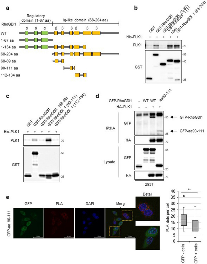
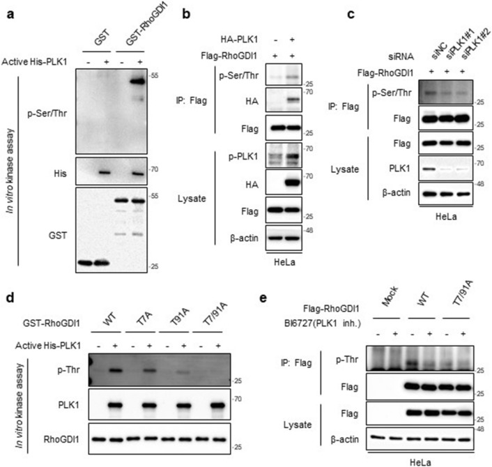
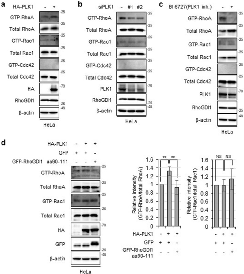
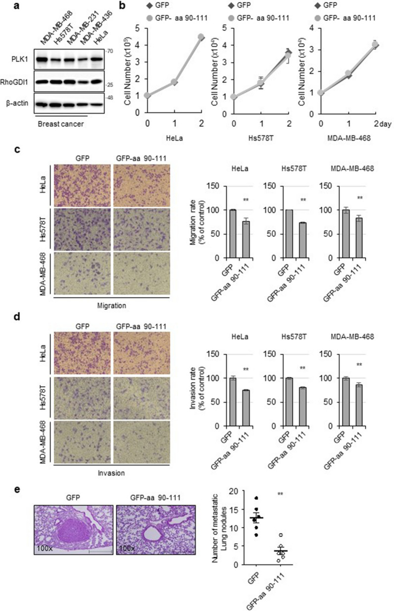
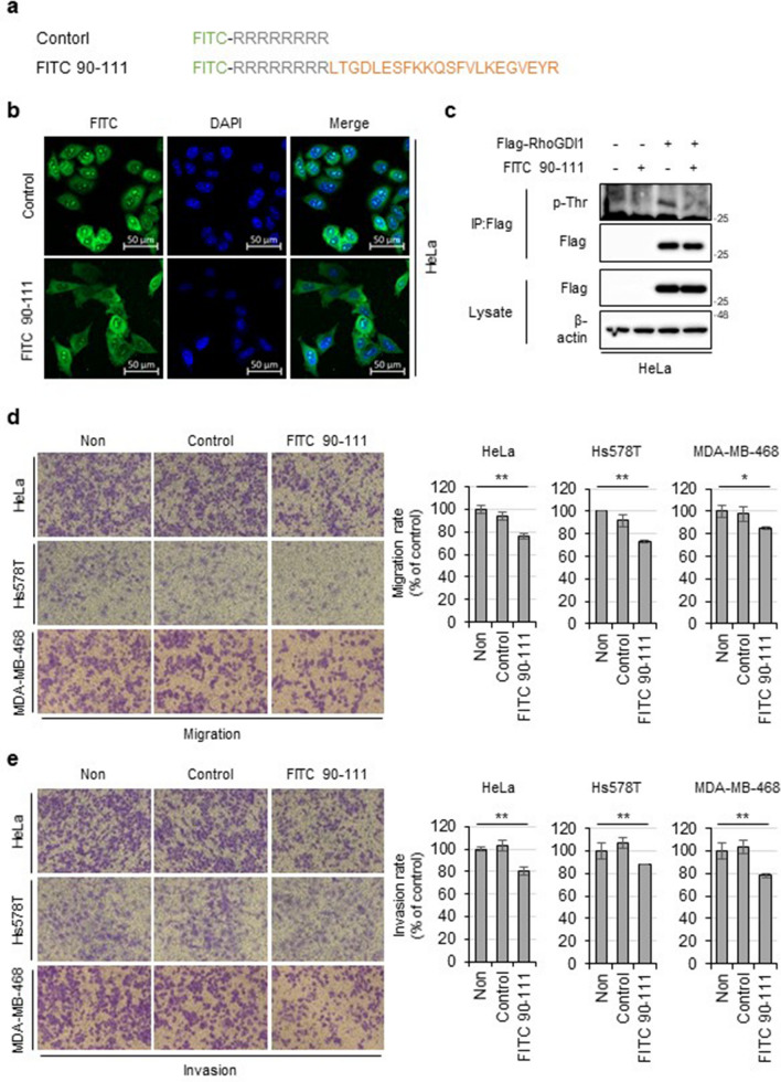
Results: GST pull-down assays and PLA showed that PLK1 directly interacted with RhoGDI1 in vitro and in vivo. Truncation mutagenesis revealed that aa 90-111 of RhoGDI1 are critical for interacting with PLK1. We also showed that PLK1 phosphorylated RhoGDI1 at Thr7 and Thr91, which induces cell motility. Overexpression of the GFP-tagged RhoGDI1 truncated mutant (aa 90-111) inhibited the interaction of PLK1 with RhoGDI1 and attenuated RhoA activation by PLK1. Furthermore, the overexpression of the RhoGDI1 truncated mutant reduced cancer cell migration and invasion in vitro and suppressed lung metastasis in vivo.

Conclusions: Collectively, we demonstrate that the phosphorylation of RhoGDI1 by PLK1 promotes cancer cell migration and invasion through RhoA activation. This study connects the interaction between PLK1 and RhoGDI1 to the promotion of cancer cell behavior associated with malignant progression, thereby providing opportunities for cancer therapeutic interventions.

Keywords: Cancer; Migration; PLK1; RhoA; RhoGDI1.


Conflict of interest statement

The authors declare no potential conflicts of interest.

Figures

Fig. 1
Fig. 1
PLK1 directly binds to RhoGDI1 in vitro and in vivo. a 293 T cells were co-transfected with HA-tagged PLK1 and Flag-tagged RhoGDI1. Cell lysates were immunoprecipitated with HA (left) or Flag antibody (right). Immunoprecipitates and total lysates were immunoblotted with HA and Flag antibodies. b Cell lysates from HeLa were immunoprecipitated with IgG (as control) or PLK1 antibodies, and immunoblotted with PLK1 or RhoGDI1 antibodies. c Proximity ligation assay (PLA) was performed with PLK1 and RhoGDI1 antibodies in HeLa cells. Representative fluorescence images show blue (DAPI) staining nuclei and red dots (PLA signals). Scale bars: 50 μm. d His-tagged PLK1 and GST-tagged RhoGDI1 were expressed in E.coli. GST pull-down assay was performed with His-PLK1 expression lysates and purified GST-RhoGDI1. The GST pull-down products were immunoblotted with PLK1 and GST antibodies
Fig. 2
Fig. 2
Mapping the region of RhoGDI1 for the interaction with PLK1. a Schematic representation of wild-type RhoGDI1 and truncation mutants of RhoGDI1 (1-67, 1-134, 68-204, 68-89, 90-111, 112-134 amino acids). b, c GST pull-down assay was performed with His-PLK1 expression lysates and purified GST-tagged wt RhoGDI1 or truncation mutants. The GST pull-down products were immunoblotted with PLK1 and GST antibodies. d 293 T cells were co-transfected with HA-tagged PLK1 and GFP-tagged RhoGDI1 wt or mutant (aa 90-111). Cell lysates were immunoprecipitated with a HA antibody. Immunoprecipitates and total lysates were immunoblotted with HA and GFP antibodies. e The proximity ligation assay was performed with PLK1 and RhoGDI1 antibodies in HeLa cells transfected with GFP-RhoGDI1 mutant (aa 90-111). Representative images (left) display blue (DAPI) staining nuclei and red dots (PLA signals). Scale bars: 50 μm. Quantitative data plots of PLA (right) show PLA dots per GFP-RhoGDI1 aa 90-111 positive or negative cells. **P < 0.01
Fig. 3
Fig. 3
PLK1 phosphorylates RhoGDI1 at Thr7/91 residue in vitro and in vivo. a Purified GST-RhoGDI1 was subjected to in vitro kinase assay with( +) or without(-) active His-PLK1. The assay products were immunoblotted with the indicated antibodies. b Flag-RhoGDI1 was transfected with or without HA-PLK1 into HeLa cells. Cell lysates were immunoprecipitated with Flag antibody. Immunoprecipitates and total lysates were immunoblotted with the indicated antibodies. c HeLa cells were co-transfected Flag-RhoGDI1 with the control siRNA or two PLK1 siRNAs. Cell lysates were analyzed by Immunoprecipitation and immunoblotting using the indicated antibodies. d Purified His-RhoGDI1 wt and threonine-to-alanine-substituted mutants (T7A, T91A and T7/91A) were subjected to in vitro kinase assay with (+) or without (−) active His-PLK1. The assay products were immunoblotted with the indicated antibodies. e HeLa cells were transfected with Flag-RhoGDI1 wt or T7/91A and then treated with 200 nM of BI6727 (PLK1 inhibitor) for 4 h. Cell lysates were immunoprecipitated with Flag antibody. Immunoprecipitates and total lysates were immunoblotted with the indicated antibodies
Fig. 4
Fig. 4
PLK1 promotes RhoA activation by association with RhoGDI1. a HeLa cells were transfected with mock or HA-PLK1. A pull-down assay was performed using Rhotekin-agarose beads to detect active RhoA and PAK1-agarose beads to detect active Rac1/Cdc42 as described in Materials and Methods b HeLa cells were transfected with control siRNA or two PLK1 siRNAs. Cell lysates were subjected to pull-down assay. c Cells were treated with 200 nM of BI 6727 for 4 h. Cell lysates were subjected to pull-down assay. d HeLa cells were co-transfected with HA-PLK1 along with GFP or GFP-RhoGDI1 aa 90-111. Cell lysates were subjected to pull-down assay (left panel). Relative intensity graphs show mean ± S.D. (n = 3) for GTP-RhoA/total RhoA or GTP-Rac1/total Rac1 (right panel). **P < 0.01
Fig. 5
Fig. 5
Interaction between PLK1 and RhoGDI1 is required for the migration and invasion of cancer cells. a Western blot analysis of PLK1 and RhoGDI1 expression in human cancer cell lines. b Indicated cells were transfected with GFP or GFP-RhoGDI1 aa 90–111. Cell proliferation was assessed by counting the viable cells after trypan blue staining at each day point. c, d Indicated cells were transfected with GFP or GFP-RhoGDI1 aa 90–111. 24 h after transfection, cells were serum-starved overnight and then subjected to migration c and invasion d assay as described in Materials and Methods. Representative images of migrating or invading cells stained with crystal violet were displayed (left). The relative percentages of migrating or invading cells were quantified as described in Materials and Methods (right). Quantitative data display the mean ± SD (n = 3). e 2 × 10.6 HeLa cells that overexpress GFP (control) or GFP-RhoGDI1 aa 90-111 were injected via the tail vein of female BALB/c nude mice (n = 6 per group). Representative image of H&E stained lung tissue from HeLa-GFP (control) or HeLa-GFP-RhoGDI1 aa 90-111 (left). The number of metastatic nodules was counted per lung section and displayed in the scatter plots (right). Quantitative data display the mean ± SD (n = 6). Scale bar = 500 μm. **P < 0.01
Fig. 6
Fig. 6
The synthetic peptide containing RhoGDI1 aa 90-111 inhibits cancer cell migration and invasion. a FITC-labeled synthetic peptide sequence. b Cellular uptake of FITC-labeled peptides by HeLa cells. Scale bar = 50 μm. Data are representative of three independent experiments. c HeLa cells were transfected with Flag-RhoGDI1 and treated with 20 μM peptides for 12 h. The lysates were immunoprecipitated with Flag antibody. Immunoprecipitates and total lysates were immunoblotted with the indicated antibodies. d, e Indicated cells were treated with 20 μM peptides and subjected to migration (d) and Invasion (e) assay. Representative images of migrating or invading cells (left). The relative percentages of migrating or invading cells (right). Quantitative data display the mean ± S.D. (n = 3). *P < 0.05; **P < 0.01
Fig. 7
Fig. 7
The proposed model to illustrate how PLK1 promotes cell migration and invasion

Similar articles

Cited by

References

    1. Bray F, Laversanne M, Weiderpass E, Soerjomataram I. The ever-increasing importance of cancer as a leading cause of premature death worldwide. Cancer. 2021;127(16):3029–30. doi: 10.1002/cncr.33587. - DOI - PubMed
    1. van Zijl F, Krupitza G, Mikulits W. Initial steps of metastasis: cell invasion and endothelial transmigration. Mutat Res. 2011;728(1–2):23–34. doi: 10.1016/j.mrrev.2011.05.002. - DOI - PMC - PubMed
    1. Friedl P, Wolf K. Tumour-cell invasion and migration: diversity and escape mechanisms. Nat Rev Cancer. 2003;3(5):362–74. doi: 10.1038/nrc1075. - DOI - PubMed
    1. Novikov NM, Zolotaryova SY, Gautreau AM, Denisov EV. Mutational drivers of cancer cell migration and invasion. Br J Cancer. 2021;124(1):102–14. doi: 10.1038/s41416-020-01149-0. - DOI - PMC - PubMed
    1. Svensmark JH, Brakebusch C. Rho GTPases in cancer: friend or foe? Oncogene. 2019;38(50):7447–56. doi: 10.1038/s41388-019-0963-7. - DOI - PubMed

LinkOut - more resources