. 2024 Feb 14;15(1):1348.
doi: 10.1038/s41467-024-45555-x.

TGF-β blockade drives a transitional effector phenotype in T cells reversing SIV latency and decreasing SIV reservoirs in vivo

Affiliations

TGF-β blockade drives a transitional effector phenotype in T cells reversing SIV latency and decreasing SIV reservoirs in vivo

Jinhee Kim et al. Nat Commun. .

Erratum in

Abstract

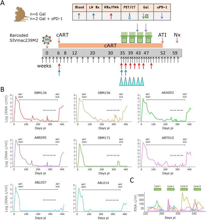
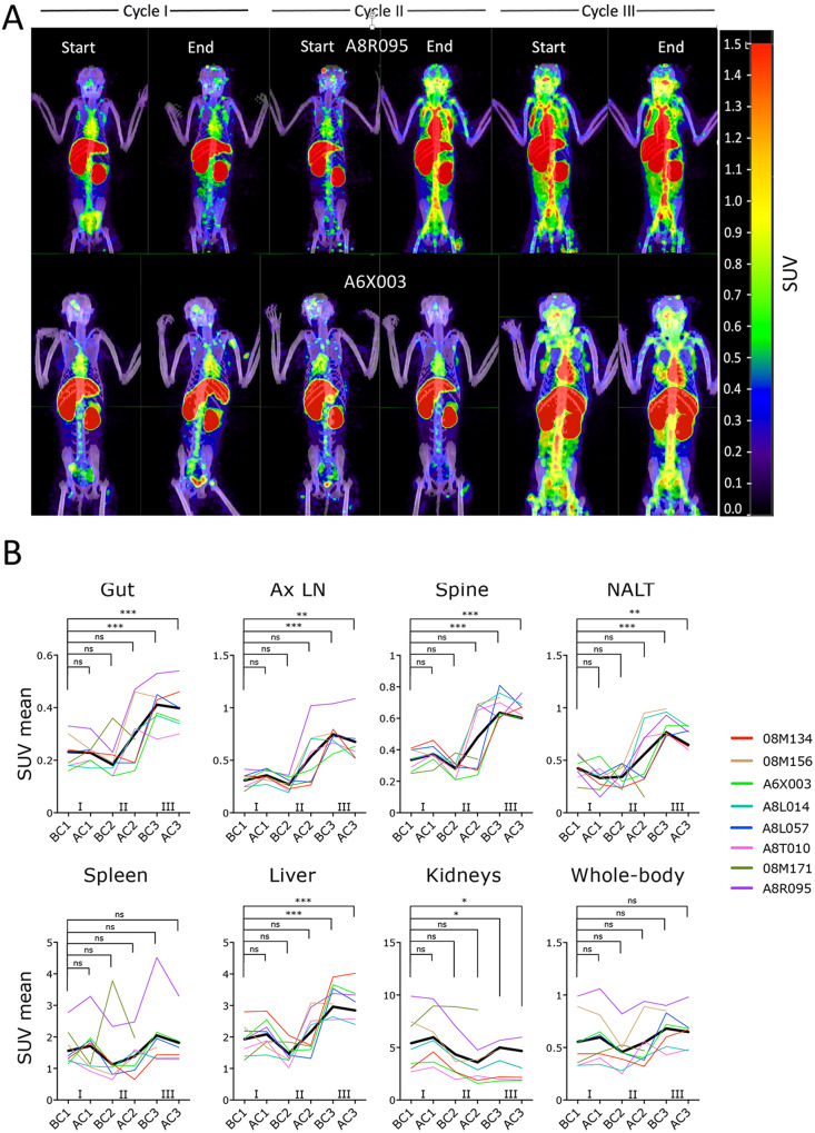
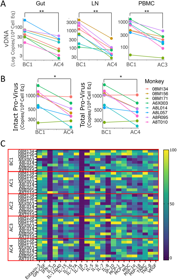
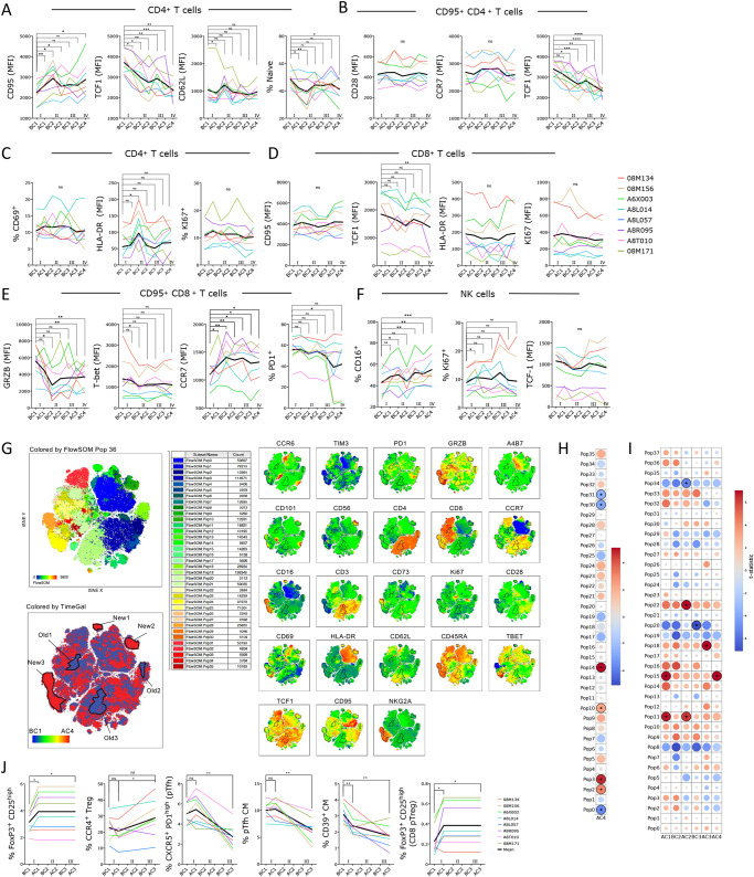
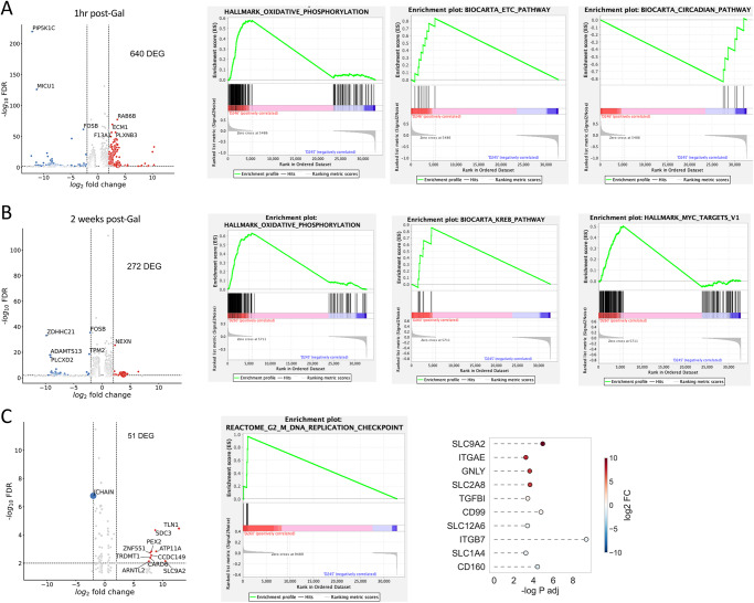
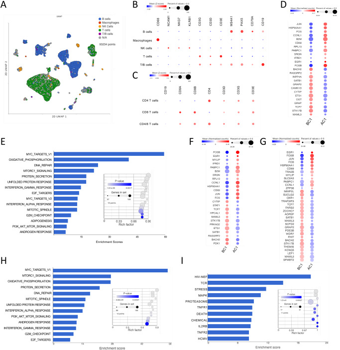
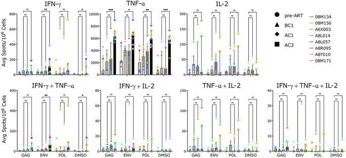
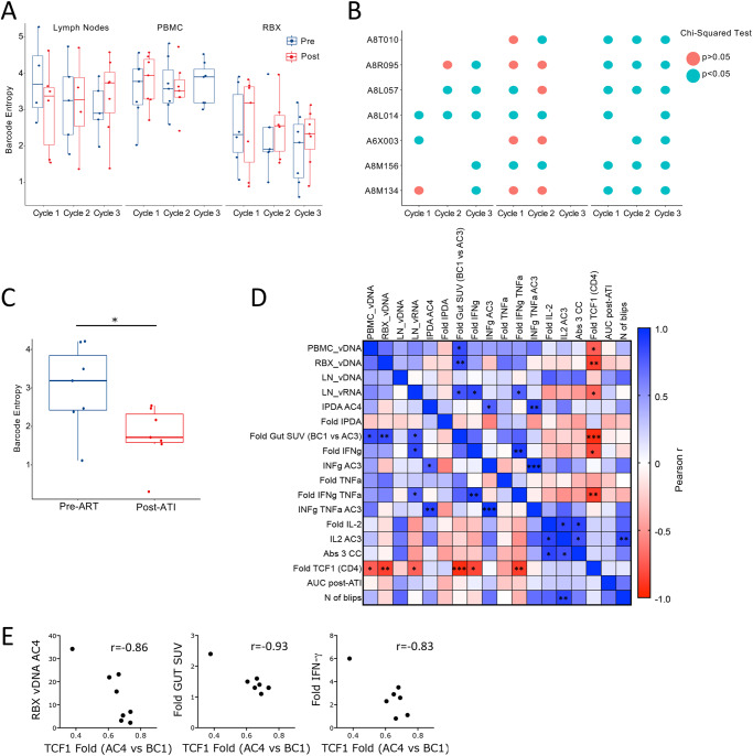
HIV-1 persistence during ART is due to the establishment of long-lived viral reservoirs in resting immune cells. Using an NHP model of barcoded SIVmac239 intravenous infection and therapeutic dosing of anti-TGFBR1 inhibitor galunisertib (LY2157299), we confirm the latency reversal properties of in vivo TGF-β blockade, decrease viral reservoirs and stimulate immune responses. Treatment of eight female, SIV-infected macaques on ART with four 2-weeks cycles of galunisertib leads to viral reactivation as indicated by plasma viral load and immunoPET/CT with a 64Cu-DOTA-F(ab')2-p7D3-probe. Post-galunisertib, lymph nodes, gut and PBMC exhibit lower cell-associated (CA-)SIV DNA and lower intact pro-virus (PBMC). Galunisertib does not lead to systemic increase in inflammatory cytokines. High-dimensional cytometry, bulk, and single-cell (sc)RNAseq reveal a galunisertib-driven shift toward an effector phenotype in T and NK cells characterized by a progressive downregulation in TCF1. In summary, we demonstrate that galunisertib, a clinical stage TGF-β inhibitor, reverses SIV latency and decreases SIV reservoirs by driving T cells toward an effector phenotype, enhancing immune responses in vivo in absence of toxicity.


Conflict of interest statement

The corresponding author’s institution, Northwestern University filed a patent application including all the data from the present manuscript. Application number: 18/515,196, Filing date: November 20, 2023. Inventor, Elena Martinelli. All other authors declare no competing interests.

Figures

Fig. 1
Fig. 1. Four 2-weeks cycles with galunisertib lead to viral reactivation in blood.
A Schematic representation of the study and sampling schedule. B Plasma VL in blood for each macaque throughout the study. The longer black line indicates the period on ART, while the 4 small black lines indicate the start and end of each galunisertib cycle. C Enlarged plasma VL for all macaques during galunisertib therapy. Green bars indicate galunisertib cycles. Source data are provided as a Source Data file. Image from BioRender.
Fig. 2
Fig. 2. Galunisertib leads to viral reactivation in tissues.
A The 64Cu-DOTA-Fab2(7D3) probe was injected ~24 h before PET/CT scan before and at the end of each of the first 3 galunisertib cycles. Representative images from the maximum intensity projections (MIP) of fused PET and CT scans are shown for a macaque with a major increase at the end of cycle 2 and one showing increase at the beginning of cycle 3. MIPs were generated using the MIM software, set to a numerical scale of 0–1.5 SUVbw, and visualized with the Rainbow color scale. B Mean SUV were calculated for each anatomical area, and values were analyzed with mixed-effect analysis. Data from the scans performed at the last 2 time points (BC3 and AC3) in 08M171 were excluded because of technical issues with the probe. Thicker black line represents the mean. P-values were calculated for comparison of each time point with the before cycle 1 time point (BC1; AC1= after cycle 1, BC2= before cycle 2; AC2= after cycle 2; BC3= before cycle 3; AC3= after cycle 3; Holm-Sidak multiple comparison correction; *p ≤ 0.05 **p ≤ 0.01 ***p ≤ 0.01). Source data are provided as a Source Data file.
Fig. 3
Fig. 3. Galunisertib decreases viral reservoir in absence of systemic inflammation.
A Levels of cell-associated (CA)-vDNA per cell equivalent are shown for the time point before cycle 1 (BC1) and at the end of cycle 3 (AC3) or 4 (AC4) for the respective tissues for all 8 macaques. B IPDA data are shown for intact and total provirus for BC1 and AC4 in PBMC for the 8 macaques. P-values are shown for Wilcoxon matched pair signed-rank non-parametric two-tailed test comparing before and after galunisertib data from the 8 macaques (*p ≤ 0.05 **p ≤ 0.01 ***p ≤ 0.01). C Heat map of cytokine concentration in plasma at the indicated time point are shown after Log transformation and normalization. Statistical analysis was run on each factor separately and together (no significant differences after multiple comparison adjustment). Source data are provided as a Source Data file.
Fig. 4
Fig. 4. Galunisertib leads toward effector in T and NK cells, increasing Treg and decreasing Tfh frequencies.
AF Geometric mean fluorescent intensities (MFI) of each marker and frequency of indicated subset within live, singlets CD3+ CD4+ T cells (A and C) or CD3+ CD4+ CD95+ T cells (B) or CD8+ or CD8+ CD95+ T cells or NK cells (NKG2A+ CD8+ CD3 cells) are shown. Thick black line represents the mean. Changes from baseline (beginning of cycle 1, BC1) are shown for graphs with at least 1 significant difference (Repeated measures ANOVA with Holm-Sidak correction for multiple comparisons; *p ≤ 0.05 **p ≤ 0.01 ***p ≤ 0.01). G tSNE of lymphocyte, live, singlets events after normalization for BC1 and AC4 (all 8 macaques) with FlowSOM 36 clusters overlaid on tSNE (top left) or heatmap of each markers MFI (right) or heatmap of time point (blue is BC1 and red is AC4; bottom left) is shown. 6 populations were manually gated on red or blue areas (red, New1-3 and blue Old1-3). H Bubble chart displaying changes in AC4 from BC1 in populations (FlowSOM clusters) characterized by markers MFI in Supplementary Fig. S8A. Color is proportional to the effect size and size to p-value (Wilcoxon sum rank non-parameter two-tailed test). I Bubble chart displaying changes from BC1 at all time points in populations (FlowSOM clusters) characterized in Supplementary Fig. S8C (ANOVA repeated measures with Holm-Sidak multiple comparisons correction; *p ≤ 0.05 **p ≤ 0.01 ***p ≤ 0.01). J Frequency of indicated subset within live, singlets CD3+ CD4+ T or CD3+ CD4+ CD95+ CD28+ T cells (CM = central memory) or within CD3+ CD8+ T cells. Changes from baseline (BC1) are shown (ANOVA repeated measures with Holm-Sidak correction for multiple comparisons; *p ≤ 0.05 **p ≤ 0.01 ***p ≤ 0.01). Source data are provided as a Source Data file.
Fig. 5
Fig. 5. OXPHOS and other metabolic pathways increase rapidly with TGF-β blockade.
Bulk RNAseq was performed with PBMC from before cycle 1 (24 h) and 1hrs after the first dose of galunisertib (A) or after the last dose of cycle 1 (B) and with rectal biopsies collected before cycle 1 (24 h) and after the last dose of cycle 1 (C). The number of differentially expressed genes (DEG) obtained by DESeq2 with an FDR < 0.05 and abs(log2FC)>2 are shown in each respective volcano plot. Enrichment plots are shown after GSEA (with all FDR < 0.05 DEGs) for significantly enriched pathways (top 1 or 2 pathway by ES). C Lollipop graph of selected DEG of interest among significantly different genes (FDR < 0.05). Source data are provided as a Source Data file.
Fig. 6
Fig. 6. scRNAseq of lymph node before and after cycle 1 confirms a switch toward effector and increased metabolism in all immune subsets with galunisertib.
A UMAP projection of 93234 cells from lymph nodes collected right before and at the end of cycle 1 from all 8 macaques (16 samples). Gene-based classification of major immune subset is overlaid on UMAP. In gray are unclassified cells. Bubble plots showing expression (mean normalized counts proportional to the color; size proportional to the percentage of cells) of each marker listed in each cell subset. Marker listed are those used for classification of major immune subsets (B) or CD4+ and CD8+ T cells (C). D Significantly different genes obtained by Hurdle model (FDR < 0.05; log2FC = 0.15) in the T cell subset are shown with color proportional to normalized counts. E Significantly enriched pathways (FDR < 0.01) in T cells DEGs within the hallmark collection. Significantly different genes (FDR < 0.05; log2FC = 0.15) in the CD4+ (F) and CD8+ (G) T cell subset. Significantly enriched pathways (FDR < 0.01) in CD4+ T cells DEGs within the hallmark (H) and biocarta (I) collections. Source data are provided as a Source Data file.
Fig. 7
Fig. 7. Galunisertib increases SIV-specific responses.
Average spots (from triplicates) per 106 PBMC at the time of ART initiation (pre-ART), before cycle 1 (BC1), after cycle 1 (AC1) and at the end of cycle 3 (AC3) with galunisertib after 24hrs ex vivo stimulation with 15-mer peptides (gag, env, pol) or mock (DMSO). Each post-galunisertib time point was compared to BC1 (Mixed effect analysis adjusted for multiple comparisons with Dunnet post-hoc p-values are shown; *p ≤ 0.05 **p ≤ 0.01 ***p ≤ 0.001). Bars represent the median with interquartile range as error bars. Source data are provided as a Source Data file.
Fig. 8
Fig. 8. TCF1 decrease associates with virological and immunological endpoints.
A Barcode diversity measure as Shannon Entropy is shown before and after each of the first 3 galunisertib cycles for LN, PBMC and colorectal biopsies. Box-and-whisker plot represents the median +/− the interquartile range of data from 4 to 8 macaques (all data from macaques with detectable barcodes were included at each time point for a given tissue; no significant differences using linear mixed effects models). Blue= before; Red= end of each cycle. B Bubble plot shows the results of statistical testing (Chi-squared) for differences in frequency distribution of barcodes before compared to after, for each of the first 3 cycles of galunisertib for each macaque in the indicated tissues. Blue indicates significant differences p ≤ 0.05. C Barcode entropy of virus isolated at the time of ART initiation compared to week 6 post-ATI in plasma (Wilcoxon matched pairs two-tailed test; *p ≤ 0.05). D Correlation matrix of several key variables of virological or immunological effect of galunisertib. Color is proportional to Pearson r coefficient. *p ≤ 0.05 **p ≤ 0.01 ***p ≤ 0.001 indicate significant correlations. E Association between fold increase in TCF1 (MFI) from BC1 to AC4 with CA-vDNA levels at AC4, change in gut SUV at AC3 compared to BC1 and fold increase in IFN-γ (AC3 vs BC1). Person r is shown. All correlations have *p ≤ 0.05. Source data are provided as a Source Data file.

Update of

References

    1. Gantner, P. et al. HIV rapidly targets a diverse pool of CD4(+) T cells to establish productive and latent infections. Immunity56, 653–668.e655 (2023). - PMC - PubMed
    1. Whitney, J. B. et al. Rapid seeding of the viral reservoir prior to SIV viraemia in rhesus monkeys. Nature512, 74–77 (2014). - PMC - PubMed
    1. Persaud, D., Zhou, Y., Siliciano, J. M. & Siliciano, R. F. Latency in human immunodeficiency virus type 1 infection: no easy answers. J. Virol.77, 1659–1665 (2003). - PMC - PubMed
    1. Margolis, D. M. & Archin, N. M. Proviral Latency, Persistent Human Immunodeficiency Virus Infection, and the Development of Latency Reversing Agents. J. Infect. Dis.215, S111–S118 (2017). - PMC - PubMed
    1. Wagner, T. A. et al. HIV latency. Proliferation of cells with HIV integrated into cancer genes contributes to persistent infection. Science345, 570–573 (2014). - PMC - PubMed

Substances