. 2024 Feb 14;10(1):35.
doi: 10.1038/s41531-024-00646-w.

Differences in brain aging between sexes in Parkinson's disease

Affiliations

Differences in brain aging between sexes in Parkinson's disease

Iman Beheshti et al. NPJ Parkinsons Dis. .

Abstract

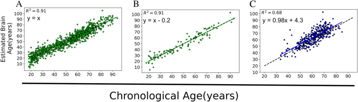
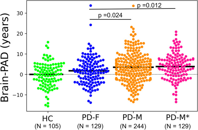
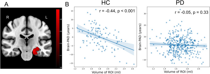
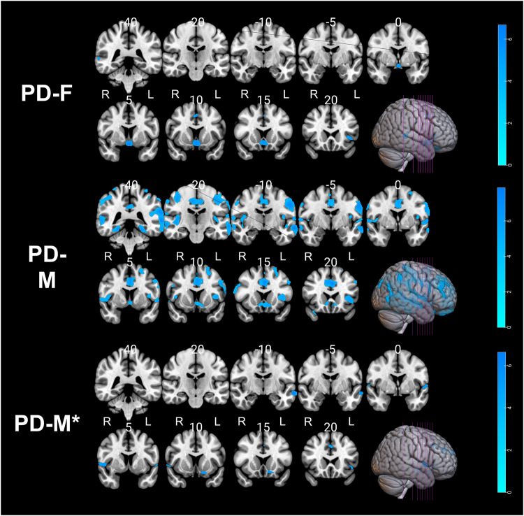
Parkinson's disease (PD) is linked to faster brain aging. Male sex is associated with higher prevalence, severe symptoms, and a faster progression rate in PD. There remains a significant gap in understanding the function of sex in the process of brain aging in PD. The structural T1-weighted MRI-driven brain-predicted age difference (i.e., Brain-PAD: the actual age subtracted from the brain-predicted age) was computed in a group of 373 people with PD (mean age ± SD: 61.37 ± 9.81, age range: 33-85, 34% female) from the Parkinson's Progression Marker Initiative database using a robust brain-age estimation framework that was trained on 949 healthy subjects. Linear regression models were used to investigate the association between Brain-PAD and clinical variables in PD, stratified by sex. Males with Parkinson's disease (PD-M) exhibited a significantly higher mean Brain-PAD than their female counterparts (PD-F) (t(256) = 2.50, p = 0.012). In the propensity score-matched PD-M group (PD-M*), Brain-PAD was found to be associated with a decline in general cognition, a worse degree of sleep behavior disorder, reduced visuospatial acuity, and caudate atrophy. Conversely, no significant links were observed between these factors and Brain-PAD in the PD-F group. Having 'older' looking brains in PD-M than PD-F supports the idea that sex plays a vital function in PD, such that the PD mechanism may be different in males and females. This study has the potential to broaden our understanding of dissimilarities in brain aging between sexes in the context of PD.


Conflict of interest statement

The authors declare no competing interests.

Figures

Fig. 1
Fig. 1. Scatter plots showing the estimated brain age versus actual age in different datasets.
A The training set (N = 949) that was evaluated through a 10-fold cross-validation strategy, B the hold-out HC (N = 105), and C PD (N = 373). The dashed black line represents the identity line (y = x).
Fig. 2
Fig. 2. Box-plots showing the grouped Brain-PAD values among the hold-out HC and PwP with respect to the sex categories.
The solid black line represents the mean Brain-PAD values of each group, while the dashed black line represents the reference line (y = 0). The statistical tests between groups were performed using a student t-test.
Fig. 3
Fig. 3. Negative association between gray and white matter volumes and Brain-PAD scores in the healthy control and PD groups.
The p values were corrected using FWE at a threshold of p < 0.05, and the cluster extent threshold was set to a minimum of 100. Labels "R" and "L" stand for the right and left hemispheres, respectively.
Fig. 4
Fig. 4. Reduced correlation between brain-PAD and regional GM volume in PD.
A The brain region shows a significant difference in regression slopes between the HC and PD groups in GM. B The correlation between GM volumes in the region that was identified to be significant and Brain-PAD within each group. Labels ‘R’ and ‘L’ stand for the right and left hemispheres, respectively. The color bar corresponds to t-test values.
Fig. 5
Fig. 5. Negative association between gray matter volumes and Brain-PAD scores in the PD group within each sex.
The p values were corrected using FWE at a threshold of p < 0.05, and the cluster extent threshold was set to a minimum of 100. Labels "R" and "L" stand for the right and left hemispheres, respectively.

References

    1. Mu J, et al. Parkinson’s disease subtypes identified from cluster analysis of motor and non-motor symptoms. Front. Aging Neurosci. 2017;9:301. doi: 10.3389/fnagi.2017.00301. - DOI - PMC - PubMed
    1. Han JW, et al. Psychiatric manifestation in patients with Parkinson’s disease. J. Korean Med. Sci. 2018;33:e300. doi: 10.3346/jkms.2018.33.e300. - DOI - PMC - PubMed
    1. Georgiev D, Hamberg K, Hariz M, Forsgren L, Hariz GM. Gender differences in Parkinson’s disease: a clinical perspective. Acta Neurol. Scand. 2017;136:570–584. doi: 10.1111/ane.12796. - DOI - PubMed
    1. Elbaz A, et al. Risk tables for Parkinsonism and Parkinson’s disease. J. Clin. Epidemiol. 2002;55:25–31. doi: 10.1016/S0895-4356(01)00425-5. - DOI - PubMed
    1. Cholerton B, et al. Sex differences in progression to mild cognitive impairment and dementia in Parkinson’s disease. Parkinsonism Relat. Disord. 2018;50:29–36. doi: 10.1016/j.parkreldis.2018.02.007. - DOI - PMC - PubMed