. 2024 Feb 14;14(1):3740.
doi: 10.1038/s41598-024-54295-3.

Activin is a neural inducer of a male-specific muscle in Drosophila

Affiliations

Activin is a neural inducer of a male-specific muscle in Drosophila

Ken-Ichi Kimura et al. Sci Rep. .

Abstract

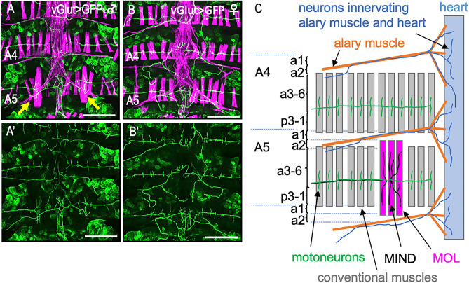
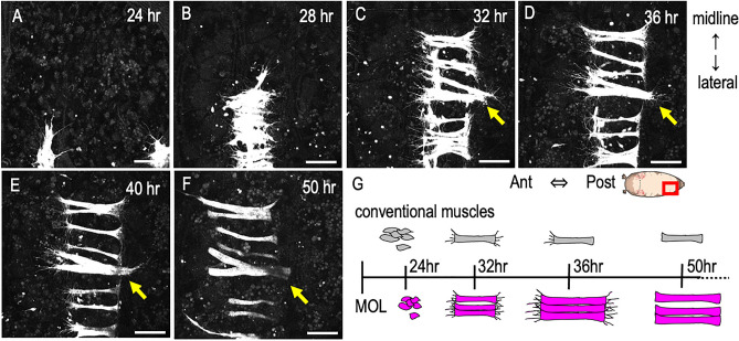
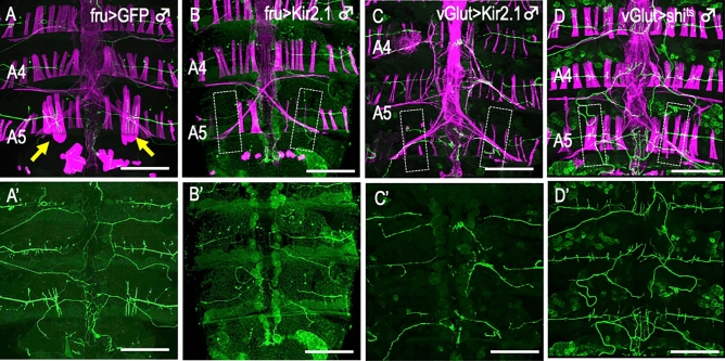
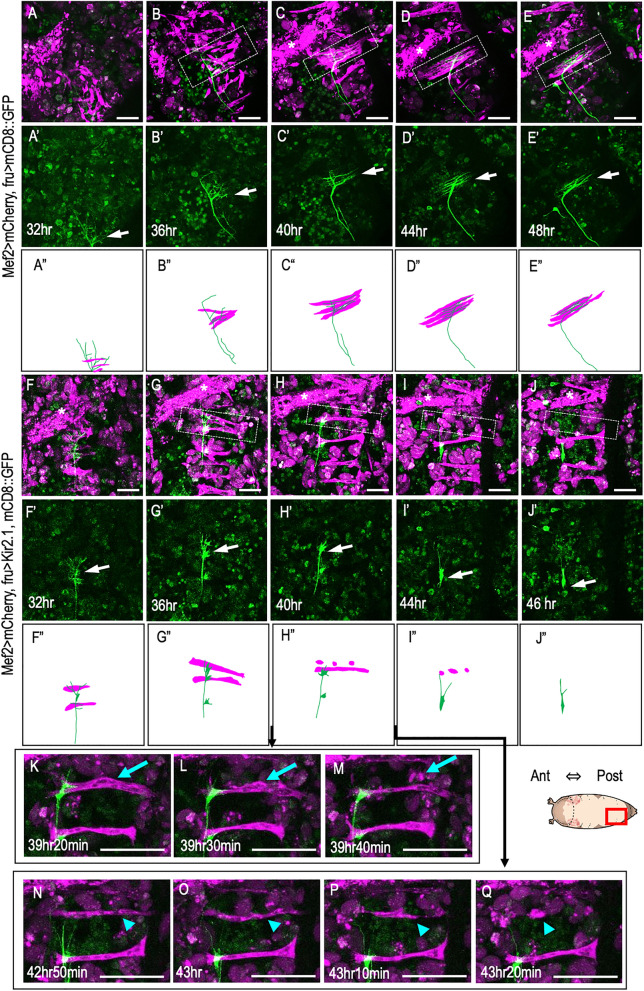
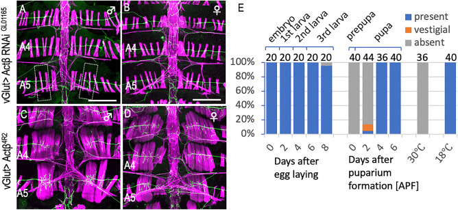
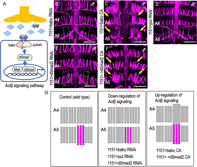
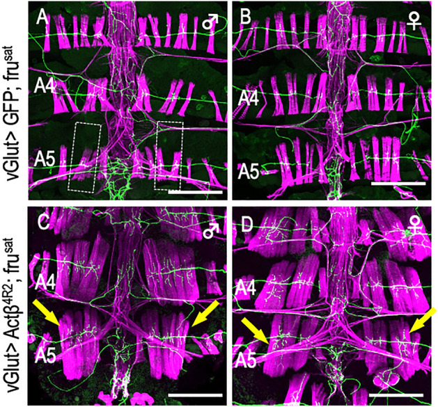
Drosophila melanogaster has a pair of male-specific muscles called the muscle of Lawrence (MOL) in abdominal segment 5 (A5) of adult flies. The MOL is produced only when its innervating motoneuron expresses FruitlessM (FruM) neural masculinizing proteins. We show that MOL induction is hampered by: (1) silencing electrical activities in the motoneuron, (2) blocking vesicular release from the motoneuron, and (3) knocking down Activin ß (Actß) in the motoneuron or knocking down Actß signaling pathway components in the myoblasts. Our timelapse live imaging of the developing neuromuscular system reveals that, upon contact with the presumptive MOL, the motoneuronal axon retracts concomitant with the progression of MOL degeneration resulting from neural silencing. We conclude that MOL formation depends on the bidirectional trophic interactions between pre- and postsynaptic cells, with motoneuron-derived Actß playing an inducing role in MOL formation.


Conflict of interest statement

The authors declare no competing interests.

Figures

Figure 1
Figure 1
The male-specific muscle MOL and its innervation by the Mind neuron. (AB’) Dorsal musculatures (A and B; magenta) and innervating nerves (A-B’; green) of adult male (A, A’) or female (B, B’) abdominal segments 4 (A4) and 5 (A5). (C) A schematic drawing of the nerve and muscle layout is shown for the A4 and A5 hemi-segments. Cuticular subdomains (a1 to p3-1) are also indicated. The MOLs are indicated by yellow arrows in (A). The MOLs varied in appearance around their attachment sites from individual to individual, particularly when observed with high contrast imaging: some appeared to be split into a few bundles. The MOLs are indicated by yellow arrows. Muscles were stained with phalloidin and nerves were labeled with GFP. Note that the phalloidin-labeled fibers which run across the segments are heart muscles, the phalloidin-labeled elements that irradiate diagonal thin fibers are of alary muscles, and fat bodies are occasionally labeled with GFP. The genotype of the flies is y hs-flp; vGlutOK371-GAL4 UAS-mCD8::GFP. Scale bar: 200 µm.
Figure 2
Figure 2
Dynamics of the developing MOL. (AF) Snapshots of myogenesis in A5 captured every 4 h between 24 and 50 h apf. The midline is on the upper side, while the lateral edge is on the lower side. Proliferated myoblasts move toward the midline and fuse with each other to form an elongated myotube (A,B). The myotube actively protrudes filopodia-like structures while extending toward an apodeme (C). Conventional muscles extend to the apodeme at their posterior end, where they cease protruding filopodia-like structures (D). Subsequently, the anterior end extends to the apodeme (E). The MOL continues to grow, attaining its large size (F). (G) Schematic of the steps through which conventional muscles and the MOL form. The genotype of flies is 1151-GAL4/Y; UAS-mCD8::GFP. Scale bar: 50 µm.
Figure 3
Figure 3
Lack of neural activity or synaptic transmission results in the loss of the MOL. (AD’) Transgenic expression of Kir2.1 to silence neural activity (BC’) or shits to block synaptic transmission (D,D’) impaired the MOL formation, while the MOLs were intact when GFP was expressed (A, yellow arrows). The boundaries of anticipated position of the MOL or of presumptive MOL substitutes are delineated by broken lines in (BD). To silence neural activity with Kir2.1, Tub-GAL80ts was inactivated after the white pupa stage by a temperature increase from 25 °C to 30 °C (C), and synaptic transmission was blocked with shits by a temperature increase from 25 °C to 30 °C at 24 h apf and thereafter (D). The severity of the effect of shits expression varied depending on the shits fly line used. In this study, a shits line with the strongest effect was used. To drive UAS transgenes, fru-GAL4 (A,B) or vGlut-GAL4 (C,D) was used. Innervating motoneurons were labeled with GFP and muscles were visualized with phalloidin (magenta). The genotypes of the flies are y hs-flp;UAS-mCD8::GFP; fru-GAL4/TM6B (A,A’) and y hs-flp; UAS-mCD8::GFP/ Mef2-LexA, 13xLexAop2-6xmCherry-HA; fru-GAL4/UAS-Kir2.1 (B,B’), y hs-flp; vGlutOK371-GAL4 UAS-mCD8::GFP/G13 Tub-GAL80ts; UAS-Kir2.1-GFP/ + (C,C’) and y hs-flp; vGlutOK371-GAL4 UAS-mCD8::GFP/ + ; UAS- × 20 shits/ + (D, D’). Scale bar: 200 µm.
Figure 4
Figure 4
Neural activity silencing causes Mind axon retraction and MOL degeneration. (AE”) Normal formation of the Mind neuron‒MOL synapse. The axon tip (magenta) together with myoblasts (green) migrate toward the midline (the upper side) by 36 h apf, when fine processes of the growth cone extend along the MOL (boxed with broken lines), establishing synapses on it 40–48 h apf. The growth cone of the Mind neuron is indicated by white arrows. Asterisks indicate larval muscles. Images composed of muscles and nerves (AE), nerve only (A’E’) and schematic drawings of the above images (A”E”) are shown. (FJ”) Loss of the synapse upon neural silencing with Kir2.1. By 36 h apf, the Mind neuron axon extends processes on the MOL even in the absence of neural activity. Subsequently, the Mind axon tip loses fine processes at 36 h apf and is then withdrawn back to the soma at 40–46 h apf. The nerve and muscle patterns are schematically shown in (F”J”). (KQ) Enlarged images of the contact point of the Mind axon (green) with the MOL (magenta), captured every 10 min between 39 h 20 min and 43 h 20 min apf. Myotubes undergoing fragmentation are indicated by blue arrows and arrowheads. The genotypes of flies are y hs-flp; Mef2-LexA, 13xLexAop2-6xmCherry-HA/ UAS-mCD8::GFP; fru-GAL4/ + for (AE’) and y hs-flp; UAS-mCD8-GFP/ Mef2-LexA, 13xLexAop2-6xmCherry-HA; fru-GALl4/UAS-Kir2.1-GFP for (FQ). Scale bar: 50 µm.
Figure 5
Figure 5
Effects of knockdown and overexpression of Actß on MOL formation. (A,B) Knockdown of Actß in motoneurons impaired the MOL formation in the male A5 segment (A) without affecting the female musculatures (B). (C,D) Overexpression of Actß induces enlargement of the MOL in male A5 and the MOL-like muscles ectopically in other abdominal segments in both sexes. (E) The proportion of flies exhibiting MOLs (ordinate) is shown against time (hours after egg-laying and hours after puparium formation [apf] as indicated below the bars on the abscissa). This time period involves a temperature shift from 18 to 30 °C, followed by a reversion from 30 to 18 °C. The two rightmost bars represent the values obtained from flies that experienced no temperature changes; i.e., consistently maintained at either 30 °C or 18 °C. The numerals above the bars indicate the numbers of hemisegments examined. The genotypes of files are vGlutOK371 > Actß RNAiGL01165 (y hs-flp; vGlutOK371-GAL4 UAS-mCD8::GFP/ + ; UAS-Actß RNAiGL01165/ +) for (A,B), vGlutOK371 > Actß4R2 (y hs-flp; vGlutOK371-GAL4 UASmCD8::GFP/ + ; UAS-Actß4R2/ +) for (C,D), y hs-flp; vGlutOK371-GAL4/ + , UAS-mCD8-GFP/ + ; Tub-GAL80ts/UAS-Actß RNAiGL01165 for (E). Scale bar: 200 µm.
Figure 6
Figure 6
Involvement of Actß signal transduction in MOL formation. (A) Schematic of Actß signaling. (BF) Knockdown of either of babo (B) or put (D), the genes for two Actß receptor components, or of the gene for dSmad2 (E), a downstream transcription factor results in the loss of the MOL, whereas overexpression of their constitutively active form (C,F) promotes the formation of the MOL, accompanied by ectopic induction of the MOL-like muscles in abdominal segments other than A5 in males. The MOL and MOL-like muscles are indicated by yellow arrows. Schematic drawings of the muscle structure under three different conditions are shown in (G). The genotypes of the flies are 1151-GAL4/Y; UAS-mCD8::GFP/UAS-babo RNAi10E2/6E2 (B), 1151-GAL4/Y; UAS-mCD8::GFP/ + ; UAS-babo CA/ + (C), 1151-GAL4/Y; UAS-mCD8::GFP /UAS-put RNAi7904R-3 (D), 1151-GAL4/Y; UAS-mCD8::GFP/ + ; UAS-sMad2 RNAiJF02320/ + (E) and 1151-GAL4/Y; UAS-mCD8::GFP/ + ; UAS-sMad2SDVD (3)/ + (F). Scale bar: 200 µm.
Figure 7
Figure 7
Overexpression of Actß bypasses the need of fru in MOL induction. Motoneuronal overexpression of Actß in a frusat homozygous male (A) and female (B) that otherwise lacks the MOL induced the MOL ((C) and (D); yellow arrows). The genotypes of flies were y hs-flp; vGlutOK371-Gal4 UAS-mCD8::GFP/CyO; frusat/ frusat for (A, B) and y hs-flp; vGlutOK371-Gal4 UAS-mCD8::GFP/ UAS-Actß4R2; frusat/ frusat for (C,D). Scale bar: 200 µm.

Similar articles

References

    1. Shah NM, Sanes JR. Sexual differentiation of the nervous system (Chapter 51) In: Kandel ER, Koester JD, Mack SH, Siegelbaum SA, editors. Principles of Neural Science. 6. McGraw-Hill; 2021. p. 1646.
    1. Gorski RA, Gordon JH, Shryne JE, Southam AM. Evidence for a morphological sex difference within the medial preoptic area of the rat brain. Brain Res. 1978;148:333–346. doi: 10.1016/0006-8993(78)90723-0. - DOI - PubMed
    1. Nottebohm F, Arnold AP. Sexual dimorphism in vocal control areas of the songbird brain. Science. 1976;194:211–213. doi: 10.1126/science.959852. - DOI - PubMed
    1. Arnold AP. Sex chromosome and brain gender. Nat. Rev. Neurosci. 2004;5:701–708. doi: 10.1038/nrn1494. - DOI - PubMed
    1. Yang CF, Shah NM. Representing sex in the brain, one module at a time. Neuron. 2014;82:261–278. doi: 10.1016/j.neuron.2014.03.029. - DOI - PMC - PubMed

LinkOut - more resources