. 2024 Feb 14;7(1):31.
doi: 10.1038/s42004-024-01107-4.

Catalyst-free late-stage functionalization to assemble α-acyloxyenamide electrophiles for selectively profiling conserved lysine residues

Affiliations

Catalyst-free late-stage functionalization to assemble α-acyloxyenamide electrophiles for selectively profiling conserved lysine residues

Yuanyuan Zhao et al. Commun Chem. .

Abstract

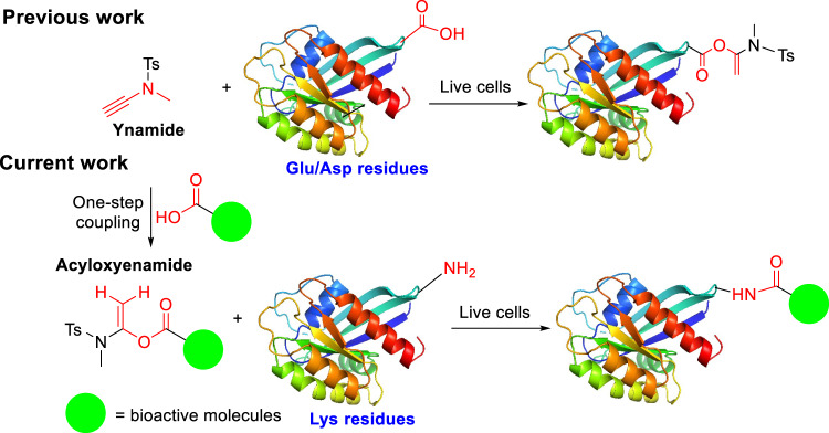
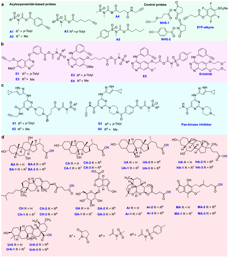
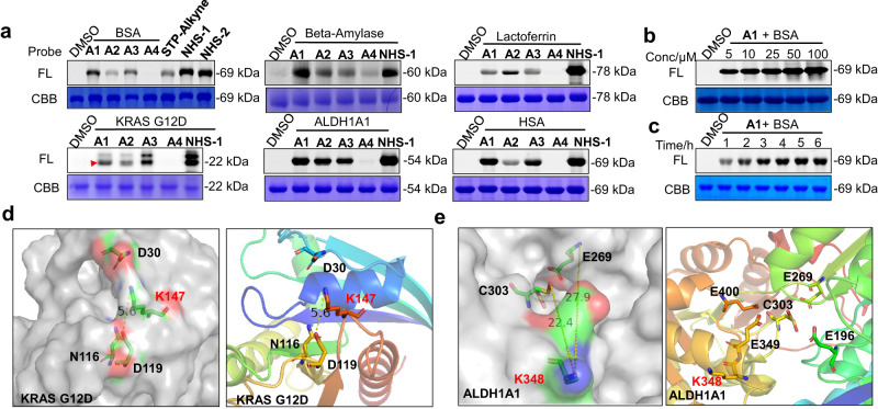
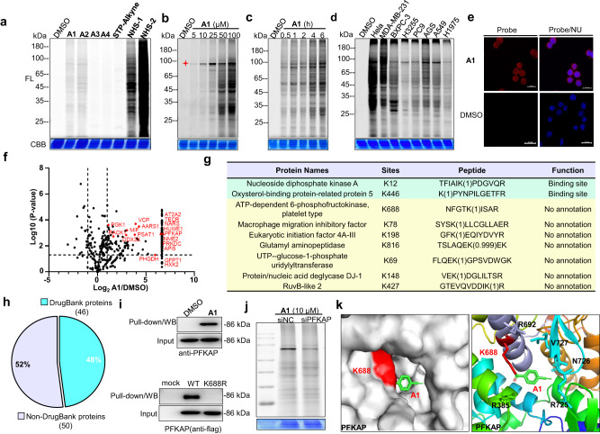
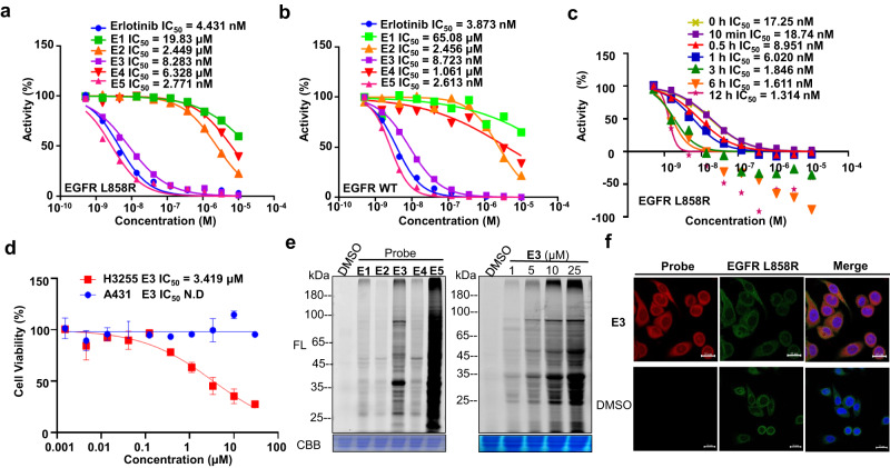
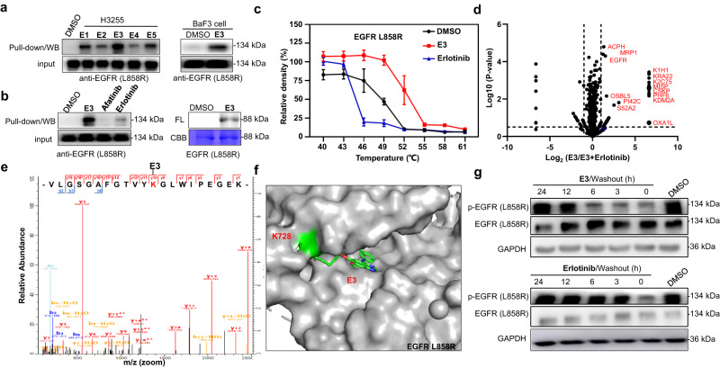
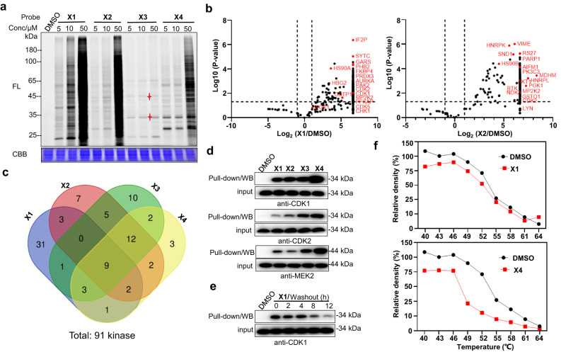
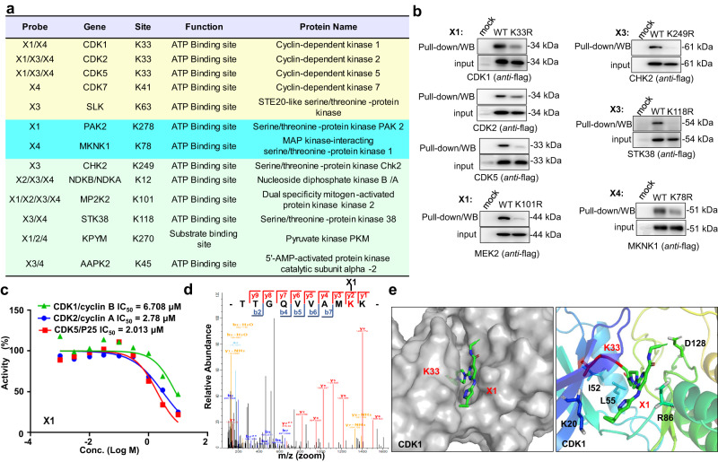
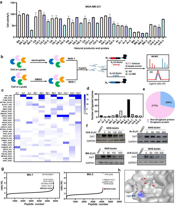
Covalent probes coupled with chemical proteomics represent a powerful method for investigating small molecule and protein interactions. However, the creation of a reactive warhead within various ligands to form covalent probes has been a major obstacle. Herein, we report a convenient and robust process to assemble a unique electrophile, an α-acyloxyenamide, through a one-step late-stage coupling reaction. This procedure demonstrates remarkable tolerance towards other functional groups and facilitates ligand-directed labeling in proteins of interest. The reactive group has been successfully incorporated into a clinical drug targeting the EGFR L858R mutant, erlotinib, and a pan-kinase inhibitor. The resulting probes have been shown to be able to covalently engage a lysine residue proximal to the ATP-binding pocket of the EGFR L858R mutant. A series of active sites, and Mg2+, ATP-binding sites of kinases, such as K33 of CDK1, CDK2, CDK5 were detected. This is the first report of engaging these conserved catalytic lysine residues in kinases with covalent inhibition. Further application of this methodology to natural products has demonstrated its success in profiling ligandable conserved lysine residues in whole proteome. These findings offer insights for the development of new targeted covalent inhibitors (TCIs).


Conflict of interest statement

The authors declare no competing interests.

Figures

Fig. 1
Fig. 1. Proposed mechanism of protein labeling by α-acyloxyenamide intermediate at lysine residues.
α-acyloxyenamide electrophiles covalently bind to lysine residues and selectively label conserved lysine residues in live cells.
Fig. 2
Fig. 2. The chemical structures of probes.
a Aromatic and aliphatic probes A1, A2, A3, A4, and A5 with an α-acyloxyenamide reactive group and the chemical structures of control probes, aliphatic or aromatic probes NHS-1/2, STP-alkyne. b Erlotinib and Erlotinib-based probes E1, E2, E3, E4, and E5 with an α-acyloxyenamide embedded into different positions. c α-acyloxyenamide was embedded into a pan-kinase inhibitor to form the probes X1, X2, X3, and X4 and the parent inhibitor. d Structure of natural products and probes by incorporation with NHS and α-acyloxyenamide for comparison. BA cholic acid, CA chenodeoxycholic acid, UA ursodeoxycholic acid, HA hyodeoxycholic acid, CH cholesteryl hemisuccinate, GA geniposidic acid, Ar artesunate, MA mycophenolic acid, UrA ursolic acid. The synthesis was presented in supplementary scheme S1–S41 and the NMR spectra of the probes were in Supplementary data 2.
Fig. 3
Fig. 3. In vitro proteome labeling and modification sites analysis.
a Labeling profiles of A1, A2, A3, A4, STP-alkyne, NHS-1, NHS-2 (50 μM) with BSA. And labeling profiles of A1, A2, A3, A4, NHS-1/-2 (50 μM) with β-Amylase, Lactoferrin, KRAS G12D, ALDH1A1, HSA. FL = in-gel fluorescence scanning. CBB = Coomassie gel. b Concentration- and c Time-dependent labeling profiles of A1 (50 μM) with BSA. d Crystal structure of the K147 site in Kras G12D (PDB code: 4LPK). e K348 in ALDH1A1(PDB code: 5L2M).
Fig. 4
Fig. 4. In situ protein labeling and modification site analysis.
a Proteome reactivity profiles of A1, A2, A3, A4, STP-alkyne and NHS-1/2 (100 μM) with MDA-MB-231 live cells. FL = in-gel fluorescence scanning. CBB = Coomassie gel. b Concentration- (4 h incubation) and c Time-dependent labeling profiles of A1 (100 μM) with MDA-MB-231 live cells. d Proteome reactivity profiles of A1 (100 μM) with different cancer cells. e Cellular imaging of A1 (50 μM) in MDA-MB-231 live cells. NU = nucleus, scale bar = 20 μm, DMSO-treated samples were used as controls. f Protein hits identified by pull-down/LC−MS with A1 (10 μM), DMSO-treated sample was used as a negative control. g Subset of residues identified by A1 (100 μM) with MDA-MB-231 live cells. h Among the proteins with lysine modification by A1, the fraction found in DrugBank. i Target validation by pull-down/WB with A1 (10 μM), the western blots used PFKAP antibodies (upper gel). Pull-down/WB of wild type and K688R PFKAP with A1 (10 μM), the western blots were confirmed by using flag antibodies (lower gel). j Labeling of target protein PFKAP by probe A1 after gene knockdown. k Docking experiments to predict the binding mode of A1 with PFKAP at K688 (PDB code: 4WL0).
Fig. 5
Fig. 5. The activity of erlotinib-based probes.
a Inhibitory activity of Erlotinib, E1, E2, E3, E4 and E5 against EGFR L858R mutants. b Inhibitory activity of Erlotinib, E1, E2, E3, E4 and E5 against EGFR WT. c Time-dependent inhibition assay of E3 against EGFR L858R. d Cellular inhibition assay of E3 against H3255 cancer cells and A431 cancer cells. e Proteome reactivity profiles of E1, E2, E3, E4, and E5 (10 μM) with H3255 cells (left gel). Concentration-dependent labeling profiles of E3 (2 h incubation) with H3255 cells (right gel). FL = in-gel fluorescence scanning. CBB = Coomassie gel. f Cellular imaging of E3 (10 μM) and immunofluorescence (IF) in H3255 live cells. scale bar = 20 μm, DMSO-treated samples were used as controls.
Fig. 6
Fig. 6. E3 probe covalently binds to EGFR L858R.
a Pull-down/WB validation of EGFR L858R in H3255 cells with E1, E2, E3, E4 and E5 (10 μM) (left); Pull-down/WB validation of EGFR L858R in BaF3 cells with E3 (10 μM) (right). EGFR antibodies were used in western blots. b Competitive labeling of endogenous EGFR (L858R) with H3255 live cells in the presence of afatinib or erlotinib (5×) as a competitor (left). Labeling of EGFR L858R protein (0.1 µg/µL) with E3 (10 μM) (right). FL = in-gel fluorescence scanning. CBB = Coomassie gel. c Cellular thermal shift assay performed in H3255 cells with E3 (10 μM), and EGFR L858R kinase is the target protein. d Mass spectrometry-based profiling of E3/(E3+1×Erlotinib) binding proteins (10 μM probe concentration) from H3255 cells. e Mass spectrum of binding site of EGFR L858R at K728 modified by E3, identified modification site by MS2 spectra was highlighted (red). f Docking experiment to predict the binding mode of E3 with EGFR L858R (PDB code: 4LQM). g H3255 cells were treated with E3 or Erlotinib (10 μM) for 2 h, followed by compound wash-out for the indicated time. Effect on the phosphorylation level of EGFR was tested.
Fig. 7
Fig. 7. Proteome reactivity profiles of α-acyloxyenamide-based pan-kinase inhibitor.
a Concentration-dependent labeling profiles of X1, X2, X3, and X4 (10 μM) with K562 cells. FL = in-gel fluorescence scanning. CBB = Coomassie gel. b MS-spectrometry-based profiling of X1 and X2 binding proteins (10 μM), DMSO-treated sample was used as a negative control, the data of X3 and X4 were presented in Fig. S22 (Supporting information). c Venn diagram explains the coincidence and difference of X1, X2, X3, and X4 kinase targets. d Target validation by pull-down, WB with X1, X2, X3, and X4 and the corresponding antibodies. e K562 cells were treated with X1 (10 μM, 2 h), followed by compound washout for the indicated times. Pull-down/WB validation with a CDK1 antibody. f Cellular thermal shift assay performed in K562 cells with X1 and X4 (10 μM), and CDK1 kinase is the target protein.
Fig. 8
Fig. 8. Modification site analysis of X1, X2, X3, and X4 in whole proteome.
a Subset of residues identified by X1, X2, X3, or X4 (100 μM) with K562 cell lysates. b Pull-down/WB of wild type and mutant proteins with X1, X3, and X4 (10 μM); The western blots were confirmed by using flag antibodies. c Inhibitory activity of X1 against CDK1, CDK2, CDK5. d Mass spectrum of modification site of CDK1 at K33 by X1, identified modification site by MS2 spectra was highlighted (red). e Docking experiment to predict the binding mode of X1 with CDK1 (PDB code: 4Y72).
Fig. 9
Fig. 9. Evaluation of the performance of α-acyloxyenamide- and NHS-based probes derived from natural products.
a Antiproliferation activity was tested against MDA-MB-231 cells with natural products and electrophilic probes. b General process to identify the binding sites of natural product-based probes in live cells or cell lysates by using light and heavy reagents (iso-TOP) and LC-MS/MS, DMSO- (heavy, or red) versus fragment-treated (light, or bule) samples. R value ≥4 was used to define a fragment binding toward a quantified lysine. c Heat map presents the liganded targets by natural product-based probes bearing various electrophilic warheads, and RH/L ≥4 sites are considered to be targets of natural product ligands (Data processing is to take the mean value of two or more groups of R, and CV ≤ 40 was used as a cutoff value). d Plot comparison of the number of lysine residues for each probe. e Drugbank database analysis of protein hits identified by the different electrophilic probes (29% are druggable proteins and 71% are undruggable proteins). f Competitive labeling of OAT target by pull-down/WB experiment with NHS-biotin probe (100 µM) in the presence of Ar-2, MA-2, UrA-2, UrA-3. g Protein hits and binding sites of MA-1/3 identified from MDA-MB-231 breast cancer cells by iso-TOP experiments, red-colored IMPDH2 is a known target of mycophenolic acid, K438 is the binding site by both covalent probes MA-1 and MA-3. MYH9 K856, TPR K364 are identified unknown targets. h Docking experiments to predict the binding mode of MA-3 with IMPDH2 at K438 (PDB code: 6U8E). All error bars represent standard deviation (n = 3), there is no statistical significance between the samples (p > 0.1).

Similar articles

Cited by

References

    1. Lagoutte R, Patouret R, Winssinger N. Covalent inhibitors: an opportunity for rational target selectivity. Curr. Opin. Chem. Biol. 2017;39:54–63. doi: 10.1016/j.cbpa.2017.05.008. - DOI - PubMed
    1. Péczka N, Orgován Z, Ábrányi-Balogh P, Keserű GM. Electrophilic warheads in covalent drug discovery: an overview. Expert Opin. Drug Discov. 2022;17:413–422. doi: 10.1080/17460441.2022.2034783. - DOI - PubMed
    1. Abdeldayem A, Raouf YS, Constantinescu SN, Moriggl R, Gunning PT. Advances in covalent kinase inhibitors. Chem. Soc. Rev. 2020;49:2617–2687. doi: 10.1039/C9CS00720B. - DOI - PubMed
    1. Lu X, Smaill JB, Patterson AV, Ding K. Discovery of cysteine-targeting covalent protein kinase inhibitors. J. Med. Chem. 2022;65:58–83. doi: 10.1021/acs.jmedchem.1c01719. - DOI - PubMed
    1. Chaikuad A, Koch P, Laufer SA, Knapp S. The cysteinome of protein pinases as a target in drug development. Angew. Chem. Int. Ed. 2018;57:4372. doi: 10.1002/anie.201707875. - DOI - PubMed